
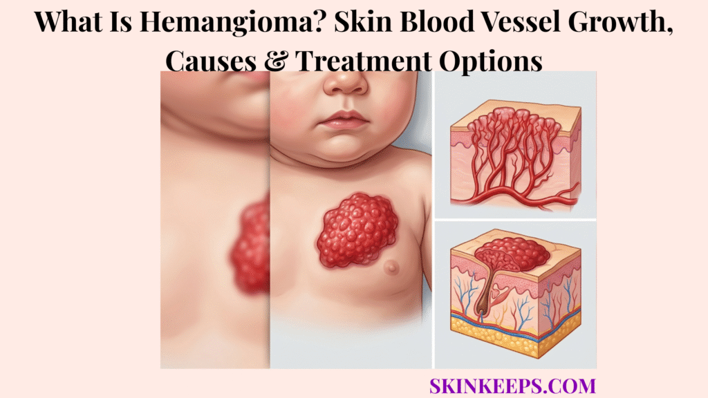
A hemangioma is a benign growth formed by abnormal proliferation of blood-vessel cells. This page focuses mainly on infantile hemangioma, also called infantile haemangioma, strawberry hemangioma, strawberry birthmark or strawberry nevus.
Infantile hemangioma is usually absent or faint at birth, becomes noticeable during the first weeks of life, grows rapidly during early infancy and later involutes. Superficial lesions look bright red, deep lesions may look blue-purple or skin-coloured, and high-risk locations can require treatment before natural shrinkage begins.
Which Skin Blood-Vessel Growth Does “Hemangioma” Usually Describe?
In baby skin care, “hemangioma” usually refers to infantile hemangioma, a true benign vascular tumour.
Modern vascular-anomaly terminology separates vascular tumours from vascular malformations. Infantile hemangioma, congenital hemangioma, pyogenic granuloma, cherry angioma, port-wine birthmark and venous malformation are not interchangeable labels.
| Lesion | Birth Timing | Postnatal Growth | Involution | Typical Context |
|---|---|---|---|---|
| Infantile hemangioma | Usually absent or faint | Rapid early growth | Usually gradual | Infancy |
| Congenital hemangioma | Fully formed | No classic IH growth | Rapid, partial or none | Birth |
| Port-wine birthmark | Present | Proportional growth | No spontaneous involution | Birth onward |
| Pyogenic granuloma | Usually later | Rapid reactive growth | No IH cycle | Any age |
| Cherry angioma | Later life common | Slow/local | Persistent | Adults |
How Can You Recognize an Infantile Hemangioma on the Skin?
Infantile hemangioma can appear as a red raised surface mark, a blue-purple swelling under the skin, or a mixed lesion with both components.
Early clues include a faint pale, pink or red patch, visible small vessels, a bruise-like area, subtle swelling or a mark that enlarges rapidly during early infancy.
Superficial lesions are bright red, soft or rubbery and often raised or lobulated. Deep lesions create smooth blue, purple or skin-coloured swelling and can be larger beneath the skin than the visible mark suggests.
Mixed hemangiomas combine surface redness with deeper bulk. Colour alone does not measure depth or complication risk.
| Type | Visible Clue | Depth | Risk Note |
|---|---|---|---|
| Superficial | Bright red raised lesion | Upper skin | Risk depends on site and growth |
| Deep | Blue-purple or skin-coloured swelling | Under-skin bulk | Full extent may be hidden |
| Mixed | Red surface plus deeper swelling | Both levels | May have greater tissue volume |
Figure 1. Infantile hemangiomas may be superficial, deep or mixed; colour alone does not measure risk, because location, hidden depth, growth pattern and complications determine referral urgency.
Where Can Infantile Hemangiomas Develop?
Infantile hemangiomas can develop on the face, scalp, trunk, limbs, diaper area, or less visibly inside the mouth, airway, parotid gland or liver.
Common skin locations include the scalp, forehead, eyelids, nose, lips, cheeks, ears, neck, chest, back, arms, legs and perineal region.
Location can matter more than diameter. A small eyelid, nasal-tip, lip, ear-canal or airway lesion can be higher risk than a larger low-risk trunk lesion.
| Location | Main Concern | Direction |
|---|---|---|
| Eyelid or orbit | Vision development | Early eye and hemangioma review |
| Nose or columella | Obstruction and distortion | Early specialist review |
| Lip or mouth | Feeding and ulceration | Early treatment assessment |
| Beard area | Possible airway involvement | Breathing and ENT assessment |
| Ear canal | Hearing obstruction | ENT or specialist review |
| Diaper or perineal area | Ulceration and infection | Early skin-integrity care |
| Liver | Heart and thyroid complications | Imaging when indicated |
How Does an Infantile Hemangioma Change From Birth Through Childhood?
What Happens During the Early Hemangioma Growth Phase?
An infantile hemangioma may appear during the first weeks of life and enlarge rapidly, with the fastest growth commonly concentrated in the first few months.
Deep and segmental lesions may continue enlarging longer. Waiting several months on a high-risk lesion can allow visual, airway, feeding or structural injury before treatment begins.
Potentially problematic lesions should be referred early, ideally by about one month of age.
What Happens During the Hemangioma Plateau Phase?
During the plateau phase, rapid enlargement slows and the lesion may remain relatively stable.
Stability does not remove risk when the lesion already blocks vision, distorts a structure or has ulcerated.
What Happens During Hemangioma Involution?
During involution, the lesion gradually softens, fades and loses volume over several years.
Bright red may become dull red, grey or pale, and the centre may fade before the edges.
Which Skin Changes Can Remain After Hemangioma Involution?
Loose skin, fibrofatty tissue, visible vessels, colour change, texture irregularity, ulcer scars or distortion of the nose, lip, ear or eyelid may remain.
Natural involution does not guarantee restoration of normal skin or anatomy.
Figure 2. Infantile hemangioma usually progresses from a faint precursor through rapid early proliferation, plateau and years-long involution; focal, segmental and multifocal patterns carry different complication and screening implications.
What Causes an Infantile Hemangioma to Develop?
The exact initiating cause of infantile hemangioma is not fully understood.
Endothelial and related vascular cells proliferate abnormally during infancy. Local oxygen signalling, angiogenic pathways and developmental vascular factors may contribute.
Infantile hemangioma is not caused by infection, poor hygiene, childbirth injury, pregnancy diet or something a parent did. It is not contagious and is usually sporadic.
| Supported Biology | Not a Cause |
|---|---|
| Abnormal vascular-cell proliferation | Poor hygiene |
| Developmental vascular growth pathways | Infection |
| Angiogenic and oxygen signalling | Childbirth injury |
| Local tissue signalling | Pregnancy diet or parental behaviour |
| Usually sporadic occurrence | Contagious contact |
Which Babies Are More Likely to Develop Infantile Hemangiomas?
Infantile hemangiomas are reported more often in girls, premature or low-birth-weight babies, multiple pregnancies and some placental or pregnancy contexts.
A family history may be present, but most lesions are sporadic. Risk factors do not predict whether a particular lesion will threaten function or require treatment.
After a lesion appears, its location, depth, growth speed, pattern and complications matter more than demographic associations.
| Factor | Association | Cannot Predict |
|---|---|---|
| Female sex | Higher reported frequency | Complication risk |
| Prematurity | Higher reported frequency | Treatment need |
| Low birth weight | Higher reported frequency | Severity |
| Multiple pregnancy | Association reported | Outcome |
| Family history | Sometimes present | Recurrence certainty |
How Do Focal, Segmental and Multifocal Hemangiomas Differ?
What Is a Focal Infantile Hemangioma?
A focal infantile hemangioma is a localized round, oval or discrete lesion arising from one main point.
Many focal lesions have lower syndrome risk, but location, size and complications still determine management.
What Is a Segmental Infantile Hemangioma?
A segmental infantile hemangioma is a broad plaque-like lesion following a developmental territory.
Segmental lesions carry greater risk of ulceration, functional impairment, disfigurement and PHACE or lower-body structural associations.
What Does Multifocal Infantile Hemangioma Mean?
Multifocal infantile hemangioma means several separate skin lesions are present.
The visible lesions should be counted. Five or more cutaneous infantile hemangiomas can prompt consideration of liver ultrasound because of possible hepatic involvement.
Which Infantile Hemangiomas Are Considered High Risk?
Infantile hemangiomas are high risk when they may threaten life, function, skin integrity, permanent appearance or signal underlying structural abnormalities.
- Eyelid or periocular lesion.
- Nasal tip, columella, lip, mouth, ear or ear canal lesion.
- Lower-face or beard-distribution lesion.
- Neck, diaper, perineal or genital lesion.
- Large segmental facial or scalp lesion.
- Lumbosacral or perineal segmental lesion.
- Airway or liver involvement.
- Rapid early growth or large hidden deep component.
- Surface whitening, ulceration or bleeding.
- Interference with vision, breathing, feeding or hearing.
- Five or more separate skin lesions.
| Pattern | Threat | Possible Complication | Urgency |
|---|---|---|---|
| Eyelid/periocular | Vision | Amblyopia or astigmatism | Early specialist |
| Lip/mouth | Feeding | Ulceration and poor intake | Early specialist |
| Beard/lower face | Airway | Stridor or obstruction | Prompt assessment |
| Diaper/perineal | Skin integrity | Ulcer and infection | Early specialist |
| Segmental face/scalp | Syndromic risk | PHACE | Specialist workup |
| Lumbosacral/perineal segment | Structural risk | LUMBAR/PELVIS/SACRAL | Targeted imaging |
| Five or more lesions | Internal disease | Liver, cardiac or thyroid issues | Screening consideration |
How Can an Eyelid Hemangioma Affect Vision?
An eyelid or orbital hemangioma can block or distort visual input while the infant’s visual system is developing.
It may cover the visual axis, change corneal shape and cause astigmatism, ptosis, eye displacement, strabismus or amblyopia. Deep orbital extension can rarely compress the optic nerve.
A baby may not show obvious visual difficulty while permanent visual development is being affected, so periocular lesions need early ophthalmology and hemangioma assessment.
Vision pathway: eyelid or orbital growth → blocked or distorted input → amblyopia → possible permanent visual deficit.
How Can a Facial or Beard-Area Hemangioma Affect the Airway?
A segmental hemangioma across the chin, lower lip, jaw or front of the neck can be associated with airway hemangioma.
Visible beard-distribution lesions do not prove airway involvement, but they lower the threshold for pediatric and ENT assessment.
- Noisy breathing.
- Stridor.
- Persistent hoarse cry.
- Breathing difficulty that worsens with crying or respiratory illness.
- Feeding difficulty linked to breathing.
Urgent: breathing difficulty, stridor, blue lips or respiratory distress needs emergency assessment rather than routine follow-up.
How Can a Lip or Mouth Hemangioma Affect Feeding?
A lip or mouth hemangioma can distort the feeding seal, cause pain, ulcerate or bleed in a moist high-friction area.
Poor sucking, reduced intake and inadequate weight gain can follow. Persistent structural distortion may later affect dental development or speech.
Lip hemangiomas often warrant early treatment because rapid growth can alter anatomy and ulceration can be extremely painful.
- Lip-shape distortion.
- Difficulty forming a seal.
- Pain during sucking.
- Ulceration or bleeding.
- Reduced intake.
- Poor weight gain.
Why Do Some Infantile Hemangiomas Ulcerate?
Some infantile hemangiomas ulcerate when rapid growth, friction, moisture or pressure breaks down the skin surface.
Ulceration is more likely during the proliferative phase and at the lip, neck, folds, nasal tip and diaper or perineal region.
A pale or white surface change may precede opening. Ulcers can cause severe pain, bleeding, infection and permanent scarring.
Ulceration pathway: rapid growth or friction → surface whitening → skin breakdown → pain, bleeding and scarring.
How Should an Ulcerated Hemangioma Be Cared For?
An ulcerated hemangioma needs prompt assessment because surface wound care and treatment of the active tumour may both be required.
- Clean gently.
- Use a non-adherent dressing.
- Apply clinician-approved barrier ointment.
- Provide appropriate pain control.
- Reduce friction, moisture and pressure.
- Assess for bacterial infection.
- Treat the underlying growing hemangioma when indicated.
- Monitor feeding and hydration for lip or mouth lesions.
Do not scrub, pierce, cauterize or apply harsh antiseptics to an open vascular lesion.
When Can Multiple Hemangiomas Signal Internal Involvement?
Five or more separate infantile hemangiomas on the skin can signal possible liver involvement.
Hepatic hemangiomas may be focal, multifocal or diffuse. Extensive disease can increase cardiac workload and contribute to consumptive hypothyroidism.
Possible signs include abdominal enlargement, rapid breathing, sweating during feeding, poor intake, poor weight gain or other heart-failure signs.
Liver ultrasound is commonly considered at the five-lesion threshold, while cardiac and thyroid assessment depends on hepatic findings and symptoms.
Screening route: count lesions → five or more → liver ultrasound consideration → cardiac and thyroid assessment if hepatic disease is found.
When Is a Large Facial Hemangioma Associated With PHACE Syndrome?
A large segmental facial or scalp infantile hemangioma can be associated with PHACE syndrome.
- P: posterior-fossa brain abnormalities.
- H: large segmental hemangioma.
- A: arterial abnormalities.
- C: cardiac defects or coarctation of the aorta.
- E: eye abnormalities.
Assessment may include brain and arterial MRI/MRA, cardiac examination and echocardiography, ophthalmology and a vascular-anomalies team.
A small focal facial lesion does not automatically indicate PHACE.
When Is a Lower-Body Hemangioma Associated With LUMBAR or PELVIS Features?
A large segmental hemangioma over the lower back, pelvis, perineum or genital region can be associated with underlying structural abnormalities.
Possible concerns include spinal dysraphism, tethered cord, urogenital or renal abnormalities, anorectal malformations, abnormal arteries and bone or limb differences.
Large plaque-like distribution, buttock or perineal involvement, skin pits or tags, asymmetry, bladder, bowel or lower-limb symptoms increase the value of targeted imaging.
Not every small focal lower-body hemangioma requires structural imaging.
How Is an Infantile Hemangioma Different From a Congenital Hemangioma?
Infantile hemangioma usually appears and grows after birth, while congenital hemangioma is fully developed at birth.
| Feature | Infantile Hemangioma | Congenital Hemangioma |
|---|---|---|
| Birth appearance | Absent or subtle | Fully formed |
| Growth | Rapid after birth | Main growth before birth |
| Involution | Gradual after proliferation | Rapid, partial or none |
| Subtypes | Focal, segmental, multifocal | RICH, PICH, NICH |
| Propranolol response | Often responds | Generally not the same response |
| Histology | Typically GLUT1-positive | Different GLUT1 pattern |
How Do RICH, PICH and NICH Congenital Hemangiomas Differ?
What Is a Rapidly Involuting Congenital Hemangioma?
A rapidly involuting congenital hemangioma is fully developed at birth and begins shrinking soon afterwards.
It may flatten substantially during infancy but can leave indentation, loose skin or visible vessels.
What Is a Partially Involuting Congenital Hemangioma?
A partially involuting congenital hemangioma shrinks initially but stops before disappearing.
A persistent vascular component may require later surgical or cosmetic assessment.
What Is a Non-Involuting Congenital Hemangioma?
A non-involuting congenital hemangioma is fully formed at birth, grows proportionately with the child and does not enter a meaningful shrinking phase.
RICH, PICH and NICH do not generally respond to propranolol in the same way as infantile hemangioma.
How Is a Hemangioma Different From a Port-Wine Birthmark?
Infantile hemangioma is a proliferating vascular tumour, while a port-wine birthmark is a capillary vascular malformation.
| Feature | Infantile Hemangioma | Port-Wine Birthmark |
|---|---|---|
| Category | Vascular tumour | Capillary malformation |
| Onset | Often after birth | Present at birth |
| Texture | May become raised | Usually flat early |
| Growth | Proliferates then involutes | Grows proportionately |
| Spontaneous fading | Often involutes | Does not usually involute |
| Main treatment | Beta-blocker if high risk | Vascular laser when treated |
How Is a Hemangioma Different From Other Red Skin Growths?
How Does a Hemangioma Differ From Nevus Simplex?
Nevus simplex is a flat pink or red capillary mark present at birth that often darkens when a baby cries and commonly fades without a raised proliferative phase.
How Does a Hemangioma Differ From Pyogenic Granuloma?
Pyogenic granuloma is a fragile rapidly growing red papule that bleeds easily but does not follow the infantile hemangioma growth and involution cycle.
How Does an Infantile Hemangioma Differ From a Venous Malformation?
A venous malformation is present from birth, grows proportionately and is often blue, soft and compressible, with swelling during dependency, crying or straining.
How Does a Hemangioma Differ From a Bruise?
A bruise changes colour and fades over days or weeks, while a deep hemangioma persists and may enlarge according to its biological growth phase.
| Lesion | Onset | Growth Pattern | Bleeding | Pathway |
|---|---|---|---|---|
| Infantile hemangioma | First weeks | Proliferates then involutes | If ulcerated | Observe or treat by risk |
| Nevus simplex | Birth | Often fades | Not typical | Usually observe |
| Pyogenic granuloma | Later | Rapid reactive growth | Common | Removal and pathology |
| Venous malformation | Birth | Proportional growth | Variable | Vascular-anomalies care |
| Bruise | After trauma | Fades over days or weeks | No tumour bleeding | Assess if unexplained |
How Do Clinicians Diagnose an Infantile Hemangioma?
Clinicians usually diagnose infantile hemangioma from appearance, age of onset and growth history.
- Age when the mark appeared.
- Whether it was fully formed at birth.
- Rate and direction of growth.
- Colour, surface and depth.
- Compressibility and warmth.
- Focal, segmental or multifocal distribution.
- Number of lesions.
- Functional effects.
- Ulceration or bleeding.
- Serial photographs and measurements.
Routine imaging or biopsy is unnecessary for a typical low-risk lesion.
When Is Ultrasound or MRI Needed for a Hemangioma?
Ultrasound or MRI is used when imaging answers a specific question about diagnosis, depth, internal involvement, syndromic risk or treatment planning.
| Clinical Concern | Ultrasound | MRI/MRA | Purpose |
|---|---|---|---|
| Uncertain or deep lesion | Initial depth and flow | If anatomy is complex | Define extent |
| Five or more lesions | Liver screening | If hepatic anatomy is complex | Internal involvement |
| Segmental facial lesion | Limited role | MRI/MRA | PHACE assessment |
| Lumbosacral/perineal segment | Initial structural assessment | MRI when needed | Spine and pelvis |
| Airway, orbit or parotid concern | Sometimes | Often useful | Map deeper tissue |
| Surgical planning | Sometimes | Yes | Define anatomy |
Imaging is not routinely required for every small superficial lesion.
When Is a Hemangioma Biopsy or Genetic Test Needed?
Biopsy or genetic testing is not routine for an ordinary isolated infantile hemangioma, but it may help when the diagnosis remains uncertain.
- Atypical growth pattern.
- A fully formed birth lesion that does not behave as expected.
- Concern for a malignant, borderline or locally aggressive vascular tumour.
- Unusual firmness, ulceration or rapid change.
- Imaging that does not establish the diagnosis.
Infantile hemangiomas are typically GLUT1-positive, while congenital hemangiomas have a different pattern. Selected atypical tumours or syndromic malformations may require specialist molecular testing.
Does Every Infantile Hemangioma Require Treatment?
Most small, low-risk infantile hemangiomas do not require active medical treatment.
Treatment is considered when a lesion threatens vision, breathing, feeding, hearing, skin integrity, heart function, permanent appearance or associated structures.
The statement “it will shrink” is not enough when the early growth phase can cause irreversible damage.
| Situation | Direction | Reason |
|---|---|---|
| Small low-risk stable lesion | Active observation | Low complication risk |
| Rapid early growth | Early specialist review | Short treatment window |
| Eye, lip, nose or ear lesion | Refer early | Function and appearance risk |
| Beard-area lesion | Airway assessment threshold | Possible airway association |
| Ulcerated lesion | Treat | Pain, infection and scarring |
| Five or more lesions | Screening consideration | Possible liver involvement |
| Uncertain diagnosis | Specialist assessment | Avoid wrong classification |
How Should a Low-Risk Hemangioma Be Monitored?
Low-risk hemangioma monitoring should be active, documented and more frequent during early growth.
- Confirm the diagnosis.
- Record location, dimensions, colour and depth.
- Take standardised photographs from the same angle.
- Review more frequently during early proliferation.
- Ask about feeding, breathing, vision, hearing, pain and sleep.
- Watch for whitening, crusting, ulceration or bleeding.
- Reassess if growth accelerates.
- Continue through involution to evaluate residual skin change.
| Date | Size | Thickness | Colour | Surface Integrity | Functional Symptoms |
|---|---|---|---|---|---|
| Baseline | |||||
| Follow-up |
How Does Oral Propranolol Treat Infantile Hemangiomas?
Oral propranolol is the first-line systemic treatment for infantile hemangiomas that require medical therapy.
This beta-blocker narrows vessels within the lesion and reduces signals that support vascular growth. Colour may fade, the lesion may soften and growth may stop or reverse.
Treatment works best during active proliferation and often continues for months, but dose and duration must be determined by the treating specialist.
Effect pathway: active vascular growth → beta-blockade → reduced flow and growth signalling → softening and involution.
Which Safety Checks Are Needed During Propranolol Treatment?
Propranolol requires safety screening and feeding precautions because it can affect heart rate, blood pressure, breathing and blood glucose.
- Medical and cardiac history.
- Heart rate and blood pressure.
- Cardiac examination.
- Wheezing or bronchospasm history.
- Feeding pattern and fasting risk.
- Blood-glucose risk.
- Weight review as the infant grows.
- Illness and vomiting safety plan.
Important risks include hypoglycaemia, bradycardia, hypotension, bronchospasm, sleep disturbance and cool hands or feet.
The medicine is generally given during or just after feeding. The prescribed plan should explain withholding doses during vomiting or poor intake and when urgent assessment is needed.
Emergency: extreme sleepiness, poor responsiveness, seizure or collapse during beta-blocker treatment may indicate severe hypoglycaemia.
When Can Topical Timolol Treat a Hemangioma?
Topical timolol may be considered for selected small, thin, superficial infantile hemangiomas that do not threaten major function.
It is not an adequate substitute for systemic assessment of large, deep, rapidly growing, ulcerated, mucosal or function-threatening lesions.
Systemic absorption is possible, especially through ulcerated skin or mucosa, so application must follow clinician direction.
| Lesion | Timolol Fit? | Better Direction |
|---|---|---|
| Small thin superficial lesion | Possible | Clinician-directed topical care |
| Deep lesion | No | Systemic or specialist assessment |
| Large lesion | No | Early specialist review |
| Ulcerated lesion | High caution | Wound and systemic assessment |
| Eye, mouth or mucosa | High caution | Specialist plan |
| Function-threatening lesion | No | Early systemic evaluation |
When Are Corticosteroids Used for Infantile Hemangioma?
Corticosteroids are no longer routine first-choice treatment but may be considered when propranolol is contraindicated, not tolerated or ineffective.
Systemic steroids can cause growth suppression, immune suppression, hypertension, elevated glucose, mood or sleep effects and adrenal suppression.
Intralesional corticosteroids may be used for carefully selected focal bulky lesions or critical locations under specialist care.
When Can Laser Treatment Help a Hemangioma?
Laser treatment may help selected superficial or residual vascular changes, but it is not a universal substitute for propranolol.
| Target | Possible Benefit | Main Limitation |
|---|---|---|
| Residual surface redness | May help | Several sessions may be needed |
| Telangiectasia | May help | Residual-change role |
| Very thin superficial lesion | Selected use | Limited depth |
| Ulcer pain or healing | Selected support | Specialist decision |
| Deep hemangioma | Limited | Systemic pathway usually more relevant |
| Darker skin tones | Possible with caution | Pigment alteration risk |
Laser should not delay systemic treatment for a rapidly growing high-risk lesion.
When Is Surgery Used for a Hemangioma?
Surgery is used selectively when diagnosis, function, persistent ulceration, residual deformity or a non-involuting congenital lesion justifies an operation.
- Diagnostic uncertainty.
- Localized lesion suitable for safe removal.
- Failure or contraindication of medical therapy.
- Immediate functional correction.
- Persistent ulceration in selected cases.
- Residual fibrofatty tissue or loose skin.
- Nose, lip, eyelid or ear distortion.
- Scar revision.
- Persistent PICH or NICH.
Early surgery can produce blood loss and scarring, while delayed surgery may allow natural shrinkage. Timing is individualized.
Which Hemangioma Treatment Mistakes Should Be Avoided?
Treatment mistakes can cause bleeding, scarring, delayed referral or beta-blocker harm.
| Mistake | Why It Fails | Potential Harm | Safer Response |
|---|---|---|---|
| Delayed high-risk referral | Misses early growth window | Vision, feeding or appearance damage | Refer early |
| Caustic home products | Wrong lesion type | Burns and scars | Medical assessment |
| Piercing, squeezing or cutting | Vascular tissue can bleed | Infection and scarring | Do not manipulate |
| Scrubbing an ulcer | Damages the wound | Pain and infection | Gentle wound care |
| Tight dressings | Friction and pressure | Ulcer worsening | Non-adherent protection |
| Abrupt propranolol stop | Rebound or safety issue | Growth return | Follow specialist taper |
| Dose during poor intake | Hypoglycaemia risk | Collapse or seizure | Follow illness plan |
| Timolol on large ulcer | Absorption risk | Systemic effects | Clinician direction |
| Laser delay | Wrong priority | Functional damage | Systemic or specialist care |
| Misreading bleeding as malignancy | Misreads ulcer or trauma | Panic or wrong care | Pressure and assessment |
How Long Does an Infantile Hemangioma Take to Shrink?
Infantile hemangioma shrinkage usually happens slowly after the early growth and plateau phases.
Involution occurs over years rather than days. Deep lesions can shrink more slowly, and visible change may continue into school age.
Residual loose skin, fibrofatty tissue, vessels, colour or texture change can remain after vascular tissue shrinks.
No fixed birthday guarantees complete disappearance.
Can a Hemangioma Return After Treatment?
A hemangioma can show rebound growth after propranolol is stopped, especially if treatment ends while proliferation remains active.
Renewed redness, warmth, thickness or enlargement may occur. Deep or segmental lesions may need longer monitoring and some children require treatment adjustment.
Rebound does not mean the lesion has become cancerous. Medication tapering and duration must follow the specialist plan.
Rebound route: treatment response → medication withdrawal → growth monitoring → specialist adjustment if meaningful regrowth occurs.
What Complications Can an Infantile Hemangioma Cause?
Most infantile hemangiomas do not cause severe complications, but high-risk lesions can affect skin integrity, function, appearance or internal organs.
| Location or Pattern | Mechanism | Consequence | Specialist |
|---|---|---|---|
| Eyelid or orbit | Blocks or distorts vision | Amblyopia | Ophthalmology and dermatology |
| Beard or airway | Possible deep extension | Stridor or obstruction | ENT and paediatrics |
| Lip or mouth | Friction and distortion | Feeding difficulty or ulcer | Dermatology and feeding support |
| Diaper or perineal | Moisture and friction | Ulcer and infection | Dermatology and wound care |
| Five or more lesions | Possible hepatic disease | Heart strain or hypothyroidism | Paediatrics and imaging |
| Segmental face | PHACE association | Brain, artery, heart or eye abnormality | Vascular-anomalies team |
| Lumbosacral segment | Structural association | Spine or pelvic abnormality | Imaging and specialists |
Other complications include pain, bleeding, scarring, hearing impairment, nasal obstruction, permanent facial distortion and psychosocial effects.
When Should a Hemangioma Be Checked Urgently?
A hemangioma should be checked early when it is rapidly growing, ulcerating, near a high-risk structure, segmental, multiple or diagnostically uncertain.
- Lesion near the eye, nose, lip, mouth, ear or ear canal.
- Beard-distribution lesion.
- Large segmental facial or scalp lesion.
- Lumbosacral or perineal segmental lesion.
- Five or more skin lesions.
- Rapid early growth.
- Whitening, ulceration, pain or bleeding.
- Uncertain diagnosis.
Urgent or emergency care: breathing difficulty, stridor, blue lips, inability to feed, markedly reduced intake or urine, visual-axis obstruction, sudden major swelling, uncontrolled bleeding despite firm pressure, fever with pus or spreading redness, extreme sleepiness, poor responsiveness, seizure, collapse, or heart-failure signs.
Figure 3. Diagnosis begins with timing, depth, pattern and risk; low-risk lesions need documented monitoring, while high-risk lesions require early specialist-directed treatment and medication safety planning.
What Should You Remember About Hemangiomas?
Hemangioma is a vascular-tumour term, not a label for every red birthmark.
- This page focuses mainly on infantile hemangioma affecting the skin.
- Infantile hemangioma is a benign vascular tumour.
- It usually appears shortly after birth, grows rapidly and later involutes.
- Superficial lesions look bright red; deep lesions may look blue or skin-coloured.
- Most small low-risk lesions can be actively monitored.
- Location and growth behaviour determine risk more reliably than colour alone.
- Eye, lip, nose, airway, segmental and ulcerated lesions need early attention.
- Five or more lesions may prompt liver screening.
- Oral propranolol is first-line systemic therapy for high-risk infantile hemangioma.
- Topical timolol is limited to selected thin superficial lesions.
- Congenital hemangiomas are fully formed at birth and behave differently.
- Involution may leave scars, loose skin or structural distortion.
What Questions Do People Ask About Hemangiomas?
Is a hemangioma a tumour or a birthmark?
Infantile hemangioma is a benign vascular tumour. It may look like a birthmark, but it behaves differently from vascular malformations such as port-wine birthmarks.
Is an infantile hemangioma usually present at birth?
It is usually absent or faint at birth and becomes noticeable during the first weeks of life, unlike congenital hemangioma, which is fully formed at birth.
Why does a hemangioma grow rapidly during early infancy?
Vascular cells proliferate during the early growth phase. Early assessment protects function and anatomy during this growth window.
What is the difference between superficial and deep hemangiomas?
Superficial hemangiomas are bright red and raised, while deep hemangiomas create blue-purple or skin-coloured swelling under the skin. Mixed hemangiomas have both features.
Is a hemangioma cancerous?
Infantile hemangioma is benign and not cancer. Atypical vascular lesions may need imaging, biopsy or specialist review when the diagnosis is uncertain.
Is a hemangioma contagious?
No. It does not spread through touch, towels, clothing, breastfeeding or caregiving.
Does every infantile hemangioma eventually disappear?
Most shrink gradually, but not every lesion disappears completely. Loose skin, fibrofatty tissue, visible vessels, colour or texture change, scarring or structural distortion may remain.
Can a hemangioma leave a permanent scar or loose skin?
Yes. Residual change is more likely when the lesion is large, thick, segmental, ulcerated or distorts the nose, lip, ear or eyelid.
Why do some hemangiomas ulcerate?
Rapid growth, friction, moisture and pressure can break down the surface. Ulceration may cause pain, bleeding, infection and scarring.
Can a hemangioma affect vision or breathing?
Yes. Eyelid or orbital lesions can affect visual development, while lower-face or beard-distribution lesions may be associated with airway involvement.
Why are five or more skin hemangiomas important?
Five or more separate skin hemangiomas can raise concern for liver hemangiomas, so clinicians may consider liver ultrasound and further assessment if hepatic involvement is found.
What is the difference between infantile and congenital hemangiomas?
Infantile hemangioma usually appears and grows after birth. Congenital hemangioma is fully formed at birth and may rapidly involute, partially involute or not involute.
How is a hemangioma different from a port-wine birthmark?
Infantile hemangioma is a vascular tumour that proliferates then involutes. A port-wine birthmark is a capillary malformation present at birth that grows proportionately and does not usually involute.
When does a hemangioma need propranolol?
Propranolol may be used when an infantile hemangioma threatens vision, breathing, feeding, hearing, skin integrity, appearance or another high-risk outcome. It requires clinician prescribing and monitoring.
Can topical timolol treat every hemangioma?
No. Topical timolol is considered only for selected small, thin, superficial lesions. It is not adequate for large, deep, ulcerated, mucosal or function-threatening hemangiomas.
Can a hemangioma grow again after propranolol is stopped?
Rebound growth can occur, especially when treatment ends while the lesion remains biologically active. Monitoring and dose changes must follow the specialist plan.
When should a baby with a hemangioma see a specialist urgently?
Rapid growth, ulceration, eye, nose, lip, ear or beard-area involvement, large segmental or lumbosacral lesions, five or more lesions, uncertain diagnosis, breathing or feeding problems, visual obstruction, infection, uncontrolled bleeding or severe beta-blocker safety symptoms need prompt assessment.
Which Sources Support This Hemangioma Guidance?
ISSVA — Classification for Vascular Anomalies 2025 — Current distinction between vascular tumours and vascular malformations and modern terminology.
DermNet — Infantile Haemangioma: Definition and Pathogenesis — Definition, superficial, deep and mixed appearance, growth phases, patterns, risk factors, diagnosis and GLUT1 context.
DermNet — Infantile Haemangioma: Complications and Treatment — Ulceration, high-risk locations, eye, airway, lip, ear and liver complications, monitoring and treatment roles.
American Academy of Pediatrics — Clinical Practice Guideline — Early referral, high-risk classification, imaging thresholds, propranolol, timolol, laser and surgery recommendations.
PubMed — AAP Guideline Abstract — Early growth window, referral by about one month and systemic-treatment summary.
Royal Children’s Hospital — Infantile Haemangioma Pre-Referral Guidance — Practical high-risk referral criteria, five-lesion liver concern and topical-treatment boundaries.
Great Ormond Street Hospital — Congenital Haemangioma — RICH, PICH and NICH timing, involution patterns and lack of typical propranolol response.
FDA — HEMANGEOL Prescribing Information — Systemic-therapy indication, feeding and vomiting precautions, hypoglycaemia, heart-rate, blood-pressure and bronchospasm warnings.
This SkinKeeps article is educational and does not diagnose or replace paediatric dermatology, paediatrics, ophthalmology, ENT, vascular-anomalies, imaging, wound, pathology, emergency or medication care. Rapid growth, ulceration, eye, nose, lip, ear, beard, airway, diaper, segmental or multiple lesions, feeding or breathing problems, vision obstruction, infection or uncontrolled bleeding need prompt assessment. Do not cut, squeeze, pierce, freeze, acid-treat or tightly cover a hemangioma, and never start, stop or change propranolol, timolol or corticosteroids without clinician guidance.



