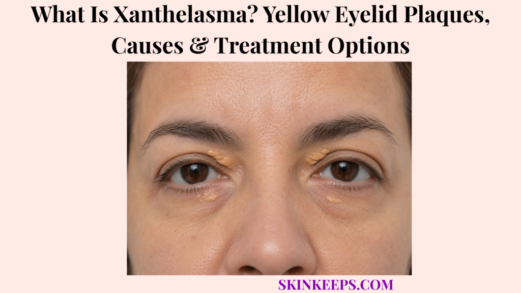
Xanthelasma, or xanthelasma palpebrarum, is a benign eyelid condition in which lipid-filled macrophages collect in superficial skin and form soft yellow or yellow-white plaques, usually near the inner corners of the eyes. These plaques are the most common cutaneous xanthoma and are usually painless, slow-growing and mainly cosmetic.
Abnormal cholesterol or other metabolic disorders can coexist with xanthelasma, but normal routine lipid levels do not exclude the diagnosis and the plaque does not by itself diagnose cardiovascular disease. Typical lesions are usually recognized clinically; removal is optional, recurrence is possible and atypical bleeding, ulcerated or destructive eyelid growths need a separate diagnostic pathway.
Medical note: This article is for educational purposes only. A rapidly growing, painful, ulcerated, bleeding, crusted or lash-destroying eyelid lesion, or one affecting eyelid movement or vision, should be assessed by a qualified healthcare professional.
What Is Xanthelasma and What Do Its Yellow Eyelid Plaques Look Like?
Xanthelasma is a benign accumulation of lipid-filled macrophages in eyelid skin that produces soft, yellow or yellow-white plaques, usually near the inner corners of the eyes.
What Builds Up Inside a Xanthelasma Plaque?
Xanthelasma contains macrophages that have accumulated lipid and become foam cells within superficial eyelid tissue. The core sequence is lipid deposition → macrophage uptake → foam-cell accumulation → visible yellow plaque.
Why Does Xanthelasma Look Yellow?
The yellow color reflects cholesterol-rich lipid material stored inside foam cells in superficial eyelid skin. The visible color can range from yellow-white to yellow-orange, so exact shade is less important than the typical plaque morphology and location.
Is Xanthelasma a Skin Tumor or Cancer?
No; ordinary xanthelasma is a benign lipid deposit rather than a malignant growth, and it has no established malignant transformation pathway. That distinction matters because classic plaques do not require excision for cancer prevention.
Does Xanthelasma Affect Vision?
Typical superficial xanthelasma is mainly a cosmetic condition and generally does not interfere with vision. Very extensive or anatomically unusual eyelid disease can create local functional concerns, but this is not the usual presentation.
What Does a Typical Xanthelasma Plaque Look Like?
A typical plaque is yellow to yellow-white, flat or slightly raised, soft or semisolid, well defined, painless and slowly enlarging. It usually looks more like a plaque than a discrete inflamed lump.
Where Does Xanthelasma Usually Develop?
Xanthelasma most often appears on the medial eyelids—the upper or lower eyelid skin nearest the nose. This nasal-side or medial-canthus distribution is one of the highest-value recognition clues.
Can Xanthelasma Affect Both Eyes?
Yes; xanthelasma frequently develops on both sides and can become relatively symmetrical around the inner eyelids. Multiple lesions are common, although a person can first notice a single plaque.
Does Xanthelasma Hurt or Itch?
Typical xanthelasma is usually painless and asymptomatic, so marked tenderness, inflammation, persistent crusting or ulceration should prompt reconsideration of the diagnosis. Symptoms that suggest tissue destruction do not fit the classic benign pattern.
What Causes Xanthelasma and Is It Always Related to High Cholesterol?
Xanthelasma reflects lipid deposition within eyelid macrophages, but abnormal blood lipids are present in only some patients, so the plaque does not by itself prove high cholesterol or cardiovascular disease.
How Is High Cholesterol Related to Xanthelasma?
Abnormal lipid profiles occur in a meaningful proportion of people with xanthelasma, making cholesterol assessment clinically relevant in many patients. The correct relationship is association rather than diagnosis: xanthelasma can coexist with dyslipidemia, but the plaque alone cannot confirm it.
Can Xanthelasma Occur With Normal Cholesterol?
Yes; clinically typical xanthelasma can occur even when routine blood cholesterol levels are normal. A normal lipid result therefore does not invalidate a classic medial-eyelid plaque diagnosis.
Can Familial Hypercholesterolemia Be Associated With Xanthelasma?
Yes; inherited severe LDL elevation can be associated with xanthomas including xanthelasma, which makes unusually young-onset disease more important to investigate. The plaque itself is not enough to diagnose familial hypercholesterolemia, so age and family history must remain part of the assessment.
Which Other Conditions Can Be Associated With Xanthelasma?
Reported associations include diabetes, thyroid dysfunction, selected cholestatic liver disorders and other lipid abnormalities, but none should be assumed to be present in every patient. These are reasons for targeted clinical consideration rather than universal causes.
Does Xanthelasma Automatically Mean Someone Has Heart Disease?
No; xanthelasma does not diagnose cardiovascular disease. Research on its value as an independent cardiovascular-risk marker has been mixed and is still evolving, so the practical response is to assess conventional cardiometabolic risk appropriately rather than convert an eyelid plaque into a heart-disease diagnosis.
How Is Xanthelasma Diagnosed and Distinguished From Other Eyelid Growths?
Xanthelasma is usually diagnosed clinically from its soft yellow plaque morphology, medial-eyelid location and gradual painless course, while blood testing assesses selected metabolic associations and atypical eyelid lesions may need biopsy.
How Does a Clinician Diagnose Xanthelasma?
Typical xanthelasma is diagnosed clinically from yellow coloration, plaque-like morphology, characteristic inner-eyelid distribution and slow painless progression. Routine biopsy is unnecessary when the appearance is classic and the clinical course is typical.
Which Blood Tests May Be Considered?
Clinical evaluation commonly considers a lipid profile and may add glucose, thyroid or liver testing according to age, symptoms, family history and overall metabolic context. The same laboratory panel is not required for every patient.
Why Is Lipid Testing Especially Important in a Young Patient?
Xanthelasma appearing unusually early increases concern for inherited lipid disorders and makes family history and lipid assessment particularly important. Young age raises the index of suspicion, but appearance alone still cannot diagnose an inherited dyslipidemia.
How Is Xanthelasma Different From Milia?
Xanthelasma forms broader soft yellow plaques, while Milia are tiny discrete firm keratin cysts that often look like individual white or pearly bumps. Their size, firmness and cyst-like morphology make them a different eyelid lesion.
How Is Xanthelasma Different From a Chalazion or Stye?
Xanthelasma is a chronic superficial yellow plaque, while a chalazion is a deeper localized eyelid lump and a stye is typically a tender inflammatory lesion near an eyelid gland or lash. Pain and acute inflammation point away from classic xanthelasma.
When Can an Eyelid Growth Be Basal Cell Carcinoma Rather Than Xanthelasma?
An ulcerated, crusted, bleeding or destructive eyelid lesion—especially with lash loss or abnormal tissue architecture—should not be assumed to be xanthelasma and may require evaluation for Basal Cell Carcinoma or another malignancy.
When Can Squamous Cell Carcinoma Mimic an Eyelid Plaque?
A rapidly enlarging, firm, ulcerated, bleeding or otherwise atypical eyelid lesion can require assessment for Squamous Cell Carcinoma rather than being treated cosmetically as presumed xanthelasma.
When Is Biopsy Needed?
Biopsy is considered when an eyelid lesion is atypical, infiltrative, ulcerated, rapidly changing or otherwise inconsistent with classic xanthelasma. Its purpose is diagnostic clarification, not routine confirmation of every soft yellow medial plaque.
How Is Xanthelasma Treated and Which Removal Options Are Available?
Xanthelasma does not require removal for health reasons when it is typical and asymptomatic, but cosmetic treatment can use surgery, laser, clinician-applied chemical destruction, cryotherapy or radiofrequency according to plaque size, depth and eyelid anatomy.
Does Xanthelasma Have to Be Removed?
No; xanthelasma is benign, so removal is usually optional and performed mainly when the patient wants cosmetic improvement. Observation is a valid choice when the plaque is classic, stable and not functionally troublesome.
Can Lowering Cholesterol Remove Existing Xanthelasma?
Not reliably; treating abnormal cholesterol is important for systemic health when dyslipidemia is present, but established plaques often persist and may need separate cosmetic treatment if removal is desired. Systemic lipid management and plaque removal are different treatment tasks.
How Does Surgical Excision Remove Xanthelasma?
Surgical excision physically removes the plaque and may suit selected larger, deeper or anatomically favorable lesions. Because the eyelid is delicate, scarring, pigment change and eyelid distortion must be considered when choosing surgery.
How Do Laser Treatments Remove Xanthelasma?
Laser therapy destroys or vaporizes lipid-rich plaque tissue and can be selected according to lesion depth, size, skin characteristics and operator expertise. Carbon-dioxide and other laser systems have been studied, but no laser is automatically best for every plaque.
How Is Trichloroacetic Acid Used?
Clinician-applied trichloroacetic acid can chemically destroy superficial xanthelasma tissue, but its use near the eye requires careful control and should never be attempted as a home peel. Uncontrolled chemical injury can cause burns, scarring, pigment change or ocular injury.
Can Cryotherapy or Radiofrequency Be Used?
Yes; cryotherapy, radiofrequency and selected electrosurgical techniques can treat certain plaques, with the method chosen according to lesion depth, size, eyelid anatomy and scarring or pigment risk. These are clinician-performed procedures rather than home treatments.
Which Xanthelasma Treatment Is Best?
There is no universally best removal method because treatment choice depends on plaque size, depth, eyelid anatomy, skin characteristics, recurrence risk, clinician expertise and patient preference. The best-fit technique is therefore lesion-specific rather than brand- or device-specific.
Can Xanthelasma Return, and When Should an Eyelid Plaque Be Medically Reassessed?
Xanthelasma can recur after successful removal, so long-term care focuses on appropriate metabolic assessment, realistic expectations and prompt reassessment of lesions that become painful, destructive, bleeding or otherwise atypical.
Can Xanthelasma Return After Removal?
Yes; recurrence is possible after treatment because removing a plaque does not eliminate the underlying tendency to form new lipid deposits. A technically successful procedure therefore does not equal permanent cure.
Does Treating High Cholesterol Prevent Every Recurrence?
No; treating dyslipidemia is appropriate when present, but xanthelasma can occur with normal lipids and can recur despite successful removal. Lipid treatment remains worthwhile for its own medical indication rather than as a guarantee against plaque recurrence.
When Should New Xanthelasma Prompt Lipid or Metabolic Evaluation?
Lipid or metabolic assessment becomes particularly important when xanthelasma appears at a young age, is extensive, occurs with a strong family history or develops in someone whose lipid status is unknown. Diabetes, thyroid or liver testing can be added when the broader clinical picture supports it.
When Should a Plaque Be Checked Instead of Assumed to Be Xanthelasma?
A lesion should be reassessed if it grows rapidly, becomes painful, bleeds, crusts, ulcerates, changes markedly, causes lash loss or interferes with normal eyelid function. These findings are not part of the usual slow, painless xanthelasma pattern.
When Does an Eyelid Lesion Need Broader Skin-Cancer Assessment?
An atypical destructive or non-healing eyelid lesion should follow a Skin Cancer diagnostic pathway rather than cosmetic xanthelasma treatment. Malignancy is not inferred from xanthelasma itself; the concern comes from atypical behavior and tissue destruction.
Why Should Xanthelasma Removal Near the Eye Be Professionally Performed?
Eyelid skin is thin and anatomically delicate, so poorly controlled chemical, thermal or destructive treatment can injure the skin, distort the eyelid or damage nearby ocular structures. Burns, scarring, pigment change, eyelid distortion and ocular injury are reasons to reject home destructive treatment.
What Should You Remember About Xanthelasma?
Xanthelasma is a benign yellow eyelid plaque caused by lipid-filled macrophages, and its presence should prompt appropriate metabolic consideration without being treated as automatic proof of high cholesterol or cardiovascular disease.
- Xanthelasma palpebrarum is the most common cutaneous xanthoma.
- Lipid accumulates inside macrophages, producing lipid-laden foam cells in superficial eyelid skin.
- Typical plaques are yellow or yellow-white, soft, well defined, flat or slightly raised and usually painless.
- The medial upper or lower eyelid is the characteristic location, and bilateral relatively symmetrical disease is common.
- Classic superficial xanthelasma is benign, non-neoplastic and generally does not impair vision.
- Abnormal cholesterol can coexist, but high cholesterol is not required and normal routine lipids do not exclude xanthelasma.
- Young-onset disease raises concern for inherited dyslipidemia but does not diagnose familial hypercholesterolemia by appearance alone.
- Diabetes, thyroid dysfunction and selected liver disorders are associations rather than universal causes.
- Xanthelasma does not by itself diagnose cardiovascular disease; overall cardiometabolic risk still has to be assessed conventionally.
- Diagnosis is usually clinical, with lipid testing and other metabolic tests selected according to patient context.
- Milia, chalazion, stye and malignant eyelid lesions can enter the differential when morphology or behavior is atypical.
- Biopsy is selective and mainly used when the diagnosis is uncertain or the lesion is destructive or atypical.
- Removal is optional and mainly cosmetic for typical asymptomatic plaques.
- Lowering abnormal cholesterol treats systemic risk but does not reliably remove an established plaque.
- Surgery, laser, clinician-applied TCA, cryotherapy and radiofrequency are recognized removal options.
- No procedure is universally best; plaque size, depth, anatomy, skin characteristics and clinician expertise guide selection.
- Recurrence can occur after any removal approach.
- Rapid growth, pain, bleeding, crusting, ulceration, lash loss or eyelid destruction requires diagnostic reassessment.
- Home chemical or destructive treatment near the eye is unsafe.
Frequently Asked Questions About Xanthelasma
The main xanthelasma questions concern cholesterol, spontaneous disappearance, recurrence, cancer risk and safe eyelid removal.
Does Xanthelasma Always Mean You Have High Cholesterol?
No; abnormal cholesterol occurs in some people with xanthelasma, but the plaques can also develop when routine lipid levels are normal.
Can Xanthelasma Disappear if Cholesterol Levels Are Lowered?
Not reliably; lowering abnormal cholesterol is important for systemic health, but established eyelid plaques often require separate treatment if cosmetic removal is desired.
Can Xanthelasma Come Back After Laser or Surgery?
Yes; recurrence can occur after any removal method because treatment clears the existing plaque without guaranteeing that new lipid deposits will not form.
Is Xanthelasma Dangerous or Cancerous?
Typical xanthelasma is benign and non-cancerous, although atypical eyelid lesions that bleed, ulcerate, grow rapidly or destroy surrounding tissue require reassessment.
What Is the Safest Way to Remove Xanthelasma From the Eyelid?
Removal should be performed by an appropriately trained clinician who can select surgery, laser, chemical treatment or another technique according to plaque depth, size and eyelid anatomy.
DermNet — Xanthomas: xanthelasma classification, macrophage lipid accumulation, medial-eyelid morphology, normal-lipid possibility and systemic associations.
NCBI Bookshelf / StatPearls — Xanthelasma Palpebrarum: updated clinical presentation, foam-cell pathology, metabolic evaluation, benign nature, treatment options, complications and recurrence.
MSD Manual Professional — Eyelid Growths: typical yellow-white medial-eyelid plaque appearance, benign nature, clinical diagnosis and optional cosmetic removal.
Plastic and Reconstructive Surgery Global Open — Practical Review of Xanthelasma Management: systematic review of surgery, laser, chemical peeling, electrosurgery, cryotherapy, complications and lesion-specific treatment selection.
European Journal of Preventive Cardiology — Xanthelasma and Cardiovascular Events: systematic review showing uncertainty around cardiovascular-event prediction and the need for cautious interpretation.
Ophthalmic Plastic & Reconstructive Surgery — Eyelid Xanthelasma and Cardiovascular Outcomes: 2026 propensity-matched evidence of association with hyperlipidemia and cardiovascular events, supporting overall risk assessment without making the plaque itself diagnostic.