
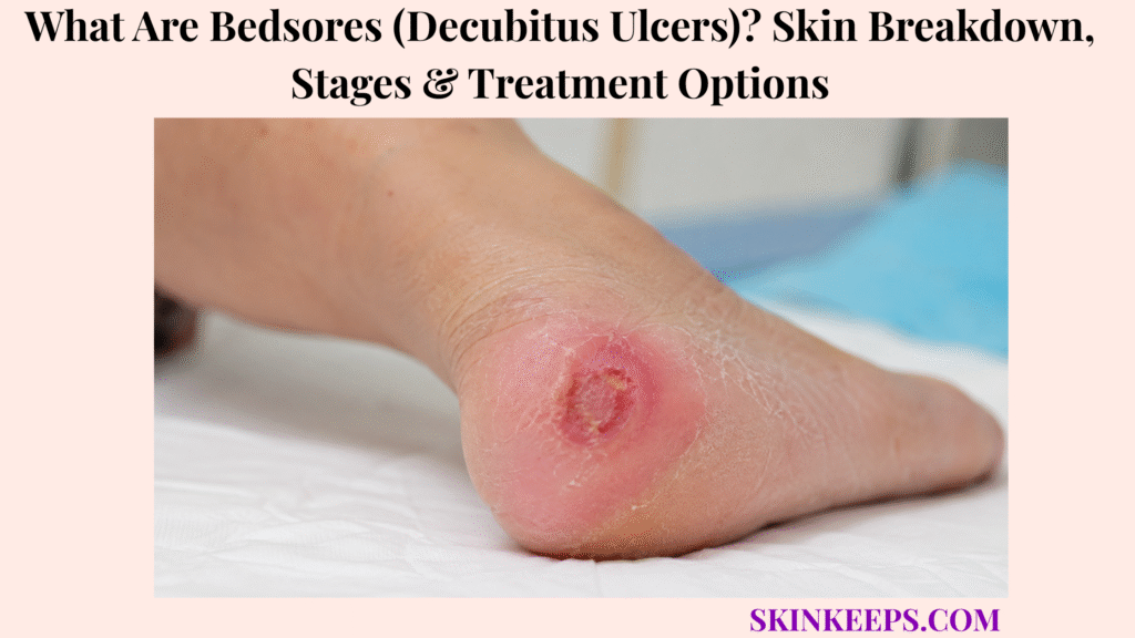
Bedsores, also called decubitus ulcers or pressure injuries, are areas of skin and soft-tissue damage caused by sustained pressure, shear, friction, or pressure from medical devices.
This page explains warning signs, body sites, stages, causes, risk factors, diagnosis, treatment options, prevention, mistakes, and urgent signs. Pressure relief is central to care because dressings or creams cannot work well while the same pressure source keeps damaging tissue.
What Are Bedsores and Why Are They Called Pressure Injuries?
Bedsores are pressure injuries, meaning localized damage to skin and underlying soft tissue caused by prolonged pressure, pressure with shear, or pressure from a medical device.
The modern term pressure injury is useful because damage can begin before the skin opens. A person may first notice discoloration, pain, warmth, firmness, sponginess, or a texture change over a pressure area.
Older terms such as pressure ulcer, pressure sore, and decubitus ulcer are still commonly used. The core problem is the same: tissue is being damaged by pressure, pressure plus shear, or device force.
Why Does Pressure Damage Skin and Deeper Tissue?
Pressure damages skin and deeper tissue by compressing blood vessels, reducing oxygen delivery, and lowering tissue tolerance until skin or soft tissue breaks down.
Body weight can press soft tissue between bone and a bed, chair, wheelchair, mattress, cushion, cast, brace, oxygen tubing, or other device. The bony prominence concentrates the force.
Shear adds risk when a person slides down in bed or a chair. Moisture, poor perfusion, poor nutrition, fragile skin, fever, severe illness, and reduced sensation can lower the skin’s ability to tolerate that force.
Why Is “Bedsore” Too Narrow?
“Bedsore” is too narrow because pressure injuries can happen in beds, chairs, wheelchairs, hospital settings, home care, or under medical devices.
Oxygen tubing, masks, braces, casts, splints, catheters, compression devices, wheelchair parts, and other equipment can create device-shaped pressure injuries. Prevention must happen wherever pressure occurs, not only in bed.
Practical rule: Bedsores are pressure injuries, so care starts by removing pressure from the injured area before expecting wound dressings, creams, or nutrition support to help.
What Bedsore Warning Signs Show Early Skin Breakdown?
Early bedsore warning signs can include skin discoloration that does not blanch, warmth, firmness, sponginess, pain, itching, swelling, or texture change over a pressure area.
Blanching means a skin color change briefly fades when pressed and released. Non-blanching discoloration does not fade with pressure and can signal early tissue injury.
What Do Early Bedsores Look Like on Lighter and Darker Skin?
Early bedsores may look red or pink on lighter skin, while brown or black skin may look purple, blue, darker, shiny, or simply different from the surrounding skin.
Redness-only checks can miss early pressure injury in darker skin tones. Pain, warmth, coolness, firmness, bogginess, sponginess, swelling, itching, or a texture change can be as important as color.
What Warning Signs Suggest a Bedsore Is Getting Worse?
A bedsore may be getting worse when the area blisters, opens, drains, smells bad, becomes more painful, deepens, or develops yellow, black, or dead-looking tissue.
Yellow slough, black eschar, foul odor, pus, increasing drainage, exposed fat, tendon, muscle, or bone are not routine irritation. Do not cut away yellow or black tissue at home.
| Skin Sign | What the Reader May Notice | Why It Matters |
|---|---|---|
| Non-blanching discoloration | Color does not fade when pressed | Early pressure injury clue |
| Red/pink area | Common on lighter skin | May signal Stage 1 if pressure-related |
| Purple/blue/darker/shiny area | Common warning pattern on darker skin | Redness-only checks may miss it |
| Warmth/coolness | Temperature differs from nearby skin | Tissue stress clue |
| Firm/boggy/spongy feel | Texture change under pressure point | Early breakdown risk |
| Pain/itch | Discomfort over pressure area | Can appear before open wound |
| Blister/open sore | Skin breakdown has started | Needs wound-care assessment |
| Drainage/odor/pus | Fluid, smell, or pus | Possible infection |
| Black/yellow tissue | Eschar or slough may obscure depth | Needs expert assessment |
Where Do Bedsores Usually Form on the Body?
Bedsores usually form over bony areas where pressure is concentrated, especially when a person lies, sits, or uses medical devices for long periods.
Common pressure areas include the sacrum, tailbone, buttocks, hips, heels, ankles, elbows, shoulder blades, back of head, ears, spine, and knees. Device-related injuries can appear under oxygen tubing, masks, braces, casts, catheters, splints, or wheelchair equipment.
High-risk skin should be inspected daily because early changes can be subtle, especially on darker skin or under devices.
| Position / Device | Pressure Area to Inspect | What to Look For |
|---|---|---|
| Lying on back | Sacrum, tailbone, heels, elbows, shoulder blades, back of head | Color change, pain, warmth, blister, open sore |
| Lying on side | Hip, ankle, knee, ear, shoulder | One-sided pressure marks |
| Wheelchair sitting | Buttocks, tailbone, thighs, heels, back | Discoloration, soreness, device marks |
| Oxygen tubing / mask | Ears, cheeks, nose bridge | Device-shaped pressure injury |
| Cast / brace / splint | Edges and contact points | Pain, rubbing, skin breakdown |
| Catheter / tubing | Contact lines and trapped areas | Moisture, pressure mark, erosion |
What Are the Stages of Bedsores?
Bedsores are staged by depth, tissue involvement, and wound appearance, and staging should be done by trained clinicians because it guides treatment.
Stages are not a home-diagnosis label from a photo. They help the care team match offloading, dressings, infection review, debridement, nutrition support, pain control, and specialist care to the wound.
What Is Stage 1 Pressure Injury?
Stage 1 pressure injury means the skin is still intact, but there is localized non-blanching discoloration or redness over a pressure area.
On darker skin, Stage 1 may not look red. It may appear purple, blue, darker, shiny, warmer, cooler, firmer, softer, painful, or different from nearby skin.
The priority is immediate offloading and protection. Stage 1 is not an open ulcer.
What Is Stage 2 Pressure Injury?
Stage 2 pressure injury means partial-thickness skin loss with exposed dermis, often appearing as a shallow open wound or serum-filled blister.
The wound bed may look pink or red and moist. Fat, slough, eschar, muscle, tendon, or bone should not be visible in Stage 2.
Stage 2 should not be used to label moisture damage, skin tears, burns, or abrasions that are not caused by pressure or shear.
What Are Stage 3 and Stage 4 Pressure Injuries?
Stage 3 and Stage 4 pressure injuries are deeper wounds that require professional wound care because full-thickness tissue loss is present.
Stage 3 means full-thickness skin loss; visible fat may be present, and slough, rolled edges, undermining, or tunneling may occur.
Stage 4 means full-thickness skin and tissue loss with exposed or palpable fascia, muscle, tendon, ligament, cartilage, or bone. These wounds have serious infection and deep-structure risk and should not be treated at home without wound-care support.
What Are Unstageable and Deep Tissue Pressure Injuries?
Unstageable and deep tissue pressure injuries need expert assessment because the true depth may be hidden or deeper damage may be present beneath intact or blistered skin.
Unstageable pressure injury means full-thickness damage is hidden by slough or eschar. Deep tissue pressure injury may appear as persistent deep red, maroon, purple discoloration, or a blood-filled blister.
Pressure injuries do not always progress neatly from Stage 1 to Stage 4, and healing wounds should not be described as moving backward through the stages. Stable heel eschar should not automatically be softened or removed without expert judgment.
| Stage / Type | Main Clue | Tissue Depth | Safety Priority |
|---|---|---|---|
| Stage 1 | Intact skin with non-blanching discoloration | Surface warning; skin not open | Offload pressure now |
| Stage 2 | Partial-thickness wound or blister with exposed dermis | Dermis exposed | Protect wound and control moisture |
| Stage 3 | Full-thickness skin loss with visible fat possible | Deeper wound | Wound-care team |
| Stage 4 | Muscle, tendon, ligament, cartilage, fascia, or bone exposed/palpable | Deep tissue loss | Specialist care |
| Unstageable | Depth hidden by slough/eschar | Unknown full-thickness damage | Expert assessment; no DIY debridement |
| Deep tissue pressure injury | Deep red, maroon, purple area, or blood-filled blister | Deep damage suspected | Urgent monitoring and offloading |
What Causes Bedsores to Develop?
Bedsores develop when pressure, shear, friction, or moisture repeatedly stresses the same skin area and lowers the tissue’s ability to tolerate damage.
Pressure remains the central cause, but wounds often worsen because several risks act together: immobility, reduced sensation, moisture, fragile skin, poor circulation, poor nutrition, and device pressure.
How Does Pressure Cause Bedsores?
Pressure causes bedsores when body weight presses soft tissue between bone and a surface long enough to reduce blood flow and oxygen delivery.
This can happen in a bed, chair, wheelchair, operating table, hospital bed, home-care bed, or under a device. Tissue damage can begin before the skin opens.
How Do Shear and Friction Worsen Bedsores?
Shear and friction worsen bedsores when skin and deeper tissues are pulled, stretched, or rubbed during sliding, dragging, or repositioning.
Sliding down in bed or a chair can stretch deeper tissue layers while the skin stays against the surface. Dragging during transfers can rub fragile skin, especially at the heels and sacrum.
Lift or assist repositioning instead of dragging. Do not massage red, discolored, painful, or damaged pressure areas.
How Does Moisture Increase Bedsore Risk?
Moisture increases bedsore risk because sweat, urine, stool, or wound drainage can soften skin and make it break down faster under pressure or friction.
Incontinence-associated moisture damage can overlap with pressure injury, but moisture damage alone should not be staged as Stage 2 pressure injury. Care should address both moisture control and pressure relief when both are present.
| Pressure Factor | Skin / Tissue Effect | Prevention Action |
|---|---|---|
| Sustained pressure | Reduced blood flow and tissue stress | Reposition and offload |
| Shear | Deeper tissue stretching | Prevent sliding; use proper transfer help |
| Friction | Surface rubbing and skin weakening | Lift instead of dragging; protect skin |
| Moisture | Skin softening and maceration | Keep skin clean and dry; use barrier care |
| Poor perfusion | Less oxygen delivery | Medical or circulation assessment |
| Poor nutrition | Lower repair capacity | Nutrition and hydration plan |
| Medical devices | Local device-shaped pressure | Check and reposition devices |
Who Is Most at Risk for Bedsores?
Bedsore risk increases when someone has limited mobility, poor sensation, poor circulation, fragile skin, moisture exposure, poor nutrition, severe illness, surgery, medical devices, or a history of pressure injuries.
Risk is not a blame issue. A person can develop pressure injury because illness, immobility, surgery, wheelchair use, devices, frailty, or reduced sensation makes it difficult to shift pressure often enough.
How Are Bedsores Different From Other Skin Wounds?
Bedsores are pressure-related injuries, while other wounds may be driven mainly by moisture, diabetes, poor vein return, poor artery flow, infection, burns, surgery, or trauma.
The cause and location matter because treatment changes. A wound over a pressure point needs offloading, while a venous, arterial, diabetic, traumatic, or infection-driven wound may require a different care plan.
How Are Bedsores Different From Moisture-Associated Skin Damage?
Bedsores are caused by pressure and shear, while moisture-associated skin damage is linked mainly to urine, stool, sweat, trapped moisture, or wound drainage.
Both can overlap. Skin exposed to urine or stool can break down faster under pressure, so prevention needs moisture control, barrier protection, and pressure relief together.
How Are Bedsores Different From Diabetic Foot Ulcers?
Bedsores are different from diabetic foot ulcers because diabetic ulcers often involve neuropathy and weight-bearing foot areas, while bedsores follow pressure-point or device-pressure logic.
Diabetes can also increase pressure-injury risk through neuropathy, circulation problems, and healing difficulty. Foot wounds in a person with diabetes need separate foot-care evaluation.
How Are Bedsores Different From Venous or Arterial Ulcers?
Bedsores are different from venous or arterial ulcers because pressure injuries follow pressure-site logic, while venous and arterial ulcers reflect circulation problems.
Venous ulcers often involve the lower leg or ankle with swelling and vein-related skin changes. Arterial ulcers may involve painful toes or feet, cool skin, poor pulses, and poor blood flow. Compression should not be used unless a clinician confirms it is appropriate.
| Condition | Main Driver | Common Location | Why It Matters |
|---|---|---|---|
| Bedsore / pressure injury | Pressure and shear | Bony areas, device sites | Offloading is central |
| Moisture-associated damage | Urine, stool, sweat, trapped moisture | Folds, perineal area, moist contact areas | Moisture and barrier care |
| Diabetic foot ulcer | Neuropathy, pressure, poor healing | Foot sole, toes, pressure points | Diabetic foot evaluation |
| Venous ulcer | Poor venous return | Lower leg or ankle | Circulation and compression plan |
| Arterial ulcer | Poor arterial flow | Toes, feet, pressure points | Vascular evaluation |
| Skin tear | Trauma or friction on fragile skin | Arms, hands, legs | Different wound care |
| Cellulitis | Bacterial skin infection | Warm, swollen, painful spreading area | Antibiotic or urgent evaluation |
How Are Bedsores Diagnosed and Assessed?
Bedsores are assessed by examining the skin, wound depth, stage, drainage, tissue type, pain, infection signs, nutrition, mobility, support surfaces, and the pressure source.
Staging and wound assessment should be clinician-led because depth, tissue type, infection risk, and pressure source determine the treatment plan.
What Does a Clinician Check in a Bedsore?
A clinician checks a bedsore by assessing its location, stage, size, depth, drainage, odor, tissue type, pain, surrounding skin, infection signs, and pressure source.
The care team may also review mobility, turning routine, mattress or cushion quality, device pressure, moisture exposure, nutrition, hydration, medical conditions, and caregiver support.
When Might Tests Be Needed?
Tests may be needed when a bedsore is infected-looking, non-healing, deep, recurrent, associated with poor nutrition, or suspected to involve bone or poor circulation.
Blood tests may review general health or nutrition when appropriate. A wound culture may be used when infection is suspected, but wounds can be colonized, so culture results need clinical interpretation.
Imaging may be considered if bone infection is suspected. Vascular assessment may be needed if circulation is poor. Stage 3, Stage 4, unstageable, deep tissue, recurrent, infected, heel, tunneling, or non-healing pressure injuries need wound-care specialist review.
What Treatment Options Help Bedsores Heal?
Bedsore treatment usually involves reducing pressure, caring for the wound, controlling pain, preventing or treating infection, improving nutrition, and sometimes using advanced wound care or surgery for deep or non-healing ulcers.
The same wound dressing will not work well if pressure, shear, moisture, malnutrition, infection, or poor support surfaces are not addressed.
How Does Pressure Relief Treat Bedsores?
Pressure relief treats bedsores by removing the force that caused the injury, so the damaged tissue has a chance to recover.
Pressure relief may include repositioning, turning schedules, heel offloading, pressure-redistributing mattresses, wheelchair cushions, device repositioning, padding, and safer transfer methods that reduce friction and shear.
Offloading must continue after dressings are applied. A dressing does not replace pressure relief.
How Are Bedsore Wounds Cleaned and Dressed?
Bedsore wounds are cleaned and dressed based on wound depth, drainage, infection risk, and surrounding skin condition.
If the skin is not broken, gentle cleansing and protection may be used. Open wounds may be cleaned with water or saline when appropriate, and dressings are chosen to support a moist wound-healing environment while protecting the surrounding skin.
Dressing choices can include foam, film, gauze, hydrogel, alginate, hydrocolloid, or other dressings depending on depth, drainage, infection risk, and skin condition. Random creams that hide wound changes should be avoided.
When Is Debridement Needed for Bedsores?
Debridement may be needed when dead, damaged, or infected tissue blocks healing, but it should be done by trained clinicians rather than attempted at home.
Slough or eschar can hide wound depth or infection risk. Clinicians may select sharp, mechanical, enzymatic, autolytic, or other methods based on the wound. Stable heel eschar needs expert judgment.
Do not cut, scrape, peel, or pull away dead tissue at home.
When Are Antibiotics, Advanced Wound Care, or Surgery Used?
Antibiotics, advanced wound care, or surgery may be needed when a bedsore is infected, deep, non-healing, recurrent, or large enough that standard wound care is not enough.
Antibiotics are not for every pressure injury. They may be needed for spreading infection, cellulitis, bacteremia, osteomyelitis, or other clinician-confirmed infection contexts.
Advanced wound care, negative pressure wound therapy in selected clean wounds, or flap surgery may be considered for some large, deep, or non-healing ulcers. Offloading and nutrition still must be addressed for advanced care to work.
| Bedsore Situation | Main Treatment Direction | Key Caution |
|---|---|---|
| Stage 1 | Pressure relief, skin protection, monitoring | Do not massage damaged skin |
| Stage 2 | Offloading, appropriate moist dressing, moisture control | Rule out moisture damage or skin tear |
| Stage 3 / Stage 4 | Wound-care team, dressings, debridement when needed | Infection, tunneling, and deep tissue risk |
| Unstageable | Expert assessment of slough/eschar | Do not DIY debride |
| Deep tissue pressure injury | Immediate offloading and close monitoring | Can evolve quickly |
| Infected ulcer | Culture or medical review; antibiotics when indicated | Avoid random antibiotics |
| Non-healing deep ulcer | Advanced wound care or surgery in selected cases | Offloading and nutrition must be addressed |
How Do Nutrition and Hydration Support Bedsore Healing?
Nutrition does not replace offloading or wound care, but poor nutrition and dehydration can delay bedsore healing and increase the risk of further skin breakdown.
Wound healing needs enough protein, calories, fluids, and vitamins or minerals when a deficiency is present. People with weight loss, low body weight, poor appetite, swallowing problems, diabetes, kidney disease, or deep wounds may need an individualized plan.
A dietitian can help when wounds are deep, recurrent, slow-healing, or linked with poor intake. Supplements should not be treated as a cure without clinical assessment.
How Can Bedsores Be Prevented or Stopped From Getting Worse?
Bedsore prevention depends on regular repositioning, support surfaces, daily skin checks, moisture control, activity when possible, medical-device checks, and adequate nutrition.
Change position regularly based on the person’s risk, surface, mobility, and care plan. Use pressure-redistributing mattresses or cushions when needed, but do not treat support surfaces as a replacement for repositioning.
Offload heels, protect bony areas, keep skin clean and dry, manage urine, stool, sweat, and drainage moisture, and avoid dragging during transfers. Check oxygen tubing, masks, braces, casts, catheters, splints, and wheelchair equipment for pressure marks.
What Bedsore Mistakes Should You Avoid?
The biggest bedsore mistake is putting cream on the sore while leaving the same pressure, shear, moisture, or device problem in place.
Do not keep pressure on the sore. Do not massage red, discolored, painful, or damaged pressure areas. Do not use donut cushions by default because they can concentrate pressure unless a clinician specifically advises them.
Do not cut away dead tissue at home. Do not apply random antibiotic creams, harsh antiseptics, powders, bleaching products, or wound products that hide the wound from assessment.
| Mistake | Why It Is Dangerous | Better Action |
|---|---|---|
| Cream without offloading | Pressure keeps damaging tissue | Relieve pressure first |
| Massage red/damaged skin | Can worsen tissue injury | Protect and offload |
| Donut cushion by default | May concentrate pressure | Use clinician-recommended support |
| DIY debridement | Bleeding and infection risk | Wound-care clinician |
| Random antibiotics | Wrong treatment and resistance risk | Medical review for infection signs |
| Ignoring odor/pus/fever | Infection can spread | Urgent medical review |
| No support surface plan | Recurrence risk stays high | Mattress or cushion plan |
| No nutrition plan | Healing may slow | Dietitian or clinician review |
When Should Bedsores Be Checked Urgently?
Bedsores need urgent care when there is pus, hot or swollen skin, spreading redness or purple/blue discoloration, high temperature, severe or worsening pain, foul odor, rapidly worsening tissue damage, black tissue with inflammation, or exposed deeper structures.
Urgency is higher in older, frail, immobile, diabetic, immunosuppressed, spinal-cord-injured, poor-circulation, or severely ill people because infection and deeper tissue damage can become serious.
Which Infection Signs Need Urgent Medical Review?
Infection signs that need urgent medical review include pus, foul odor, increasing drainage, spreading warmth, swelling, redness or purple/blue color change, fever, chills, confusion, or severe worsening pain.
A pressure injury with spreading warmth, swelling, pain, fever, or rapidly expanding redness may need evaluation for cellulitis or another bacterial complication.
Black tissue with surrounding redness, drainage, or pain should be treated as a warning sign, not a normal scab.
Which Bedsore Features Need Wound-Care Specialist Review?
Wound-care specialist review is needed for Stage 3, Stage 4, unstageable, deep tissue, heel, recurrent, tunneling, undermined, infected, exposed-tissue, or non-healing pressure injuries.
Specialist review is also important when a wound is not improving despite offloading, or when the person has diabetes, poor circulation, immune suppression, spinal cord injury, severe frailty, exposed fat, tendon, muscle, or bone.
Seek urgent medical care if a bedsore has:
What Should You Remember About Bedsores?
The most important thing to remember about bedsores is that they are pressure injuries, so the central treatment logic is pressure relief first, then stage-appropriate wound care, infection control, nutrition, and prevention.
Early discoloration, pain, warmth, firmness, sponginess, or texture change over a pressure area should not be ignored. Open, deep, infected, unstageable, recurrent, heel, or non-healing pressure injuries need professional care.
Frequently Asked Questions About Bedsores
Are bedsores the same as pressure ulcers?
Yes. Bedsores, decubitus ulcers, pressure ulcers, pressure sores, and pressure injuries are related terms. “Pressure injury” is often preferred because damage can exist before the skin opens.
What is the first sign of a bedsore?
The first sign may be non-blanching discoloration, warmth, firmness, sponginess, pain, itching, or texture change over a pressure area. On darker skin, early changes may look purple, blue, darker, shiny, or different from surrounding skin.
What are the stages of bedsores?
Stages include Stage 1, Stage 2, Stage 3, Stage 4, unstageable, and deep tissue pressure injury. Stage is a treatment-guide category, not a safe home-diagnosis label from a photo.
What is the best treatment for bedsores?
There is no one best treatment for every bedsore. Treatment depends on stage, depth, location, infection, drainage, nutrition, mobility, and pressure source. Core care includes offloading, support surfaces, wound cleaning, dressings, pain control, infection management, nutrition, and sometimes debridement or surgery.
Can bedsores heal at home?
Very early pressure injury may improve with prompt offloading and skin protection, but open, deep, infected, recurrent, unstageable, or non-healing wounds need clinician or wound-care support.
When is a bedsore infected?
A bedsore may be infected when there is pus, foul odor, increasing drainage, warmth, swelling, spreading redness or purple/blue change, fever, chills, worsening pain, confusion, or poor healing.
Should you massage a bedsore?
No. Red, discolored, painful, or damaged pressure areas should not be massaged because rubbing can worsen tissue injury.
How can bedsores be prevented?
Prevention includes regular repositioning, offloading, pressure-redistributing mattresses or cushions, daily skin checks, moisture control, device checks, safe movement, and nutrition and hydration support.
Sources & Evidence About Bedsores
Revised National Pressure Ulcer Advisory Panel Pressure Injury Staging System was used for the modern pressure injury terminology, bony prominence and device-related definition, Stage 1–4, unstageable, deep tissue pressure injury, and Stage 2 distinction from moisture-associated skin damage.
Mayo Clinic — Bedsores Diagnosis and Treatment was used for diagnosis, staging, care-team roles, pressure reduction, support surfaces, wound cleaning, dressings, debridement, pain care, nutrition, and flap surgery for large non-healing ulcers.
NHS — Pressure Ulcers / Pressure Sores was used for symptoms, bony body sites, urgent signs, risk factors, prevention steps, home-care risk assessment, and the point that pressure ulcers can sometimes appear over a few hours.
MSD Manual Professional — Pressure Injuries was used for infection signs, wound-culture caution, systemic antibiotic contexts, osteomyelitis concern, pain management, nutrition assessment, and non-healing wound complications.
AHRQ Pressure Ulcer Training Resources was used for additional stage-description support and clinician-oriented pressure injury education context.
Educational Disclaimer: This SkinKeeps article is for educational purposes only and does not diagnose, stage wounds, or replace medical care. A pressure area with pus, foul odor, fever, chills, spreading warmth, swelling, red/purple/blue color change, severe or worsening pain, black tissue, exposed fat, tendon, muscle, or bone, confusion, rapid worsening, or poor healing should be checked urgently by a qualified healthcare professional or wound-care specialist.



