
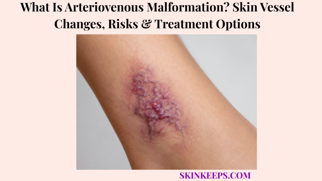
An arteriovenous malformation is an abnormal high-flow connection between arteries and veins that can create skin vessel changes, warmth, pulsation, swelling, pain, bleeding, or tissue damage. It should not be treated as a harmless surface color change when high-flow clues are present.
This page explains skin signs, high-flow behavior, body locations, risks, diagnosis, imaging, treatment options, daily protection, mistakes, and urgent warning signs. It does not diagnose the reader or present treatment as simple, cosmetic, or one-step.
What Is an Arteriovenous Malformation and Why Is It High-Flow?
An arteriovenous malformation is an abnormal connection between arteries and veins where blood bypasses the normal capillary network and moves through a high-flow vascular channel.
Arteries normally carry higher-pressure blood into smaller vessels, then into capillaries, before blood returns through veins. In an AVM, the capillary bridge is bypassed, so arterial blood can move directly into veins through an abnormal shunt or nidus.
The nidus is the central abnormal vessel network where feeding arteries and draining veins connect. Because blood flow is fast and pressurized, an AVM can create warmth, pulsation, a bruit, a thrill, swelling, bleeding, or tissue stress.
Why Are AVMs Different From Low-Flow Vascular Malformations?
AVMs are different from low-flow vascular malformations because arterial blood can pass directly into veins under higher pressure.
Venous and lymphatic malformations are usually low-flow. AVMs are high-flow, so exam and treatment planning focus on flow speed, feeding arteries, draining veins, tissue involvement, and bleeding risk.
Warmth, pulsation, a clinician-heard bruit, or a vibration-like thrill can point toward high-flow behavior. These clues should not be treated like ordinary superficial vessels.
Why May an AVM Not Be Obvious at Birth?
An AVM may not be obvious at birth because some congenital vascular malformations become visible or symptomatic later as the body grows or after triggers such as puberty, pregnancy, trauma, or procedures.
Some AVMs are quiet early. Later growth, hormonal change, injury, surgery, or other procedures can make a lesion more visible, warmer, more pulsatile, painful, swollen, bleeding, or ulcerated.
This does not mean every AVM rapidly worsens. It means a changing warm or pulsing vascular area should be assessed by clinicians who understand high-flow vascular anomalies.
Practical rule: A warm, pulsing, enlarging, painful, bleeding, or ulcerated vascular lesion needs flow-based evaluation, not surface-only assumptions.
What Skin Vessel Changes Can an Arteriovenous Malformation Cause?
A skin or soft-tissue arteriovenous malformation may appear as a red, pink, purple, bluish, or bruised-looking area that can feel warm, pulse, swell, hurt, or bleed.
Color alone cannot diagnose an AVM. The stronger clues are behavior: warmth compared with nearby skin, pulsation, vibration-like sensation, enlargement, bleeding, ulceration, pain, or function change.
What Does a Superficial AVM Look Like on Skin?
A superficial AVM may look like a red, pink, purple, bluish, bruised, swollen, or vessel-rich area on or under the skin.
Visible enlarged vessels, soft-tissue fullness, shiny skin, thinning skin, or a non-healing sore may appear when the lesion affects skin and nearby tissue. A flat red mark without warmth or pulse may have a different cause.
What Does an AVM Feel Like?
An AVM may feel warmer than nearby skin, pulsatile, tender, painful, or vibration-like when high-flow blood moves through the abnormal vessels.
A clinician may feel a thrill, which is a vibration from fast flow, or hear a bruit, which is a flow sound heard with a stethoscope. Readers should not press hard, massage, puncture, or manipulate a suspected AVM.
| Skin Sign | What the Reader May Notice | Why It Matters |
|---|---|---|
| Warmth | Lesion feels hotter than nearby skin | Suggests high-flow activity |
| Pulsation | Pulse felt or seen in the area | Strong AVM clue |
| Bruit / thrill | Clinician hears sound or feels vibration | Indicates fast blood flow |
| Red / purple / bluish color | Vascular-looking patch or bump | Needs pattern and flow assessment |
| Swelling | Soft-tissue fullness or limb enlargement | May show deeper involvement |
| Bleeding | Spontaneous or trauma-related bleeding | Higher urgency |
| Ulceration | Open sore over lesion | Tissue damage and infection risk |
| Pain | Tenderness, pressure, or severe pain | May reflect progression or tissue involvement |
Where Can Skin or Peripheral Arteriovenous Malformations Appear?
Skin or peripheral arteriovenous malformations can appear outside the brain in the face, scalp, head and neck, arms, hands, legs, feet, trunk, mucous membranes, genital areas, or deeper soft tissues.
Location changes risk. AVMs near the eye, mouth, airway, hand, foot, or genitals can affect function, bleeding risk, tissue health, and treatment planning.
This page focuses on skin and peripheral AVM patterns. It is not a brain AVM or spinal AVM guide, although the shared vascular concept is abnormal artery-to-vein flow.
| Location | Possible Skin Clue | Functional Risk |
|---|---|---|
| Face / lips | Warm red-purple swelling or pulsing area | Appearance, bleeding, mouth function |
| Eye area | Redness, swelling, visible vessels | Vision and pressure concerns |
| Scalp / head | Warm vascular patch, prominent vessels, pulse | Bleeding and deeper anatomy concerns |
| Arm / hand | Swelling, pain, weakness, pulse | Grip, motion, nerve or tissue function |
| Leg / foot | Pain, swelling, ulcer, warmth | Walking, tissue health, bleeding risk |
| Trunk | Vascular lump, warmth, bruised area | Deeper soft-tissue involvement |
| Genital / mucosal | Bleeding, swelling, pain | Sensitive location; specialist evaluation important |
What Risks Can an Arteriovenous Malformation Create?
An arteriovenous malformation can cause pain, swelling, bleeding, ulceration, tissue damage, functional limitation, disfigurement, and in advanced high-flow cases, strain on the heart.
Risk depends on the lesion’s size, location, depth, flow rate, symptoms, skin condition, and nearby structures. A small stable lesion is not the same as a rapidly growing, bleeding, painful, or ulcerated lesion.
Why Can AVMs Bleed or Ulcerate?
AVMs can bleed or ulcerate when high-flow pressure stresses fragile vessels and the overlying skin becomes thin, damaged, or easily injured.
Minor trauma can trigger bleeding when a high-flow lesion is close to the surface. Ulceration can appear when abnormal flow, tissue stress, and skin breakdown create an open sore over the lesion.
Why Can AVMs Cause Pain, Swelling, or Weakness?
AVMs can cause pain, swelling, or weakness when abnormal blood flow affects tissue pressure, oxygen delivery, nearby nerves, muscles, joints, or limb function.
Some lesions can pull blood through the abnormal shunt instead of supporting nearby tissue normally. This vascular steal pattern can contribute to tissue ischemia, pain, weakness, numbness, or motion limitation in selected cases.
Why Can Large AVMs Affect the Heart?
Large or extensive AVMs can affect the heart because high-flow shunting may increase the workload needed to circulate blood.
This is not expected with every AVM. The concern becomes stronger with large, extensive, symptomatic, or advanced high-flow lesions, especially if shortness of breath, severe fatigue, or signs of heart strain appear.
| Risk | Warning Clue | Why It Matters | Urgency Level |
|---|---|---|---|
| Bleeding | Bleeds easily or repeatedly | High-flow vessels may bleed significantly | Urgent if hard to stop |
| Ulceration | Open sore over lesion | Tissue damage and infection risk | Prompt specialist/wound review |
| Pain | Increasing tenderness or severe pain | Possible progression or deeper involvement | Prompt evaluation |
| Swelling | Limb or soft-tissue enlargement | Function and tissue pressure risk | Evaluation needed |
| Function loss | Weakness, limited movement, numbness | Nerve, muscle, or joint involvement | Higher urgency |
| Heart strain | Shortness of breath, severe fatigue, large/extensive lesion | Possible high-output strain | Medical review urgently if symptomatic |
How Is an Arteriovenous Malformation Different From Other Vascular Skin Conditions?
Arteriovenous malformation is different from hemangioma, capillary malformation, venous malformation, and lymphatic malformation because AVM is a high-flow artery-to-vein malformation, while many other vascular lesions are tumors or low-flow malformations.
Correct classification matters because the wrong procedure can increase bleeding risk, miss deeper disease, or fail to treat the nidus.
How Is AVM Different From a Hemangioma?
AVM is different from a hemangioma because a hemangioma is a vascular tumor, while an AVM is a structural vascular malformation with abnormal artery-to-vein flow.
Infantile hemangiomas often have a growth phase and later involution pattern. AVMs usually do not behave like self-resolving infantile hemangiomas, especially when warmth, pulse, bleeding, or expansion appears.
How Is AVM Different From a Venous Malformation?
AVM is different from a venous malformation because AVM is high-flow, while venous malformation is usually low-flow and often softer, bluish, and compressible.
Warmth, pulsation, bruit, or thrill points away from a typical low-flow venous pattern. Imaging is often needed because deeper tissue involvement may not match the surface appearance.
How Is AVM Different From a Capillary Malformation or Port-Wine Stain?
AVM is different from a capillary malformation or port-wine stain because capillary malformations are usually flat, low-flow stains, while AVMs can become warm, raised, pulsatile, painful, or bleeding.
A flat port-wine stain does not usually have high-flow pulse or thrill. A vascular area that becomes warm, raised, painful, pulsatile, bleeding, or rapidly enlarging deserves specialist review.
Fine surface vessels such as telangiectasia usually lack the deeper high-flow behavior of an AVM.
| Condition | Flow Type | Common Clue | Why AVM Matters |
|---|---|---|---|
| AVM | High-flow | Warm, pulsatile, bruit/thrill, bleeding risk | Needs specialist evaluation |
| Venous malformation | Low-flow | Blue/purple, soft, compressible swelling | Different imaging and treatment |
| Capillary malformation / port-wine stain | Low-flow | Flat red/pink stain | Usually not pulsatile |
| Hemangioma | Vascular tumor | Infant growth/involution pattern | Not the same disease process |
| Telangiectasia | Surface small vessels | Fine red surface vessels | Usually lacks high-flow warmth/pulse |
How Is an Arteriovenous Malformation Diagnosed or Checked?
AVM evaluation usually combines clinical exam with imaging to define blood-flow speed, feeding arteries, draining veins, lesion extent, and involvement of skin, muscle, bone, nerves, or deeper structures.
Visual inspection alone is not enough for a suspected high-flow vascular malformation. The surface mark may be smaller than the deeper vessel network.
What Does a Clinician Check During an AVM Exam?
A clinician checks an AVM by examining skin color, warmth, pulsation, thrill, bruit, pain, bleeding, ulceration, limb size, and functional impact.
The exam may also assess skin thickness, visible vessels, tenderness, tissue fullness, nerve symptoms, muscle or joint involvement, facial function, eye pressure concerns, mouth involvement, or airway-related risk depending on location.
Which Imaging Tests May Be Used for AVM?
Imaging tests for AVM may include Doppler ultrasound, MRI or MRA, CT or CTA in selected cases, and catheter angiography when detailed treatment planning is needed.
Doppler ultrasound helps assess blood-flow pattern. MRI or MRA helps define lesion extent and nearby anatomy. CT or CTA may help in selected cases when bone or complex anatomy matters.
Catheter angiography maps feeding arteries, draining veins, and the nidus in detail, especially when embolization or surgery is being planned. Not every patient needs every test.
- Clinical exam checks warmth, pulse, bruit, and thrill.
- Doppler ultrasound helps define flow.
- MRI or MRA helps define extent and nearby anatomy.
- CT or CTA may help when bone or anatomy detail is needed.
- Catheter angiography may be used when intervention is planned.
- Functional assessment matters for eye, mouth, airway, hand, foot, limb, genital, nerve, muscle, or joint involvement.
What Treatment Options Are Used for Arteriovenous Malformation?
Arteriovenous malformation treatment may include observation, symptom control, embolization, surgery, staged combined treatment, or supportive care, depending on location, symptoms, flow pattern, tissue risk, and treatment risk.
Treatment should be planned by clinicians experienced with high-flow vascular anomalies. The goal may be to monitor safely, reduce symptoms, control bleeding risk, protect tissue, reduce abnormal flow, or remove selected lesions when feasible.
When Might an AVM Be Monitored Instead of Treated Immediately?
An AVM may be monitored instead of treated immediately when it is small, stable, low-symptom, high-risk to treat, or located where intervention could damage important tissue.
Surveillance is not ignoring the lesion. It means tracking warmth, pulse, pain, swelling, bleeding, ulceration, growth, and function while balancing treatment benefit against risk.
How Does Embolization Treat an AVM?
Embolization treats an AVM by blocking or reducing abnormal blood flow through targeted vessels, often as part of a staged plan or before surgery.
Interventional radiology may use catheter-based techniques to reduce flow through the nidus or specific high-flow channels. Blocking only one visible vessel may not treat the true nidus, and some AVMs need staged sessions.
Embolization is not a guaranteed one-session cure. Planning must consider non-target embolization risk, tissue safety, bleeding risk, and whether surgery is part of the strategy.
When Is Surgery Used for an AVM?
Surgery may be used for selected AVMs when complete removal is feasible and the expected benefit outweighs bleeding, functional, cosmetic, or recurrence risks.
Surgery may follow embolization to reduce bleeding risk. Incomplete removal can risk recurrence or worsening, so surgical planning should involve an experienced team and detailed imaging.
Why May AVM Treatment Need a Multidisciplinary Team?
AVM treatment may need a multidisciplinary team because high-flow vascular malformations can involve skin, muscle, bone, nerves, function, appearance, bleeding risk, and imaging-guided procedures.
Care may involve dermatology, interventional radiology, vascular surgery, plastic or reconstructive surgery, wound care, pain support, and site-specific specialists such as ENT, ophthalmology, orthopedics, pediatrics, or neurosurgery when anatomy requires it.
| Treatment Option | Best-Fit Situation | Goal | Key Caution |
|---|---|---|---|
| Observation | Stable, low-symptom, high-risk-to-treat lesion | Track change and avoid unnecessary harm | Not the same as ignoring progression |
| Symptom support | Pain, wound, bleeding-risk support while planning care | Reduce complications | Does not remove AVM |
| Embolization | High-flow nidus control or pre-surgical planning | Reduce abnormal flow | May need staged sessions; non-target embolization risk |
| Surgery | Selected lesions where complete removal is feasible | Remove malformation | Requires experienced team; incomplete removal may recur |
| Combined staged care | Complex lesions | Control flow and remove/reduce lesion safely | Requires coordinated specialist plan |
| Wound care | Ulcerated or bleeding surface | Protect tissue and reduce infection risk | Needs medical guidance |
How Can Daily Care Reduce Skin Injury Around an Arteriovenous Malformation?
Daily care cannot remove an arteriovenous malformation, but it can reduce trauma, bleeding, skin breakdown, and infection risk while specialist evaluation or treatment planning is underway.
Protect the area from cuts, friction, and pressure. Do not puncture, drain, burn, freeze, aggressively massage, tattoo, pierce, inject, or use cosmetic procedures near a suspected AVM before diagnosis.
Ulcerated areas should be managed with clinician or wound-care guidance. Photos under consistent lighting can help document warmth, pulse, swelling, color change, bleeding, ulceration, and growth over time.
- Protect the area from cuts, friction, and pressure.
- Do not puncture, drain, burn, freeze, or massage the lesion.
- Track warmth, pulse, pain, swelling, bleeding, ulceration, and growth.
- Photograph changes under the same lighting.
- Follow wound-care guidance if skin is ulcerated.
- Ask before compression or padding.
- Avoid cosmetic procedures near the lesion until AVM is evaluated.
- Seek vascular-anomaly specialist review for concerning changes.
What Arteriovenous Malformation Mistakes Should You Avoid?
The biggest AVM mistake is treating a warm, pulsing, bleeding, painful, ulcerated, or enlarging vascular skin lesion as harmless without specialist evaluation.
Other mistakes include assuming every red or purple vascular mark is a hemangioma, using cosmetic laser before diagnosis, puncturing or draining a vascular lump, ignoring warmth or pulsation, relying on compression without guidance, or expecting one quick procedure to solve a complex AVM.
| Mistake | Why It Is Risky | Better Action |
|---|---|---|
| Calling it a surface mark only | Misses high-flow risk | Check warmth, pulse, growth, and bleeding |
| Cosmetic laser first | Wrong treatment may worsen risk | Get vascular diagnosis first |
| Puncturing or draining | Bleeding risk | Leave intact and seek care |
| Ignoring ulceration | Tissue damage may progress | Seek specialist/wound review |
| Relying on compression alone | AVM flow may not respond safely | Use only if advised |
| Expecting one quick fix | AVMs often need staged care | Plan with a vascular-anomaly team |
When Should an Arteriovenous Malformation Be Checked Urgently?
An AVM should be checked urgently when it bleeds, ulcerates, grows quickly, becomes very painful, becomes warmer or more pulsatile, affects movement or function, causes numbness or weakness, or appears near critical areas such as the face, mouth, eye, airway, hand, foot, or genitals.
Urgency rises when a lesion changes behavior. A stable vascular mark and a rapidly enlarging warm pulsing lesion with pain or bleeding should not be handled the same way.
Which AVM Signs Suggest Higher Risk?
Higher-risk AVM signs include recurrent bleeding, a non-healing ulcer, rapid enlargement, increasing warmth, stronger pulsation, new thrill or bruit, severe pain, skin thinning, function loss, or nerve symptoms.
Functional signs include numbness, weakness, limited movement, grip change, walking difficulty, facial function change, mouth function problems, or eye-area pressure symptoms.
When Is Emergency Care Needed?
Emergency care is needed when an AVM bleeds and does not stop with appropriate pressure, is severely injured, suddenly swells, becomes severely painful, shows infection signs around an ulcer, or affects breathing or swallowing.
Spreading warmth, redness, tenderness, fever, or infected-looking ulceration may suggest cellulitis or wound infection around a vulnerable area and should be checked promptly.
Large high-flow lesions paired with fainting, chest symptoms, severe weakness, or shortness of breath also need urgent medical review.
Seek urgent care if an AVM has:
What Should You Remember About Arteriovenous Malformation?
The most important thing to remember about an arteriovenous malformation is that it is a high-flow vascular malformation, so how the lesion behaves matters as much as how it looks.
Warmth, pulse, thrill, bruit, pain, bleeding, ulceration, rapid growth, or function change should move the lesion out of cosmetic thinking and into specialist evaluation.
Frequently Asked Questions About Arteriovenous Malformation
Is an arteriovenous malformation the same as a hemangioma?
No. A hemangioma is a vascular tumor, while an arteriovenous malformation is a high-flow vascular malformation with abnormal artery-to-vein connections.
What does a skin AVM look like?
A skin AVM may look red, pink, purple, bluish, bruised-looking, warm, swollen, tender, pulsatile, or bleeding, depending on depth and location.
Can an arteriovenous malformation be dangerous?
Yes, depending on size, location, blood-flow pattern, and symptoms. Risks can include bleeding, ulceration, pain, tissue damage, functional limitation, disfigurement, and in advanced high-flow cases, heart strain.
Can an AVM appear later even if someone was born with it?
Yes. AVMs are often congenital, but they may become more visible or symptomatic later with growth, puberty, pregnancy, trauma, or procedures.
How is an arteriovenous malformation diagnosed?
Diagnosis usually combines clinical exam with imaging such as Doppler ultrasound, MRI or MRA, CT or CTA in selected cases, and catheter angiography for detailed treatment planning.
What is the best treatment for arteriovenous malformation?
There is no one best treatment for every AVM. Treatment may include observation, embolization, surgery, staged combined care, wound care, or supportive care, and should be planned by an experienced vascular-anomaly team.
Can cosmetic laser remove an AVM?
Cosmetic laser should not be used before proper diagnosis because AVM is high-flow and may involve deeper vessels. Treatment planning should be specialist-led.
When should an AVM need urgent care?
Urgent care is needed for uncontrolled bleeding, ulceration, rapid growth, increasing warmth or pulsation, severe pain, infection signs, function loss, numbness, weakness, major trauma, or critical locations near the eye, mouth, airway, hand, foot, or genitals.
Sources & Evidence About Arteriovenous Malformation
Cleveland Clinic — Arteriovenous Malformation was used for AVM definition, missing capillaries, high-flow artery-to-vein connection, rarity context, asymptomatic cases, bleeding or tissue damage, bruit, and broad treatment goals.
Seattle Children’s — Arteriovenous Malformations was used for skin and mucous AVM signs, bleeding, pulsing, warm or tender bruised bump, limb swelling, pain, weakness, limited motion, tissue ischemia, congestive heart failure risk, and imaging pathway.
Johns Hopkins Medicine — Arteriovenous Malformations was used for high-flow anatomy, no-capillary explanation, Schöbinger-stage context, warmth, redness, pulse, pain, bleeding, ulcers, heart failure, and confusion with hemangioma or port-wine stain.
RadiologyInfo — Vascular Malformations was used for peripheral AVM symptoms, pain, swelling, bleeding, warmer skin, pulse, ulcers, tissue damage, heart workload, ultrasound, MRI/MRA, and imaging evaluation.
ISSVA — Multidisciplinary Teams was used for team-care logic and the value of multidisciplinary settings for complex vascular-anomaly management.
ISSVA — Classification was used for vascular-anomaly terminology and classification reference, including vascular malformation and high-flow lesion context.
Peripheral AVM Review Evidence was used for high-flow AVM treatment complexity, nidus-targeted embolization principles, and the caution that treating only an outflow vein or proximal feeder may be inadequate.
Imaging Pediatric Vascular Lesions Review was used for high-flow AVM complication context, including distal ischemia, pain, ulceration, soft tissue or bony change, and bleeding.
Educational Disclaimer: This SkinKeeps article is for educational purposes only and does not diagnose or replace medical care. A warm, pulsing, bleeding, ulcerated, rapidly enlarging, severely painful, infected-looking, numb, weak, function-limiting, or critical-location vascular lesion should be checked urgently by a qualified healthcare professional or vascular-anomaly specialist.



