
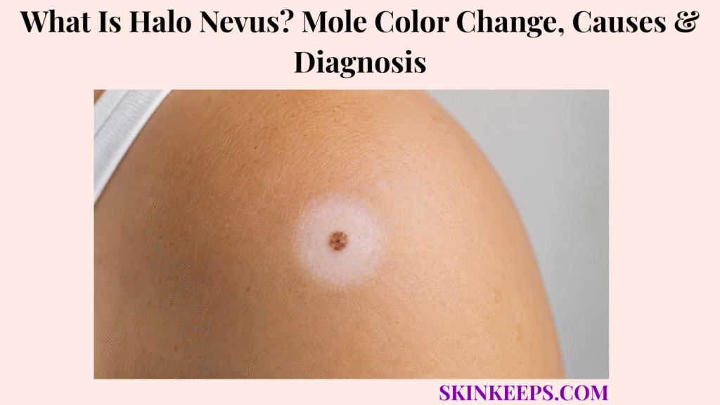
A halo nevus is a melanocytic mole surrounded by a circular or oval area of lighter or completely depigmented skin. It is also called a halo naevus, Sutton nevus, halo mole, or leukoderma acquisitum centrifugum.
Immune activity targets pigment-producing melanocytes in the mole and nearby skin, so the central mole may lighten, become pink, flatten, or disappear. Most typical lesions need observation, but irregular, uneven, rapidly changing, or adult-onset halo lesions require professional assessment to exclude melanoma.
How Can You Recognize a Typical Halo Nevus?
A typical halo nevus has a central mole surrounded by a smooth, fairly even ring of pale or white skin.
The central mole may be brown, tan, pink, or fading. The halo usually extends a relatively uniform distance around it, creating a round or oval symmetrical pattern.
One or several lesions may occur. A typical lesion has no scale, crusting, ulceration, pain, or itch and often becomes more visible when nearby skin tans.
The strongest reassuring clue is a symmetrical white halo surrounding an otherwise orderly mole. An irregular halo around an unusual central mole needs more caution.
- Central round or oval melanocytic mole.
- Brown, tan, pink, or fading mole colour.
- Smooth pale or completely white halo.
- Relatively uniform halo width.
- Symmetrical full pattern.
- One lesion or several halo nevi.
- No scale, crusting, bleeding, or ulceration.
- Usually no pain or itch.
- Gradual rather than destructive change.
Figure 1. A typical halo nevus has an orderly central mole and a smooth symmetrical white ring, then may progress through mole fading, a persistent pale patch, and gradual repigmentation.
Where Do Halo Nevi Usually Appear?
Halo nevi are seen most often on the trunk, especially the upper or lower back, but they can appear elsewhere.
The chest, abdomen, and other trunk sites are common. The head, arms, and legs are less frequent locations.
A person may have several lesions at different sites and at different stages of fading. Location supports the diagnosis but cannot prove a changing mole is benign.
| Location | How Typical? | Clinical Meaning |
|---|---|---|
| Upper or lower back | Most typical | Common site for one or several lesions |
| Chest | Typical | Truncal pattern |
| Abdomen | Typical | May coexist with back lesions |
| Other trunk areas | Common | Supports ordinary distribution |
| Head | Less common | Assess central mole carefully |
| Arms or legs | Less common | Can occur but is not the dominant pattern |
| Several body sites | Possible | Lesions may be at different stages |
How Does a Halo Nevus Change Color Over Time?
A halo nevus often follows a four-stage sequence, but not every lesion completes every stage at the same speed.
What Happens During Stage One of a Halo Nevus?
A pale or white ring develops around an existing mole while the central mole may initially remain brown.
The halo is usually round or oval and becomes more noticeable after surrounding skin tans because the depigmented area does not tan normally.
What Happens During Stage Two of a Halo Nevus?
The central mole begins to lose pigment, appearing lighter, pinker, flatter, or less prominent.
What Happens During Stage Three of a Halo Nevus?
The central mole may disappear and leave a circular or oval white patch that can resemble a small vitiligo area.
What Happens During Stage Four of a Halo Nevus?
Pigment may gradually return to the previously white skin, though repigmentation can be slow or incomplete.
The complete sequence can take months or several years, and several halo nevi may evolve at different speeds.
Why Does the Skin Turn White Around a Halo Nevus?
The skin turns white because an immune response reduces melanocytes in the mole and nearby skin.
Melanocytes produce melanin, the pigment that gives skin and moles colour. Lymphocytes, including cytotoxic T cells, participate in targeting these cells.
As melanocytes decrease, the central mole regresses and surrounding skin loses pigment. The halo is not fungus, poor circulation, or material accumulating around the mole.
Figure 2. Halo nevi form when immune cells reduce melanocytes in the central mole and surrounding skin, causing the mole to fade and a white ring to appear.
What Causes a Halo Nevus to Develop?
The exact reason the immune system begins targeting a particular mole is not fully understood.
Halo nevi are considered immune-mediated and often appear spontaneously. They are not caused by bacteria, fungi, poor hygiene, or contagious contact.
Local trauma, scratching, sunburn, broader autoimmune pigment loss, and psychosocial stress have been reported as possible preceding factors, but none is a universal cause.
| Proposed Factor | Possible Role | Universal Cause? | Practical Response |
|---|---|---|---|
| Spontaneous immune response | Central mechanism | Trigger uncertain | Monitor mole structure |
| Local trauma or scratching | Possible preceding event | No | Avoid irritating the mole |
| Sunburn | Possible preceding event | No | Protect from UV |
| Vitiligo or autoimmune pigment loss | Possible overlap | No | Assess broader signs only |
| Psychosocial stress | Reported association | No | Do not treat as direct cause |
| Bacteria, fungi, or hygiene | Myth | No | No antimicrobial treatment |
Who Is Most Likely to Develop Halo Nevi?
Halo nevi most often develop in older children, adolescents, and young adults, though they can occur later in life.
They affect all sexes and may develop around one or several existing melanocytic moles. Vitiligo or a tendency toward autoimmune pigment loss may coexist.
A first halo lesion in adulthood is less typical. A newly developing, solitary, or irregular halo in an older adult deserves closer examination rather than automatic reassurance.
| Age or Context | Usual Interpretation | Direction |
|---|---|---|
| Child or adolescent with symmetrical lesions | Usually benign pattern | Routine confirmation and monitoring |
| Young adult with several orderly halo nevi | Often benign | Full skin context if new |
| Adult with first halo lesion | More caution | Professional skin assessment |
| Older adult with atypical mole | Higher concern | Prompt dermatologist evaluation |
| Personal or family melanoma history | Higher-risk context | Specialist assessment |
Can Halo Nevi Look Different Across Skin Tones?
Halo nevi may be especially visible on brown or black skin because depigmented skin contrasts strongly with surrounding pigment.
The central mole may look brown, dark brown, pink, or skin-coloured as it fades. Early pigment loss may appear lighter before becoming completely white.
Inflammation may appear brown, purple, or grey rather than bright red. Shape, texture, symmetry, and evolution remain more important than colour contrast alone.
The depigmented halo has less melanin and may sunburn more easily.
Are Halo Nevi Related to Vitiligo?
Halo nevi and vitiligo can overlap because both involve immune-mediated melanocyte loss, but they are not the same condition.
A halo nevus has a central mole within a circular or oval depigmented ring, and the mole itself may fade.
Vitiligo patches usually develop without a central mole, may have varied shapes, can expand or join, and may affect hair pigment.
One halo nevus does not mean widespread vitiligo will inevitably follow.
| Feature | Halo Nevus | Vitiligo |
|---|---|---|
| Central mole | Present | Usually absent |
| Shape | Circular or oval around mole | Variable patch shapes |
| Main process | Mole and surrounding skin lose pigment | Skin patches lose pigment |
| Course | Mole may fade or disappear | Patches may expand or stabilise |
| Hair pigment | Not the main clue | May lighten in some patches |
| Meaning | Usually local benign process | Broader pigment condition possible |
Are Halo Nevi Associated With Thyroid or Other Autoimmune Conditions?
Halo nevi may occur alongside vitiligo or other autoimmune conditions, but one typical halo nevus does not prove thyroid disease.
Broad laboratory screening is not automatically required for every isolated typical lesion.
Symptom-guided assessment may be reasonable when there is widespread vitiligo, fatigue, unexplained weight change, temperature intolerance, or another clinical reason to suspect autoimmune disease.
| Situation | Broad Testing? | Better Direction |
|---|---|---|
| Isolated typical halo nevus | Usually no | Skin assessment and monitoring |
| Several halo nevi plus vitiligo | Context-dependent | Symptom-guided evaluation |
| Fatigue or unexplained weight change | Targeted tests may fit | Primary-care review |
| Temperature intolerance | Thyroid assessment may fit | Clinical context |
| Mole concern only | Not automatic | Focus on mole assessment |
Is a Halo Nevus Contagious or Cancerous?
A typical halo nevus is benign, not contagious, and not an infection.
It does not spread through touch, towels, clothing, or sexual contact, and most halo nevi do not become melanoma.
Melanoma can rarely show regression or surrounding pigment loss, and a halo phenomenon may rarely appear in association with melanoma elsewhere.
A symmetrical halo around an ordinary-looking mole is usually benign; an atypical mole or adult-onset halo needs careful examination.
How Is a Halo Nevus Different From a Regressing Melanoma?
A typical halo nevus is usually symmetrical and gradual, while regressing melanoma may show irregular colour, structure, borders, surface change, or rapid evolution.
Melanoma regression does not usually create a perfectly smooth circular ring. Dermoscopy and, when needed, complete excision are used because naked-eye appearance can mislead.
| Feature | Typical Halo Nevus | Possible Regressing Melanoma |
|---|---|---|
| Symmetry | Even mole and halo | Asymmetrical lesion |
| Colour | Uniform brown, pink, or fading | Several irregular colours |
| Halo pattern | Smooth circular or oval ring | Uneven or focal whitening |
| Surface | No crusting or ulceration | Bleeding, crusting, or ulceration possible |
| Age context | Children and young adults common | New older-adult lesion more concerning |
| Evolution | Gradual predictable fading | Rapid or unexplained change |
| Action | Observe if confidently benign | Dermoscopy and excision assessment |
How Is a Halo Nevus Different From Other Pale Rings Around Skin Lesions?
Several pigment and inflammatory patterns can resemble a halo nevus, but their central lesion, surface, history, and evolution differ.
How Does a Halo Nevus Differ From Post-Inflammatory Hypopigmentation?
Post-inflammatory hypopigmentation follows a rash, injury, or irritation and may lack a central melanocytic mole.
How Does a Halo Nevus Differ From a Meyerson Nevus?
Meyerson nevus is a mole surrounded by eczema, producing an itchy red or scaly ring rather than smooth pigment loss.
How Does a Halo Nevus Differ From a Pseudohalo?
A pseudohalo results when sunscreen, a plaster, or another covering prevents adjacent skin from tanning; melanocytes are not destroyed and the mole does not follow the fading stages.
How Does a Halo Nevus Differ From a Recurrent Nevus in a Scar?
A recurrent nevus is pigment that returns after partial mole removal or injury, often irregularly within scar tissue.
Previous biopsy or trauma is an important clue, and dermoscopy or complete excision may be needed if melanoma cannot be excluded.
| Condition | Central Lesion | Pale Area | Scale or Scar | Evolution | Assessment |
|---|---|---|---|---|---|
| Halo nevus | Mole present | Smooth pigment loss | No scale | Mole fades gradually | Clinical exam or dermoscopy |
| Post-inflammatory hypopigmentation | Often no mole | Matches previous rash | Prior inflammation | Slow recovery | History and exam |
| Meyerson nevus | Mole present | Eczematous ring | Itchy or scaly | Eczema fluctuates | Exam and dermoscopy |
| Pseudohalo | Mole present | Untanned protected skin | No true depigmentation | Exposure-linked | History and dermoscopy |
| Recurrent nevus | Pigment in scar | Scar-related | Scar present | Pigment returns after removal | Dermoscopy or excision |
How Do Clinicians Diagnose a Halo Nevus?
Clinicians diagnose a halo nevus by assessing age, the central mole, halo symmetry, rate of change, and the rest of the skin.
Assessment includes whether the mole existed before the ring, central-mole symmetry, border and colours, halo uniformity, scale, crusting, bleeding, ulceration, several similar lesions, vitiligo, and personal or family melanoma history.
A typical lesion is often diagnosed clinically. Adults need examination of the central mole and a broader whole-skin assessment rather than attention to the white ring alone.
- Age when the halo appeared.
- Duration and rate of change.
- Mole present before the white ring.
- Central-mole symmetry and border.
- Number and distribution of colours.
- Halo symmetry and evenness.
- Scale, crusting, bleeding, or ulceration.
- One lesion or several similar lesions.
- Personal or family melanoma history.
- Vitiligo or autoimmune context.
- Whole-body skin examination.
How Does Dermoscopy Help Evaluate a Halo Nevus?
Dermoscopy magnifies pigment and structural patterns that may not be clear to the naked eye.
A benign halo nevus may show symmetrical peripheral depigmentation and a regular globular or homogeneous central-mole pattern.
Dermoscopy can document gradual fading, and serial photography may help monitor a clearly benign evolving lesion.
Irregular structure, asymmetric colour, or melanoma-specific features increase concern. Dermoscopy does not replace biopsy when the mole remains suspicious.
Dermoscopy route: symmetrical halo and regular mole pattern → monitor; asymmetrical pigment or structure → excision assessment.
When Is a Skin Biopsy or Complete Mole Excision Needed?
Biopsy or complete excision may be needed when the central mole is irregular, rapidly changing, symptomatic, adult-onset, or diagnostically uncertain.
- Asymmetrical central mole.
- Irregular borders.
- Several irregular colours.
- Uneven or focal depigmentation.
- Bleeding, crusting, or ulceration.
- Rapid growth or rapid evolution.
- Solitary new halo lesion in an adult.
- A lesion different from the person’s other moles.
- Atypical dermoscopy.
- Uncertain diagnosis.
When melanoma is a genuine concern, examination of the entire lesion is generally more informative than sampling only a small portion. Partial biopsy may miss the most important area of an irregular regressing lesion.
Does a Typical Halo Nevus Require Treatment?
A typical halo nevus usually requires no treatment when the central mole is confidently benign.
The central mole may fade through its natural immune process, and the remaining white area may later repigment.
Removing the mole does not guarantee immediate or complete repigmentation of the halo. Surgery is mainly reserved for diagnostic uncertainty or suspicious features.
| Situation | Best Direction | Reason |
|---|---|---|
| Typical symmetrical child or teen lesion | Observe | Usually benign natural process |
| Typical but newly noticed lesion | Routine confirmation | Document baseline |
| Adult-onset halo | Full skin assessment | Less typical age context |
| Uncertain mole structure | Dermoscopy | Evaluate pigment and pattern |
| Atypical or suspicious mole | Complete excision and histology | Exclude melanoma |
| Cosmetic concern only | Reassurance | Removal may not restore pigment |
How Should Depigmented Halo Skin Be Protected From Sunlight?
Depigmented halo skin should be protected because reduced melanin makes it more vulnerable to sunburn.
- Use protective clothing when practical.
- Seek shade during strong sunlight.
- Apply broad-spectrum sunscreen to exposed halo skin.
- Reapply during prolonged outdoor exposure.
- Avoid deliberate tanning to hide the contrast.
- Do not use tanning beds.
- Keep the central mole visible enough for monitoring.
- Remember that sun protection reduces burning but does not reverse the halo.
Which Halo Nevus Treatment Mistakes Should Be Avoided?
Treatment mistakes often happen when the white ring is treated cosmetically before the central mole has been safely assessed.
| Mistake | Why It Fails | Possible Harm | Safer Response |
|---|---|---|---|
| Antifungal cream | Halo is not fungal | Delay and irritation | Assess the mole if uncertain |
| Bleaching products | Pigment loss is immune-mediated | Irritation and scarring | Do not bleach around a mole |
| Acids or wart remover | Wrong lesion type | Burns and scars | Avoid destructive OTC products |
| Home freezing | No diagnostic control | Scarring and missed melanoma | Clinician assessment |
| Cutting, shaving, or burning | Unsafe mole treatment | Infection, scar, incomplete sample | Medical removal if needed |
| Permanent covering | Blocks monitoring | Missed change | Keep visible for checks |
| Tanning the halo | Does not restore melanocytes | Sunburn and UV risk | Sunscreen and clothing |
| Assuming all halos are harmless | Melanoma can regress | Delayed diagnosis | Check warning signs |
| Partial removal of suspicious mole | May miss key area | Incomplete diagnosis | Planned complete excision |
How Long Does a Halo Nevus Take to Disappear?
A halo nevus can change slowly, and the full cycle may take months or several years.
The halo may form gradually, the central mole may fade over months, complete disappearance may take years, and the white patch may remain after the mole has gone.
Repigmentation may also be slow. Several lesions can progress at different rates, and a stable gradual course is more reassuring than rapid irregular evolution.
Natural history: halo forms → mole lightens → mole may disappear → pale patch persists → pigment may gradually return.
When Should a Halo Around a Mole Be Checked Urgently?
A halo around a mole should be checked promptly when it appears for the first time in adulthood, looks irregular, changes rapidly, or has melanoma warning signs.
- First halo lesion in adulthood.
- One solitary unusual halo lesion.
- Asymmetrical central mole.
- Irregular border.
- Several or uneven colours.
- Uneven pale area or focal white or grey regression.
- Rapid growth or change.
- Bleeding, crusting, or ulceration.
- Persistent pain or itch.
- A mole different from the person’s other moles.
- Many new halo nevi appearing suddenly in an adult.
- Personal or family melanoma history.
- No previous professional confirmation.
Safety rule: the presence of a white halo does not cancel warning signs inside the central mole.
Figure 3. Typical symmetrical halo nevi can often be observed, while adult-onset, solitary, irregular, bleeding, crusting, ulcerated, rapidly changing, or dermoscopically atypical lesions require melanoma-exclusion assessment.
What Should You Remember About Halo Nevi?
A halo nevus is a mole surrounded by symmetrical immune-mediated pigment loss.
- Other names include halo naevus, Sutton nevus, halo mole, and leukoderma acquisitum centrifugum.
- The central mole may be brown, tan, pink, fading, flatter, or eventually absent.
- Immune activity reduces melanocytes in the mole and surrounding skin.
- The white halo may remain after the mole fades and may later repigment slowly.
- Typical lesions are benign, noninfectious, and usually observed rather than treated.
- They are most common in children, adolescents, and young adults.
- A new adult-onset lesion requires more caution.
- Vitiligo may coexist but is not inevitable.
- Dermoscopy helps assess symmetry, colours, and structure.
- Suspicious lesions may need complete excision and histopathology.
- White halo skin burns more easily and needs sun protection.
What Questions Do People Ask About Halo Nevi?
Why is a white ring forming around my mole?
A white ring can form when immune activity reduces melanocytes in the mole and surrounding skin. A symmetrical ring around an orderly mole may be a halo nevus, but irregular or adult-onset changes need assessment.
Is a halo nevus always harmless?
No. A typical halo nevus is usually benign, especially in children and young adults, but a new halo around an unusual mole—particularly in an older adult—should be checked.
Can a halo nevus be a sign of melanoma?
Most halo nevi are not melanoma, but melanoma can rarely show regression or surrounding pigment loss. Asymmetry, irregular colour, bleeding, crusting, ulceration, rapid change, or adult onset requires melanoma exclusion.
Why does the central mole become pink?
The mole may look pink as immune activity reduces pigment-producing melanocytes, leaving less brown melanin within the central mole.
Does a halo nevus eventually disappear?
The central mole may fade or disappear, but not every lesion follows the same course. The pale halo can remain after the mole has faded.
How long does halo-nevus pigment loss last?
The process can last months or several years. The central mole and the white halo may change at different speeds.
Can the white skin regain its normal colour?
Yes. Pigment may gradually return, but timing varies and repigmentation cannot be promised by a fixed date.
Are halo nevi related to vitiligo?
They can occur together because both involve immune-mediated melanocyte loss. One halo nevus does not mean widespread vitiligo will inevitably follow.
Can children develop several halo nevi at once?
Yes. Children, adolescents, and young adults may develop one or several halo nevi, sometimes at different fading stages.
Is a new halo nevus in an adult concerning?
It deserves closer examination than a classic childhood halo, especially if it is solitary, irregular, rapidly changing, bleeding, crusting, painful, or different from other moles.
Can sunburn trigger a halo nevus?
Sunburn has been reported as a possible preceding factor, but it is not a universal cause. Sun protection matters because depigmented halo skin burns more easily.
Is a halo nevus contagious?
No. It is not infectious and cannot spread through touch, towels, clothing, or sexual contact.
Does a halo nevus need dermoscopy?
Dermoscopy is useful when a lesion is new, changing, adult-onset, or uncertain. Clearly typical lesions may be observed, but suspicious mole features still require biopsy or excision assessment.
When does a halo nevus require complete excision?
Complete excision may be needed when the central mole is asymmetrical, irregular, multicoloured, bleeding, crusting, ulcerated, rapidly changing, different from other moles, dermoscopically atypical, adult-onset, or uncertain.
Should sunscreen be applied to halo skin?
Yes. Depigmented halo skin has less protective melanin and may burn more easily, so exposed areas need clothing, shade, and broad-spectrum sunscreen.
Which Sources Support This Halo Nevus Guidance?
Primary Care Dermatology Society — Halo Naevus — Typical benign childhood and young-person context, adult-onset caution, and melanoma-exclusion framing.
DermNet — Halo Naevus — Definition, immune-mediated melanocyte loss, four stages, vitiligo overlap, diagnosis, whole-skin examination, excision, observation, and sun protection.
DermNet — Dermoscopy of Benign Melanocytic Lesions — Regular benign mole structures, symmetrical halo depigmentation, serial monitoring, and pseudohalo caused by protected untanned skin.
This SkinKeeps article is educational and does not diagnose or replace dermatology, primary care, dermoscopy, pathology, mole screening, or cancer evaluation. Have a new adult-onset, solitary, asymmetrical, irregularly bordered, multicoloured, unevenly depigmented, rapidly changing, bleeding, crusting, ulcerated, painful, itchy, different-looking, or previously unconfirmed halo lesion examined. Do not self-diagnose from photos or use antifungals, bleaching products, acids, wart remover, freezing, cutting, shaving, burning, tanning beds, or cosmetic destruction before the central mole is assessed.



