
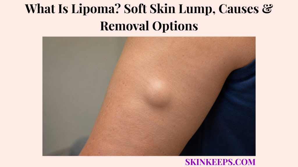
A lipoma is a common benign tumour of mature fat cells that usually appears as a soft, slowly growing and slightly mobile lump beneath the skin. Most ordinary lipomas sit in the subcutaneous layer, feel compressible or rubbery and are painless.
Most confirmed lipomas need no treatment. Removal becomes reasonable when a lipoma causes pain, pressure symptoms, movement or clothing interference, continues to enlarge, creates substantial cosmetic concern or remains diagnostically uncertain; atypical masses should be evaluated before routine removal.
A new unexplained lump deserves medical assessment, especially when it is rapidly enlarging, hard, fixed, significantly painful, deep, larger than about 5 cm, recurrent after removal or associated with numbness or movement symptoms. These features do not prove cancer, but they make further soft-tissue evaluation more appropriate.
How Can You Recognize a Lipoma?
A typical lipoma is a soft or rubbery, slightly mobile, slow-growing lump located just beneath otherwise normal-looking skin.
What Does a Lipoma Feel Like?
A typical lipoma feels soft or rubbery and can often be moved slightly beneath the skin with gentle pressure.
The lesion usually sits in subcutaneous fat and feels compressible rather than firmly attached to deeper fascia. Overlying skin is generally normal, without a central opening, drainage, ulceration or persistent redness.
Mobility is helpful but not absolute; some lipomas move less freely because of location or depth.
How Quickly Does a Lipoma Grow?
Most lipomas enlarge slowly over months or years and may remain nearly unchanged for long periods.
Continued rapid enlargement is less typical and should prompt reassessment. Growth alone does not mean a lump is malignant, but a changing pattern lowers confidence in a simple clinical diagnosis.
Where Do Lipomas Commonly Develop?
Lipomas commonly develop in subcutaneous fat on the shoulders, trunk, back, arms, buttocks and thighs.
They can occur elsewhere, including the forehead. Most are superficial, while deeper or intramuscular lipomas are less common and can be harder to classify by examination alone.
Are Lipomas Usually Painful?
Most ordinary lipomas are painless, although larger lesions can occasionally cause discomfort by pressing on nearby nerves or structures.
Pressure can produce localized discomfort, numbness or movement interference. Angiolipoma is a painful fatty-tumour variant, but pain by itself does not establish malignancy.
Figure 1. A typical superficial lipoma is soft or rubbery, slightly mobile, slow-growing and usually painless beneath normal-looking skin.
Why Do Lipomas Develop, and Why Can Some People Have Several?
Lipomas form when mature fat cells develop into a localized benign tumour, although the exact reason most individual lipomas arise remains unclear.
What Causes a Lipoma to Form?
A lipoma forms through localized benign growth of mature adipocytes, but the initiating cause is usually unknown.
Some lipomas show characteristic chromosomal or genetic alterations in the tumour cells, yet routine molecular testing is unnecessary for an ordinary superficial lesion with a typical clinical pattern.
Is a Lipoma Caused by Being Overweight?
No; an ordinary lipoma is not simply a deposit of excess body fat caused by being overweight.
A lipoma is a localized tumour rather than ordinary fat storage. Weight loss therefore does not usually make an established lipoma disappear, although the lump may become more noticeable if surrounding fat decreases.
Can Lipomas Run in Families?
Yes; most lipomas are isolated, but multiple lipomas can occur in familial or syndromic patterns.
Family history becomes more relevant when a person has numerous similar lesions or several relatives have multiple lipomas. Multiple lesions do not automatically mean a hereditary syndrome.
Are All Fatty Lumps the Same Type?
No; ordinary lipoma is the most common pattern, but other fatty tumours differ in pain, depth or histologic structure.
Angiolipoma can be painful, intramuscular lipoma extends into muscle, and spindle-cell or other variants have distinct pathologic features. These variants remain separate from the ordinary superficial lipoma that this article focuses on.
Can an Ordinary Lipoma Turn Into Liposarcoma?
An ordinary lipoma almost never transforms into liposarcoma; liposarcoma generally develops independently as a separate malignant tumour.
The clinically important problem is not whether a confirmed conventional lipoma is “turning cancerous,” but whether an unusual fatty mass was actually a different tumour from the beginning. Atypical behaviour should therefore trigger a different diagnostic pathway rather than reassurance based only on a previous assumption.
How Can You Tell a Lipoma From Other or More Concerning Lumps?
A typical lipoma is superficial, soft, mobile and slow-growing, while cysts and concerning soft-tissue masses often differ in skin attachment, firmness, depth, growth pattern or other clinical features.
How Is a Lipoma Different From an Epidermoid Cyst?
A lipoma usually moves beneath normal skin, whereas an epidermoid cyst is more closely attached to the skin and may have a visible central punctum.
An epidermoid cyst can inflame, become red or tender and release keratinous material if it opens. A lipoma usually sits deeper in subcutaneous fat, has a softer feel and moves more independently of the skin surface.
How Is a Lipoma Different From a Swollen Lymph Node?
A lipoma can occur in many fatty body areas, whereas enlarged lymph nodes occur in characteristic lymph-node regions such as the neck, armpits and groin.
Recent infection or illness can support a lymph-node explanation, but mobility alone cannot diagnose a lipoma. A persistent unexplained lump in a lymph-node region still deserves assessment.
Which Features Are Less Typical of a Simple Lipoma?
Rapid enlargement, hardness, fixation, deep location, substantial pain, recurrence or large size are less typical of an ordinary superficial lipoma and justify closer assessment.
A size around or above 5 cm is a caution feature used in soft-tissue referral guidance, not a cancer diagnosis. Smaller masses can still be atypical, and many larger lumps remain benign.
How Is a Lipoma Different From Liposarcoma?
Liposarcoma is a malignant fatty tumour that more often arises in deeper soft tissues and may behave as an enlarging, firmer or less mobile mass rather than a classic superficial lipoma.
Deep extremity or retroperitoneal masses, continued enlargement and lower mobility can raise concern, but clinical examination cannot exclude every sarcoma. Imaging and, when needed, biopsy determine the next step.
Figure 2. Rapid growth, hardness, fixation, depth, recurrence or large size does not prove cancer, but it moves the lump away from the simple-lipoma pathway and toward further evaluation.
| Feature | Lipoma | Epidermoid Cyst | Concerning Soft-Tissue Mass |
|---|---|---|---|
| Texture | Usually soft or rubbery | Often firmer | Variable; may be firm |
| Skin attachment | Usually moves independently beneath skin | Often attached to skin | Variable; fixation can be concerning |
| Central punctum | Absent | May be present | No defining pattern |
| Growth | Usually slow | Variable | Continued or rapid enlargement may occur |
| Depth | Usually subcutaneous | Skin/subcutaneous | May be deep to fascia |
| Next step | Observe if classic | Clinical cyst pathway | Imaging / specialist assessment |
How Is a Lipoma Diagnosed?
A typical superficial lipoma can often be diagnosed clinically, while ultrasound, MRI or biopsy is reserved for enlarging, deep or diagnostically uncertain masses.
Can a Doctor Diagnose a Lipoma by Examination?
Yes; a superficial, soft, mobile and slowly growing lipoma can often be diagnosed from clinical examination alone.
Examination records approximate size, depth, consistency, mobility, tenderness, growth history and relationship to surrounding structures. Atypical lumps should not be classified by touch alone.
When Is an Ultrasound Useful for a Suspected Lipoma?
Ultrasound is useful when a soft-tissue lump is enlarging or its depth and benign fatty nature are uncertain.
Ultrasound can help determine whether a lesion is superficial or deep and whether its appearance fits a benign fatty mass. NICE guidance specifically recommends considering urgent direct-access ultrasound for an unexplained adult soft-tissue lump that is increasing in size.
When Is MRI Needed for a Suspected Lipoma?
MRI is most useful for large, deep or anatomically complex masses when ultrasound or examination cannot confidently establish a simple benign lipoma.
MRI defines relationships to muscle, fascia, nerves and surrounding structures and can characterize internal tumour features before specialist planning.
When Is a Biopsy Needed for a Suspected Lipoma?
Biopsy is considered when clinical or imaging findings are atypical or when a malignant soft-tissue tumour needs to be excluded.
A classic small superficial lipoma may not require biopsy. Tissue diagnosis becomes more important when imaging is uncertain or the mass enters a malignant soft-tissue differential; an excised lesion can also be sent for histopathology.
Why Should a Suspicious Mass Be Investigated Before Routine Removal?
A suspicious soft-tissue mass should be investigated before routine removal because poorly planned excision can complicate definitive treatment if the lesion is malignant.
The safer sequence for an atypical mass is imaging first, specialist assessment where appropriate and biopsy when indicated, followed by planned definitive treatment rather than casual office removal.
Figure 3. A classic superficial lipoma may need no imaging, while enlarging, deep or uncertain masses can progress from ultrasound to MRI, specialist review and biopsy when indicated.
When Should a Lipoma Be Removed, and Which Removal Options Are Used?
A confirmed lipoma usually needs removal only when it causes symptoms, grows, interferes with function, creates substantial cosmetic concern or remains diagnostically uncertain.
Does Every Lipoma Need Removal?
No; most confirmed asymptomatic lipomas can safely be observed without treatment.
Removal becomes reasonable for pain, continued enlargement, repeated irritation, pressure on a nerve or nearby structure, movement limitation, clothing interference, substantial cosmetic concern or diagnostic uncertainty. Observation is an active management choice, not neglect.
How Is a Lipoma Removed Surgically?
Surgical excision removes the lipoma and its capsule through an incision and is the standard definitive treatment.
A small superficial lesion may be removed under local anaesthetic, while larger or deeper lipomas can require a different surgical setting. The removed tissue may be sent for pathology, particularly when diagnostic confirmation is useful.
What Are the Possible Downsides of Lipoma Excision?
The main trade-off of lipoma excision is a permanent surgical scar, with additional risks depending on the lesion’s size and location.
Bruising, bleeding, infection, fluid collection, altered sensation and injury to nearby structures are possible. Risk varies by site, size and depth rather than being identical for every lipoma.
Can Liposuction Remove a Lipoma?
Yes; liposuction can reduce selected lipomas through a smaller access site, but incomplete capsule removal can increase recurrence risk.
The smaller access incision may appeal cosmetically, but the capsule can remain and fragmented tissue provides less complete histologic assessment. Liposuction is therefore not equivalent to complete excision for every lesion.
Can Laser Lipolysis Be Used for a Lipoma?
Laser-assisted lipolysis can be used for selected lipomas, but it provides less certainty of complete capsule removal than full surgical excision.
It may offer a smaller access site in selected cosmetic cases, but incomplete removal and recurrence remain considerations. It is not the universal standard for ordinary lipoma treatment.
Can a Lipoma Return After Removal?
Yes; lipoma recurrence is possible but uncommon after complete excision.
Recurrence is more likely when part of the lesion or capsule remains. A lump that returns should be reassessed rather than automatically labelled a harmless recurrence.
When Should a Lipoma or Suspected Lipoma Be Checked Again?
A lipoma or suspected lipoma should be reassessed if it begins enlarging, becomes hard or fixed, develops persistent pain, causes nerve or movement symptoms, or returns after removal.
An unexplained enlarging adult soft-tissue mass belongs in an imaging or specialist pathway rather than being repeatedly assumed to be a lipoma.
Figure 4. Observation is reasonable for a confirmed asymptomatic lipoma. Symptoms, functional problems or cosmetic concerns can justify removal, while diagnostic uncertainty should lead to investigation before treatment.
What Should You Remember About Lipoma?
A lipoma is a benign, slow-growing fatty tumour that usually feels soft and mobile, while atypical or enlarging masses need further evaluation before they are assumed to be ordinary lipomas.
- Lipoma is benign and is composed of mature fat cells.
- Typical lesions are soft, slightly mobile, superficial and slow-growing.
- Most ordinary lipomas are painless.
- The exact cause is usually unknown.
- Being overweight is not the direct cause of an ordinary lipoma.
- Multiple or familial patterns can occur.
- Conventional lipomas almost never transform into liposarcoma.
- Rapid growth, hardness, fixation, depth, recurrence or large size changes the diagnostic pathway.
- Classic superficial lipomas often need no testing.
- Ultrasound, MRI and biopsy are used selectively.
- Most confirmed asymptomatic lipomas need no treatment.
- Complete surgical excision is the most definitive removal option.
- Recurrence after complete excision is uncommon.
Recognize → check for atypical features → confirm if needed → observe or remove → reassess recurrence or change.
Frequently Asked Questions About Lipoma
The most common lipoma questions concern why these fatty lumps form, how they are recognized, whether they can become cancerous, when removal is useful and whether they recur.
What Causes a Lipoma to Form?
A lipoma forms through localized benign growth of mature fat cells, although the exact trigger is usually unknown. Genetic changes can occur within lipomas, but most ordinary lesions do not require molecular testing.
How Can You Tell Whether a Lump Is a Lipoma?
A typical lipoma is soft, slightly mobile, slow-growing and located just beneath normal-looking skin. Rapid growth, fixation, hardness or deep location should prompt assessment rather than self-diagnosis.
Can a Lipoma Turn Into Cancer?
An ordinary lipoma almost never transforms into cancer; liposarcoma generally arises independently as a separate malignant tumour. The concern with an atypical mass is whether it was truly a conventional lipoma from the beginning.
When Should a Lipoma Be Removed?
A lipoma may be removed when it causes pain, enlarges, presses on nearby structures, interferes with function, creates significant cosmetic concern or remains diagnostically uncertain. A confirmed asymptomatic lipoma can usually be observed.
Can a Lipoma Grow Back After Removal?
Yes; recurrence can occur, but it is uncommon after complete surgical excision. A recurrent lump should be reassessed, especially if its behaviour differs from the original lesion.
Which Sources Support This Lipoma Guidance?
NHS — Lipoma — Used for the typical soft, slightly mobile, slowly growing and usually painless pattern, common body sites, clinical diagnosis and observation/removal context.
DermNet — Lipoma — Used for benign adipocyte biology, deep/intramuscular context, pressure symptoms, multiple/familial lipomas, malignant-transformation framing, excision, liposuction, laser lipolysis and recurrence.
DermNet — Lipoma Pathology — Used for mature adipocyte histology, capsule and characteristic cytogenetic alterations.
StatPearls / NCBI Bookshelf — Lipoma — Used for benign subcutaneous presentation, multiple-lipoma syndromic context and clinical differential information.
NICE — Suspected Cancer: Recognition and Referral — Used specifically for the urgent ultrasound pathway for an unexplained enlarging adult soft-tissue lump.
NHS — Tests and Next Steps for Soft Tissue Sarcoma — Used for specialist imaging, including ultrasound and MRI, and biopsy in suspicious soft-tissue masses.
UK Guidelines for the Management of Soft Tissue Sarcomas — Used for caution features such as increasing size, size above about 5 cm, deep location and pain, while preserving that these features are referral signals rather than a diagnosis of malignancy.



