
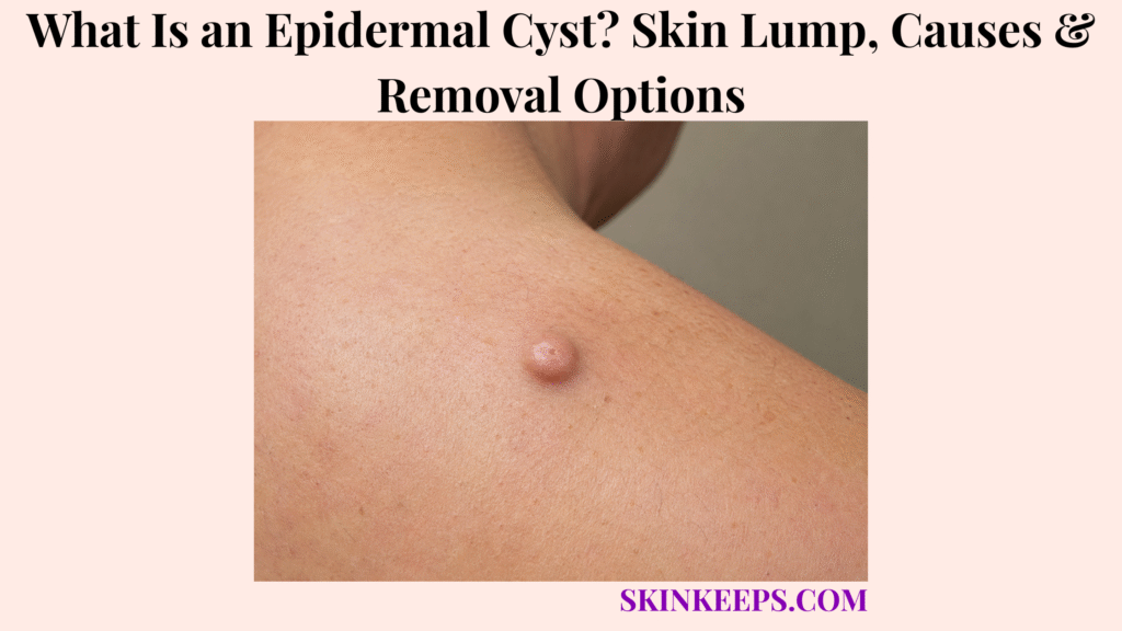
An epidermal cyst is a usually benign skin lump that forms when skin cells and keratin become trapped beneath the skin surface inside a cyst wall.
Epidermal cysts are often called epidermoid cysts or epidermal inclusion cysts, while “sebaceous cyst” is often inaccurate. This page covers appearance, causes, locations, diagnosis, observation, inflamed cyst care, infection signs, drainage, excision, recurrence, scar expectations, home care, mistakes, and doctor-warning signs.
What Is an Epidermal Cyst and What Is Inside It?
An epidermal cyst is a usually benign skin lump that forms when skin cells and keratin become trapped beneath the skin surface inside a cyst wall.
The cyst wall, lining, or capsule forms a closed sac under the skin. That lining can keep producing keratin, so the lump may slowly enlarge over time.
Keratin is not ordinary pus. It is a thick skin-cell protein material that may look yellow, cheesy, pasty, or foul-smelling if the cyst opens or drains.
Why Is Epidermal Cyst Often Called Epidermoid Cyst?
Epidermal cyst and epidermoid cyst are often used for the same common keratin-filled skin cyst.
Epidermal inclusion cyst, follicular infundibular cyst, infundibular cyst, and keratin cyst are related medical terms.
These names point to the same core idea: a skin-lined sac below the surface fills with keratin instead of disappearing like a normal shed skin cell.
Why Is “Sebaceous Cyst” Usually the Wrong Name?
“Sebaceous cyst” is usually the wrong name because most of these common cysts contain keratin from epidermal cells rather than oil from sebaceous glands.
Many people still use “sebaceous cyst” casually, but epidermal cyst, epidermoid cyst, or epidermal inclusion cyst is usually more accurate.
Using the correct term matters because safe treatment depends on the cyst wall, inflammation status, infection signs, and diagnostic certainty.
Practical rule: Do not squeeze, puncture, cut, burn, or acid-treat a cyst-like lump at home. Rupture can make inflammation, infection risk, scarring, and later removal more difficult.
What Does an Epidermal Cyst Look and Feel Like?
An epidermal cyst usually looks like a round, slow-growing lump under the skin that may feel firm, smooth, movable, and sometimes has a small central pore.
A calm cyst is often painless. The overlying skin may be skin-colored, white, yellowish, pink, brown, or darker depending on skin tone and irritation level.
What Are Common Epidermal Cyst Signs?
Common epidermal cyst signs include a round skin lump, slow growth, firm or rubbery feel, and sometimes a small pore-like opening.
The opening is called a punctum and may look like a tiny central pore or blackhead-like dot.
If the cyst opens, it may release thick keratin material with a strong odor, but forcing that material out does not remove the cyst lining.
What Symptoms Suggest Inflammation or Rupture?
Inflammation or rupture can make an epidermal cyst suddenly red, swollen, tender, painful, warm, enlarged, or draining.
An inflamed cyst can look boil-like, but inflamed does not always mean infected.
Ruptured keratin can irritate surrounding skin and create redness, pain, swelling, tenderness, and infection-like inflammation.
| Feature | Typical Epidermal Cyst Clue | When to Check |
|---|---|---|
| Growth | Slow, stable enlargement. | Rapid growth. |
| Feel | Firm or rubbery lump. | Rock-hard, fixed, very painful lump. |
| Opening | Small pore or punctum. | Ulcer, bleeding, non-healing sore. |
| Symptoms | Usually painless when calm. | Pain, warmth, fever, swelling. |
| Drainage | Thick keratin material. | Pus, foul drainage, spreading redness. |
| Skin tone | Skin-colored, pink, brown, or darker. | New color change with bleeding or ulceration. |
Where Do Epidermal Cysts Usually Appear?
Epidermal cysts can appear almost anywhere on the skin, but they are common on the face, neck, chest, upper back, shoulders, scalp, behind the ears, trunk, and genital skin.
They can also appear at sites of blocked follicles, repeated friction, acne injury, piercings, surgery scars, or earlier skin trauma.
Palms, soles, or buttocks can develop cysts through traumatic implantation of epidermal cells, even though those sites do not fit the usual follicle-based pattern.
| Body Site | Common Cyst Clue | Mimic to Consider |
|---|---|---|
| Face | Small round pore-linked bump. | Acne nodule, skin cancer, lipoma. |
| Neck | Movable skin-based lump. | Lymph node, lipoma, cyst. |
| Chest / back | Keratin-filled cyst in acne-prone area. | Acne nodule, abscess, skin cancer. |
| Scalp | Firm cyst-like lump. | Pilar cyst. |
| Behind ear | Pore-linked or inflamed lump. | Swollen node, abscess. |
| Genital skin | Small cyst-like nodule. | Folliculitis, hidradenitis, STI-related lesion. |
| Palms / soles | Trauma implantation context. | Wart, foreign body reaction, tumor. |
| Buttocks | Friction or trauma site. | Pilonidal disease, abscess. |
What Causes an Epidermal Cyst?
An epidermal cyst forms when epidermal cells move or become trapped beneath the skin surface and continue producing keratin inside a closed sac.
The cyst is not caused by dirty skin and does not spread by touch.
How Do Blocked or Damaged Follicles Cause Cysts?
Blocked or damaged follicles can cause epidermal cysts when skin cells collect below the surface and form a keratin-producing cyst wall.
The lump grows slowly as keratin builds inside the sac.
Acne-prone areas may be involved because follicle blockage and inflammation can damage the follicular opening.
How Can Injury, Surgery, or Piercing Cause an Epidermal Cyst?
Injury, surgery, or piercing can cause an epidermal cyst when epidermal cells are implanted deeper into the skin.
A cyst can appear long after a cut, puncture wound, piercing, surgery, or repeated irritation.
Not every cyst has a remembered injury, so absence of trauma does not rule it out.
Are Epidermal Cysts Caused by Poor Hygiene?
Epidermal cysts are not caused by poor hygiene.
Washing harder will not remove the cyst wall.
Scrubbing, picking, or squeezing can irritate the skin, rupture the cyst, and make a calm lump suddenly painful or inflamed.
| Cause Pathway | What Happens | Care Implication |
|---|---|---|
| Follicle damage | Skin cells become trapped below the surface. | Cyst wall forms. |
| Blocked follicle | Keratin collects inside a sac. | Slow lump growth. |
| Trauma / piercing | Epidermal cells are implanted deeper. | Cyst may form later. |
| Rupture | Keratin leaks into surrounding skin. | Red, painful inflammation. |
| Incomplete removal | Capsule remains. | Recurrence risk. |
Who Is More Likely to Get an Epidermal Cyst?
Epidermal cysts can affect almost anyone, but they are more likely after follicle damage, acne-prone skin, skin trauma, surgery, piercing, or repeated irritation.
Most people with one cyst do not have an inherited syndrome, but multiple, unusual, early-onset, or recurrent cyst patterns may need broader medical context review.
- Acne history.
- Follicle injury.
- Prior piercing.
- Prior surgery.
- Skin trauma.
- Repeated friction or irritation.
- Multiple cysts.
- Recurrent cysts.
- Unusual or early-onset cyst pattern.
Follicle blockage and inflammation may overlap with acne, but an epidermal cyst is a keratin-filled sac rather than an ordinary pimple.
How Is an Epidermal Cyst Different From Other Skin Lumps?
An epidermal cyst can resemble a pilar cyst, lipoma, abscess, boil, swollen lymph node, acne nodule, pilonidal cyst, dermoid cyst, or skin cancer, so uncertain lumps should be checked.
The goal is not to panic about every cyst-like bump, but to avoid false certainty when a lump is deep, fixed, rapidly changing, painful, bleeding, ulcerated, or repeatedly inflamed.
How Is an Epidermal Cyst Different From a Lipoma?
A lipoma is a fatty lump, while an epidermal cyst is a keratin-filled sac connected more closely with the skin surface.
Lipomas are often softer and deeper, while epidermal cysts may feel more skin-based and may have a punctum.
Both are often benign, but exam or imaging may be needed when the lump is unclear.
How Is an Epidermal Cyst Different From an Abscess or Boil?
An abscess or boil is an infected pus collection, while an epidermal cyst is usually a keratin-filled sac that can become inflamed or infected-looking.
A true infection is often painful, warm, red, swollen, or fever-associated.
Drainage may relieve pressure, but it does not always remove the cyst capsule, so the lump can come back later.
How Is an Epidermal Cyst Different From a Pilar Cyst?
A pilar cyst is another type of cyst that commonly appears on the scalp and arises from a different part of the hair follicle.
Pilar cysts may feel firm, occur in multiples, and run in families.
Scalp lumps often need distinction between epidermal cysts, pilar cysts, inflamed cysts, and less common scalp tumors.
How Is an Epidermal Cyst Different From a Swollen Lymph Node?
A swollen lymph node is usually deeper and follows lymph-node areas such as the neck, armpit, or groin, while an epidermal cyst is usually a skin-based lump.
Lymph nodes may enlarge with infection, inflammation, or malignancy.
Persistent unexplained lymph-node-like swelling, weight loss, night sweats, fever, or hard fixed swelling should be medically reviewed.
How Is an Epidermal Cyst Different From Skin Cancer?
Most epidermal cysts are benign, but a cyst-like bump that rapidly grows, bleeds, ulcerates, crusts, becomes fixed, or does not heal should be checked.
A non-healing shiny or bleeding bump may raise concern for basal cell carcinoma, while an ulcerated, crusted, thickened, or rapidly changing lump may need assessment for squamous cell carcinoma or another tumor.
Biopsy or pathology may be needed when diagnosis is uncertain.
| Lump | Main Clue | Why Confusion Happens | Safer Next Step |
|---|---|---|---|
| Epidermal cyst | Pore/punctum, keratin-filled sac. | Common skin lump. | Exam if uncertain. |
| Lipoma | Soft fatty lump. | Both can grow slowly. | Clinical exam or imaging if unclear. |
| Abscess / boil | Painful pus infection. | Inflamed cyst can mimic abscess. | Drain/culture if needed. |
| Pilar cyst | Scalp cyst-like lump. | Cyst overlap. | Dermatology/surgical exam. |
| Lymph node | Deeper node swelling. | Neck/groin lump overlap. | Check persistence/system symptoms. |
| Skin cancer | Bleeding, ulcerated, non-healing growth. | Cyst-like nodule possible. | Biopsy suspicious lesion. |
A firm skin bump that dimples when pinched may fit dermatofibroma better than an epidermal cyst, especially if there is no punctum or drainage history.
How Is an Epidermal Cyst Diagnosed?
An epidermal cyst is often diagnosed by examining the skin lump, but ultrasound, drainage evaluation, biopsy, or pathology may be used when the diagnosis is uncertain.
Diagnosis should separate a calm cyst from an inflamed cyst, infected-looking cyst, abscess, lymph node, lipoma, pilar cyst, pilonidal disease, or tumor.
What Does a Clinician Check?
A clinician checks the lump’s size, location, skin attachment, central punctum, mobility, tenderness, redness, warmth, drainage, and speed of growth.
The exam may also review prior inflammation, recurrence, trauma, acne, piercing, surgery, friction, fever, red streaking, diabetes, immune suppression, skin cancer history, and keloid tendency.
Photos showing change over time can help when a lump is slow-growing or recurrent.
When Might Ultrasound, Biopsy, or Pathology Be Needed?
Ultrasound, biopsy, or pathology may be needed when a lump is deep, fixed, rapidly growing, recurrent, atypical, ulcerated, bleeding, or difficult to identify.
Ultrasound can help characterize some soft-tissue masses when the diagnosis is unclear.
Tissue removed during excision may be sent for pathology, and drainage material may be cultured if infection is suspected.
- When the lump first appeared.
- Whether it is growing slowly or quickly.
- Pain, tenderness, warmth, redness, or swelling.
- Drainage, odor, bleeding, crusting, or ulceration.
- Whether a pore or punctum is visible.
- Prior rupture, inflammation, or recurrence.
- History of acne, injury, piercing, surgery, or friction at the site.
- Fever, red streaks, or spreading redness.
- Diabetes, immune suppression, skin cancer history, or keloid tendency.
- Photos showing change over time.
- Prior drainage, antibiotics, steroid injection, or removal.
Does an Epidermal Cyst Need Treatment?
An epidermal cyst does not always need treatment if it is small, painless, stable, and clearly benign.
Treatment may be chosen for pain, inflammation, repeated rupture, drainage, odor, cosmetic concern, friction, movement interference, or diagnostic uncertainty.
A calm cyst is usually easier to remove than an actively inflamed or ruptured cyst, so elective removal is often discussed before repeated flare-ups.
| Situation | Best Direction | Why |
|---|---|---|
| Small, stable, painless cyst | Observe. | Often harmless. |
| Cosmetic concern | Elective removal discussion. | Preference-based after diagnosis. |
| Recurrent inflammation | Consider excision. | Prevent repeated flares. |
| Painful swollen cyst | Treat inflammation/infection first. | Safer timing. |
| Uncertain lump | Diagnose, image, or biopsy. | Rule out mimics. |
| Rapid growth / bleeding | Prompt review. | Not typical cyst behavior. |
What Treatment Options Help an Inflamed or Infected Epidermal Cyst?
An inflamed epidermal cyst may need symptom treatment, steroid injection, drainage, antibiotics if true infection is present, or delayed excision after swelling settles.
Inflamed does not automatically mean infected. Ruptured keratin can create redness, swelling, tenderness, and warmth even when bacteria are not the main cause.
What Helps an Inflamed but Not Infected Cyst?
An inflamed but not infected cyst may be managed with comfort measures and clinician-directed inflammation treatment rather than automatic antibiotics.
Warm compresses may reduce mild discomfort, and a clinician may use a corticosteroid injection in selected inflamed cysts to reduce swelling.
Excision may be delayed until inflammation settles because inflamed or ruptured cysts can be harder to remove cleanly.
When Is Incision and Drainage Used?
Incision and drainage may be used when a cyst is very swollen, painful, fluctuant, draining, or abscess-like.
Drainage can ease pressure and symptoms, but it may leave the cyst wall behind.
Definitive excision may be planned later if the cyst recurs or if capsule removal is desired.
When Are Antibiotics Needed?
Antibiotics are not always needed for every red cyst, but they may be used when true infection or cellulitis is suspected.
True infection signs include spreading redness, warmth, pus, fever, red streaking, worsening pain, or cellulitis-like spread.
Culture may be needed in selected cases, especially when infection is recurrent, severe, or not responding as expected.
| Cyst State | Possible Care | Key Caution |
|---|---|---|
| Calm cyst | Observe or elective excision. | No squeezing. |
| Inflamed cyst | Compresses, injection, pain care. | Surgery may be delayed. |
| Ruptured cyst | Medical review if painful/swollen. | Looks infection-like. |
| Abscess-like cyst | Incision/drainage. | Capsule may remain. |
| True infection | Drainage ± antibiotics. | Watch cellulitis/fever. |
| Recurrent cyst | Complete excision later. | Capsule removal matters. |
What Removal Options Are Used for Epidermal Cysts?
Epidermal cyst removal aims to remove the cyst wall as well as the contents, because the cyst can return if the lining remains.
Removal is a medical procedure, not a home project. The best option depends on cyst size, location, inflammation, rupture, recurrence, scar risk, and diagnostic certainty.
What Is Complete Surgical Excision?
Complete surgical excision removes the cyst contents and capsule, which gives the best chance of preventing the same cyst from refilling.
It is often done under local anesthesia and may require stitches.
Tissue may be sent to pathology, especially if the diagnosis is uncertain, the lump is atypical, or removal is being done for recurrent or suspicious features.
What Is Minimal Excision?
Minimal excision uses a smaller opening to remove cyst contents and the capsule in selected cases.
It may reduce scar size in suitable cysts, but it is not ideal for every cyst.
Inflamed, ruptured, scarred, or recurrent cysts can be harder to remove through a small opening.
Why Is Drainage Alone Not Complete Removal?
Drainage alone is not complete removal because it empties the cyst contents but may leave the cyst lining behind.
If the lining remains, keratin can build up again and the cyst may refill.
Drainage can still be useful for acute swelling, pressure, pain, or infection-like presentation.
What Scar Expectations Should Be Discussed?
Any epidermal cyst removal can leave a scar, and scar risk depends on cyst size, location, inflammation, rupture, recurrence, and personal healing pattern.
Back, chest, shoulder, jawline, and keloid-prone areas may need extra scar discussion.
A small incision does not guarantee no scar, and cosmetic expectations should be discussed before removal.
| Option | Best-Fit Goal | Limitation |
|---|---|---|
| Observation | Small stable cyst. | Lump remains. |
| Steroid injection | Reduce inflammation. | Does not remove cyst. |
| Incision/drainage | Painful swollen drainage need. | Recurrence likely if capsule remains. |
| Minimal excision | Smaller selected cysts. | Not always suitable. |
| Complete excision | Definitive removal. | Scar/stitches possible. |
| Biopsy/pathology | Uncertain lump. | Diagnostic, not cosmetic only. |
Can an Epidermal Cyst Come Back After Removal?
An epidermal cyst can come back if the cyst wall or lining is not fully removed.
Drainage alone has a higher recurrence concern because the contents may be emptied while the capsule remains.
Ruptured or inflamed cysts can be harder to remove completely, and scar tissue from repeat inflammation or prior drainage can make later surgery more difficult.
How Should You Care for an Epidermal Cyst at Home?
At home, the safest approach is to avoid squeezing the cyst, protect the skin, use warm compresses for mild tenderness, and seek care if inflammation or infection signs appear.
Home care should never become home surgery. Needles, blades, wart acids, harsh chemicals, burning tools, or attempts to pull out the capsule can cause injury and delay diagnosis.
- Do not squeeze.
- Do not puncture with a needle.
- Do not cut the cyst.
- Do not use acids or harsh chemicals.
- Keep the area clean.
- Use warm compresses for mild discomfort.
- Reduce friction from clothing, shaving, collars, helmets, or straps.
- Cover drainage with a clean dressing and seek advice.
- Photograph changes if monitoring.
- Seek care for pain, fever, spreading redness, rapid growth, bleeding, or ulceration.
What Epidermal Cyst Mistakes Should You Avoid?
The biggest epidermal cyst mistake is squeezing or cutting the lump at home, because this can rupture the cyst, worsen inflammation, cause infection, and make later removal harder.
Do not treat it like a pimple, drain it with a needle, dig out the capsule, keep squeezing foul-smelling keratin, or assume antibiotics remove the cyst wall.
Do not remove a suspicious lump cosmetically without diagnosis, and do not delay care for fever, spreading redness, severe pain, rapid growth, bleeding, ulceration, or a hard fixed lump.
| Mistake | Why It Fails | Better Action |
|---|---|---|
| Squeezing | Rupture/inflammation risk. | Leave alone or see clinician. |
| Needle drainage | Infection/scarring risk. | Medical drainage if needed. |
| Digging out capsule | Injury and incomplete removal risk. | Clinician excision. |
| Antibiotics only | Capsule remains. | Excision if recurrent. |
| Ignoring rapid change | Missed mimic risk. | Prompt exam. |
| Removing while very inflamed | Harder surgery/scarring. | Treat inflammation first if safe. |
When Should an Epidermal Cyst Be Checked by a Doctor?
An epidermal cyst should be checked when it becomes painful, red, warm, swollen, draining, rapidly growing, recurrent, ulcerated, bleeding, fixed, or difficult to identify.
People with diabetes, immune suppression, skin cancer history, recurrent cysts, large or deep lumps, or keloid-prone skin should be more cautious.
Which Cyst Signs Need Medical Review?
Medical review is needed for sudden pain, rapid swelling, redness, warmth, pus, foul drainage, fever, red streaks, bleeding, ulceration, rapid growth, or a hard fixed lump.
Review is also useful for cysts on the face, genitals, breast, scalp, spine, or high-friction areas.
A cyst interfering with movement, sleep, shaving, clothing, or daily life may also need planned care.
Which People Should Be More Cautious?
People with diabetes, immune suppression, skin cancer history, recurrent cysts, large or deep lumps, or keloid-prone skin should be more cautious.
Babies or children with unusual lumps should be assessed because childhood lumps may have different causes.
Keloid-prone patients considering removal should discuss scar risk before any procedure.
Seek medical review if a cyst-like lump is:
- Painful.
- Red, warm, or swollen.
- Draining pus or foul material.
- Linked with fever or red streaks.
- Rapidly growing.
- Recurrent or repeatedly rupturing.
- Bleeding or ulcerated.
- Hard, fixed, or unusually deep.
- Difficult to identify.
- On the face, genitals, breast, scalp, spine, or high-friction area.
- Present in someone with diabetes, immune suppression, skin cancer history, keloid tendency, or unusual childhood lumps.
What Should You Remember About an Epidermal Cyst?
An epidermal cyst is usually a benign keratin-filled skin lump, but safe management depends on whether it is calm, inflamed, infected-looking, recurrent, or diagnostically uncertain.
The central safety rule is simple: do not squeeze or cut the cyst at home, and remember that drainage empties contents while excision targets the capsule.
- Epidermal cyst is also called epidermoid cyst or epidermal inclusion cyst.
- “Sebaceous cyst” is often inaccurate.
- The cyst contains keratin, not ordinary pus.
- Many cysts are small, slow-growing, and painless.
- Do not squeeze or cut them at home.
- Inflamed cysts can become red, swollen, painful, and boil-like.
- Inflamed does not always mean infected.
- Drainage can relieve symptoms but may not prevent recurrence.
- Complete excision removes the cyst wall and lowers recurrence risk.
- Any removal can leave a scar.
- Suspicious, rapidly growing, bleeding, ulcerated, fixed, or recurrent lumps need medical review.
Frequently Asked Questions About Epidermal Cysts
Is an epidermal cyst the same as a sebaceous cyst?
Most lumps people call “sebaceous cysts” are actually epidermoid cysts or epidermal inclusion cysts. True sebaceous cysts are different and less common.
What is inside an epidermal cyst?
An epidermal cyst contains keratin produced by epidermal cells lining the cyst wall. The material may be thick, yellowish, cheesy, or foul-smelling if the cyst opens.
Are epidermal cysts dangerous?
They are usually benign and often do not need treatment when small and painless, but painful, inflamed, infected-looking, rapidly growing, bleeding, ulcerated, fixed, or uncertain lumps should be checked.
Should I pop an epidermal cyst?
No. Squeezing can rupture the cyst wall, worsen inflammation, increase infection risk, cause scarring, and make later removal harder.
What is the best treatment for an epidermal cyst?
Treatment depends on the cyst state. Small painless cysts may be observed, inflamed cysts may need clinician-directed inflammation care, abscess-like cysts may need drainage, and definitive removal usually requires excision of the cyst and capsule.
Can an epidermal cyst come back after drainage?
Yes. Drainage removes cyst contents but may leave the cyst lining behind, so recurrence is possible.
When should an epidermal cyst be removed?
Removal may be discussed for pain, repeated inflammation, drainage, odor, cosmetic concern, friction, recurrent rupture, diagnostic uncertainty, rapid growth, or suspicious features.
When should a cyst-like lump be checked urgently?
A cyst-like lump should be checked urgently for fever, spreading redness, warmth, severe pain, red streaks, pus, rapid growth, bleeding, ulceration, fixed texture, or high-risk context such as diabetes, immune suppression, or skin cancer history.
Sources & Evidence About Epidermal Cysts
DermNet — Epidermoid Cyst was used for epidermoid cyst, epidermal inclusion cyst, follicular infundibular cyst, keratin cyst terminology, “sebaceous cyst” misnomer, clinical features, traumatic implantation, rupture inflammation, complete excision with intact capsule, and recurrence reduction.
Mayo Clinic — Epidermoid Cysts: Symptoms and Causes was used for small round bump, blackhead-like opening, thick yellow smelly drainage, slow-growing painless nature, inflammation/swelling, keratin contents, and irritation or injury cause.
Mayo Clinic — Epidermoid Cysts: Diagnosis and Treatment was used for diagnosis by skin exam, steroid injection, incision and drainage, surgery delayed if inflamed, recurrence after drainage, and warm moist cloth support.
StatPearls / NCBI Bookshelf — Epidermal Inclusion Cyst was used for rupture-related inflammation, cellulitis-like symptoms, differential diagnosis including tumors, drainage and antibiotic pathway for acute infection, and complete cyst capsule removal.
MedlinePlus — Epidermoid Cyst was used for closed-sac framing, dead-skin-cell contents, diagnosis by exam, symptom/inflammation-based treatment need, steroid injection context, and sebaceous versus epidermoid distinction.
Educational Disclaimer: This SkinKeeps article is for educational purposes only and does not diagnose or replace medical care. A painful, red, warm, swollen, draining, foul-smelling, pus-filled, fever-associated, red-streaked, rapidly growing, bleeding, ulcerated, fixed, recurrent, repeatedly ruptured, deep, unusually large, face-related, genital-related, breast-related, scalp-related, spine-related, child-related, diabetes-associated, immune-suppression-associated, skin-cancer-history-associated, keloid-prone, or uncertain cyst-like lump should be checked by a qualified healthcare professional. Do not squeeze, pop, puncture, cut, burn, acid-treat, dig out, or drain a cyst-like lump at home. Do not rely on antibiotics, cosmetic removal, or drainage alone when the diagnosis is uncertain or recurrence is likely.



