
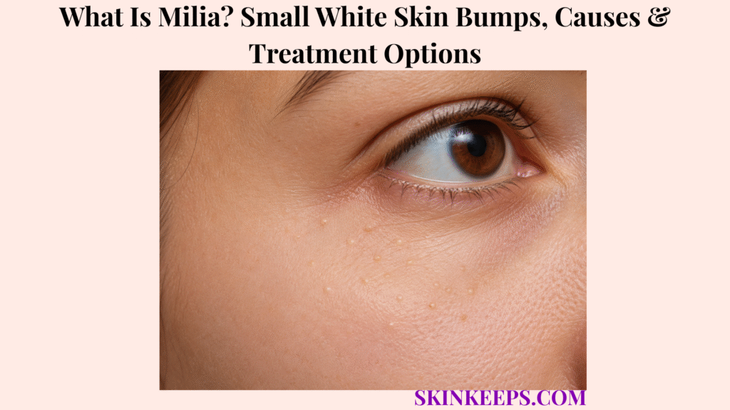
Milia are tiny benign keratin-filled cysts that appear as firm pearly-white or yellowish bumps just beneath the skin surface. Keratin is a structural protein made by epidermal skin cells, and a milium usually forms as a small dome-shaped cyst on the cheeks, eyelids or other facial skin.
Milia can affect newborns, children and adults, and many lesions resolve naturally without treatment. Adult milia can persist much longer, but they are not acne, whiteheads, trapped sebum or a contagious infection, so correct identification matters before squeezing or applying acne treatments.
An ordinary milium is harmless, but a small white or pale bump should be reassessed if it grows rapidly, becomes painful, repeatedly inflames, bleeds, crusts, changes colour or shape, appears as part of an unexplained widespread eruption, or no longer behaves like the original diagnosis. Atypical behaviour matters because not every small pale facial bump is milia.
How Can You Recognize Milia?
Milia usually appear as tiny, firm, dome-shaped pearly-white or yellowish bumps beneath otherwise normal-looking skin.
What Do Milia Look and Feel Like?
Typical milia are firm 1–2 mm dome-shaped papules that usually appear pearly white or yellowish.
The bumps are solid-feeling rather than soft or fluid-filled, and the surrounding skin often looks normal. Ordinary milia are usually painless and not significantly itchy.
Many lesions fall near the 1–2 mm range, but exact size is not a rigid diagnostic cutoff.
Where Do Milia Usually Develop?
Milia most commonly develop on the face, especially around the eyelids and cheeks, although they can occur elsewhere.
Other common facial sites include the nose and forehead. Less commonly, milia can occur on the upper trunk, limbs or genital skin.
Why Are Milia Common in Newborns?
Newborn milia are common temporary keratin cysts that usually clear spontaneously as immature skin matures.
They commonly appear on the nose, cheeks, chin, scalp and nearby facial skin during early life. Most disappear over the following weeks without scarring.
Newborn milia should not be squeezed and should not be treated as acne.
Can Adults Develop Milia?
Yes; adults can develop milia spontaneously or after skin injury, inflammation or selected treatments.
Primary adult milia can occur without a preceding injury, while secondary milia develop in a healing or inflammatory context. Adult lesions may persist for months or longer without becoming dangerous.
Persistent lesions can be removed for cosmetic reasons after the diagnosis is clear.
Figure 1. A milium forms when keratin is trapped inside a tiny superficial cyst, explaining its firm texture and why squeezing it like a pimple usually fails.
Why Do Milia Develop, and Which Types Matter?
Milia form when keratin becomes trapped in a tiny superficial cyst, either spontaneously as primary milia or during healing after skin damage as secondary milia.
What Is Inside a Milium?
A milium contains keratin, the structural protein produced by epidermal skin cells.
Keratin becomes enclosed beneath the surface and forms a small cyst. This mechanism differs from acne because milia are not sebum-filled follicular plugs.
What Are Primary Milia?
Primary milia develop spontaneously from the epidermis or pilosebaceous unit without a preceding skin injury.
This group includes neonatal milia, common facial lesions in children and spontaneous adult milia. A burn, blister or procedure is not required.
What Are Secondary or Traumatic Milia?
Secondary milia develop as keratin cysts during healing after epidermal injury or significant inflammation.
Reported contexts include burns, blistering disorders, abrasions, trauma, dermabrasion, radiotherapy and other forms of epidermal repair. Skin damage → repair → keratin trapping → secondary milia is the practical mechanism.
Not every burn, blister or procedure produces milia.
Can Medicines or Skin Treatments Cause Milia?
Selected medicines or skin treatments can be associated with secondary milia when they alter epidermal turnover or cause significant inflammation.
Medication history becomes more relevant when numerous new lesions begin after a treatment change or prolonged topical exposure. One isolated milium provides much weaker evidence for a medication association.
Prescribed medicines should not be stopped independently because of suspected milia.
What Is Milia En Plaque?
Milia en plaque is an uncommon pattern in which numerous milia cluster within an elevated abnormal plaque.
It may occur on an eyelid, behind the ear, cheek or jaw and can be associated with an underlying inflammatory skin condition. This grouped plaque pattern is different from a few ordinary isolated milia.
What Are Multiple Eruptive Milia?
Multiple eruptive milia describes the development of numerous milia over a relatively short period rather than a few isolated cysts.
The lesions can appear in crops over weeks to months and may occur without an obvious cause. Unusual widespread disease deserves dermatologic assessment rather than being reduced to a rare-syndrome checklist.
| Pattern | Main Mechanism | Typical Context | Practical Meaning |
|---|---|---|---|
| Primary milia | Spontaneous keratin trapping | Newborns, children, adults | Usually benign; observation often enough |
| Secondary milia | Keratin trapping during skin repair | Burns, blistering, trauma, procedures | Address injury/inflammatory context |
| Milia en plaque / eruptive pattern | Numerous clustered or widespread lesions | Uncommon presentation | Closer diagnostic context may be needed |
How Are Milia Different From Whiteheads and Other Small Facial Bumps?
Milia are firm keratin cysts beneath the skin surface, while whiteheads and several other facial bumps arise from different skin structures and require different treatment.
How Are Milia Different From Whiteheads?
A milium is a firm keratin-filled cyst, whereas whiteheads or closed comedones form when a hair follicle becomes blocked by sebum and keratin.
Milia are usually pearly, firm and stable and do not have the follicular acne plug found in whiteheads. This is why treating milia like pimples or trying to squeeze them often fails.
Whiteheads belong to the broader acne pathway, while milia are superficial keratin cysts.
How Are Milia Different From Sebaceous Hyperplasia?
Sebaceous hyperplasia usually forms yellowish or skin-coloured adult facial papules with a small central depression, whereas milia are typically smaller, firmer and pearly white.
Sebaceous hyperplasia commonly appears on the forehead or cheeks and arises from enlarged sebaceous glands. Milia do not characteristically have the central depression seen in sebaceous hyperplasia.
How Are Milia Different From Syringomas?
Syringomas are benign sweat-duct papules that often cluster around the lower eyelids, whereas milia are superficial keratin cysts containing a keratin pearl.
Syringomas are commonly skin-coloured or yellowish and may look less pearly than milia. The overlap can be substantial enough that patients cannot always distinguish the two visually.
Can Milia Be Diagnosed by Appearance Alone?
Yes; ordinary milia can usually be diagnosed clinically from their tiny, firm, pearly appearance and typical distribution.
Routine laboratory testing and imaging are unnecessary for a classic lesion. Clinical confidence falls when the bump changes, forms an unusual plaque or appears in an unexplained widespread pattern.
When Is a Biopsy Needed for a Suspected Milium?
Biopsy is rarely needed for ordinary milia but can be appropriate when the diagnosis is uncertain or the lesion has atypical morphology or distribution.
Possible reasons include an abnormal plaque, unusual persistent lesion, widespread unexplained eruption or a cystic or adnexal tumour differential that cannot be resolved clinically.
| Feature | Milia | Whitehead | Sebaceous Hyperplasia | Syringoma |
|---|---|---|---|---|
| Core lesion | Keratin cyst | Closed comedone | Enlarged sebaceous gland | Sweat-duct tumour |
| Texture | Firm | Follicular plug | Soft/firm papule | Firm papule |
| Colour | Pearly white/yellow | White/skin-coloured | Yellow/skin-coloured | Skin-coloured/yellow |
| Central depression | No | No | Often present | No |
| Common site | Eyelids/cheeks | Acne-prone areas | Forehead/cheeks | Lower eyelids |
| Management direction | Observe or professional extraction | Acne-specific care | Different benign-lesion pathway | Different adnexal-lesion pathway |
Do Milia Need Treatment, and How Are They Removed Safely?
Most milia do not require treatment, but persistent or cosmetically bothersome lesions can be professionally extracted or treated with selected dermatologic therapies.
Do Milia Go Away Without Treatment?
Yes; many milia resolve naturally, especially neonatal milia, although some adult lesions can persist for months or longer.
Spontaneous clearance is particularly common in newborns. Persistent adult milia remain benign, so treatment is generally optional when the diagnosis is secure.
How Does a Dermatologist Remove a Milium?
A dermatologist can remove an isolated milium by creating a tiny sterile opening and gently extracting the keratin cyst contents.
This is a clinician-controlled procedure used for a persistent lesion when the diagnosis is clear and the patient wants removal. The purpose is rapid removal without unnecessary surrounding trauma.
Why Should Milia Not Be Squeezed at Home?
Milia should not be repeatedly squeezed because the keratin cyst lies beneath intact epidermis and force may injure the surrounding skin without removing it.
Repeated squeezing or picking can cause bleeding, inflammation, infection, post-inflammatory pigmentation and scarring. Periocular skin is especially delicate, and home needle extraction should be avoided.
Can Retinoids Help Multiple or Persistent Milia?
Topical retinoids can help selected patients with multiple or persistent milia by increasing epidermal turnover.
They are unnecessary for newborn milia and usually unnecessary for one harmless isolated lesion. Irritation can occur, so periocular use requires extra caution and professional guidance.
Which Procedures Can Treat Numerous or Resistant Milia?
Selected resistant or numerous milia can be treated with procedures such as curettage, cryotherapy, electrodessication or laser, but these are not first-line choices for every isolated milium.
More intensive procedures bring greater potential for irritation, pigment alteration and scarring. Lesion burden, location and diagnosis determine whether a procedural option is reasonable.
How Are Secondary Milia Treated?
Secondary milia management should address the underlying skin injury or inflammatory disorder as well as the individual keratin cysts.
Burns, blistering diseases, trauma and procedures can create the repair environment in which secondary milia form. The practical approach is treat cause context + cyst burden rather than repeated extraction alone.
Figure 2. Newborn and mild ordinary milia are usually observed; persistent isolated lesions may be extracted professionally, while numerous, secondary or atypical lesions need a broader management decision.
Can Milia Be Prevented, and When Should They Be Rechecked?
Primary milia cannot be reliably prevented, but avoiding unnecessary skin trauma and treating underlying skin disease appropriately may reduce some secondary milia.
Can Primary Milia Be Completely Prevented?
No; primary milia can develop spontaneously, so no cleansing or skin-care routine guarantees prevention.
Newborn milia and spontaneous adult milia are not evidence of dirty skin. Milia are not caused by poor hygiene.
Can Skin Care Reduce Recurrent Adult Milia?
Gentle skin care can reduce avoidable irritation, but no universal skin-care routine reliably prevents recurrent adult milia.
Gentle cleansing and avoiding aggressive scrubbing or repeated periocular exfoliation reduce unnecessary trauma. Selected recurrent cases may benefit from dermatologist-directed turnover therapy.
How Can Secondary Milia Risk Be Reduced?
Reducing avoidable skin injury and controlling inflammatory or blistering skin disease may lower the chance of secondary milia developing during healing.
Appropriate wound care, less unnecessary trauma and control of the underlying inflammatory disease can reduce repair-related triggers. Medication relationships should be reviewed clinically rather than managed by stopping prescribed therapy independently.
When Should a “Milium” Be Reassessed?
A presumed milium should be reassessed if it grows substantially, becomes painful or inflamed, bleeds, crusts, changes colour or shape, or appears in an unusual widespread pattern.
Ordinary milia are benign; atypical behaviour is important mainly because the lesion may not actually be milia. Persistent diagnostic uncertainty should prompt reconsideration of other cystic or adnexal lesions.
Can Milia Return After Removal?
Yes; another milium can form after removal, and incomplete extraction can occasionally allow the same lesion to recur.
Recurrence can represent a new lesion, residual cyst material, ongoing skin damage or a persistent inflammatory trigger. Frequent or unusual same-site recurrence should be reassessed rather than repeatedly self-extracted.
What Should You Remember About Milia?
Milia are harmless keratin-filled cysts that usually require observation rather than treatment, while persistent lesions can be removed safely after the diagnosis is clear.
- Milia are benign keratin cysts.
- They are not acne or trapped sebum.
- Typical lesions are tiny, firm and pearly.
- The face and eyelids are common sites.
- Newborn milia usually resolve naturally.
- Adults can develop persistent lesions.
- Primary milia form spontaneously.
- Secondary milia follow skin injury or inflammation.
- Whiteheads are closed acne comedones.
- Ordinary milia are usually diagnosed clinically.
- Biopsy is rarely needed.
- Most lesions require no treatment.
- Professional extraction can help persistent isolated lesions.
- Repeated home squeezing can damage skin.
- Multiple or resistant disease may need selected treatment.
- Atypical change deserves reassessment.
Recognize → classify primary/secondary → distinguish mimics → observe if harmless → remove safely when needed → reassess atypical recurrence/change.
Frequently Asked Questions About Milia
The most common questions about milia concern what causes them, how they differ from whiteheads, whether they disappear naturally, how they are removed and whether they recur.
What Causes Small White Milia Bumps?
Milia form when keratin becomes trapped inside a tiny superficial cyst beneath the skin surface. Primary milia arise spontaneously, while secondary milia develop during healing after injury or inflammation.
Are Milia the Same as Acne Whiteheads?
No; milia are keratin-filled cysts, while whiteheads are closed acne comedones caused by blocked follicles containing sebum and keratin. Milia therefore do not behave like ordinary pimples.
Do Milia Disappear Without Treatment?
Yes; many milia clear naturally, particularly newborn milia, although persistent adult lesions may remain much longer. Treatment is optional when a lesion is harmless and the diagnosis is clear.
How Does a Dermatologist Remove Milia?
A dermatologist can remove an isolated milium by making a tiny sterile opening and extracting the trapped keratin. Professional removal avoids unnecessary squeezing and surrounding skin injury.
Can Milia Come Back After Removal?
Yes; new milia can form after previous lesions are removed, and incomplete removal can occasionally produce same-site recurrence. Frequent or unusual recurrence deserves reassessment.
Which Sources Support This Milia Guidance?
DermNet — Milium / Milia — Primary source for keratin-filled cyst definition, facial and eyelid distribution, neonatal and adult course, primary/secondary milia, traumatic milia, milia en plaque, eruptive milia, clinical diagnosis and treatment options.
StatPearls / NCBI Bookshelf — Milia — Used for benign subepidermal keratin cyst morphology, neonatal and adult presentation, secondary milia, clinical diagnosis, differential diagnosis and treatment context.
Cleveland Clinic — Milia (Milk Spots): Causes & Treatment — Used for patient-facing newborn natural history, adult persistence, whitehead distinction, avoidance of squeezing and professional treatment context.
American Family Physician — Newborn Skin: Common Rashes and Skin Changes — Used specifically for neonatal pearly-white/yellow keratin cysts, presence at birth and no-treatment natural history.
Cleveland Clinic — Whiteheads — Used specifically for the closed-comedone mechanism involving hair follicles, sebum and abnormal keratinization.
Cleveland Clinic — Syringoma — Used specifically for the distinction between periocular sweat-duct syringomas and keratin-filled milia.
American Academy of Dermatology — Epidermolysis Bullosa: Signs and Symptoms — Used specifically to support secondary milia appearing during healing after blistering skin disease.
Journal of the American Academy of Dermatology — Milia: A Review and Classification — Used as the classification evidence base for primary, secondary and uncommon milia variants.



