
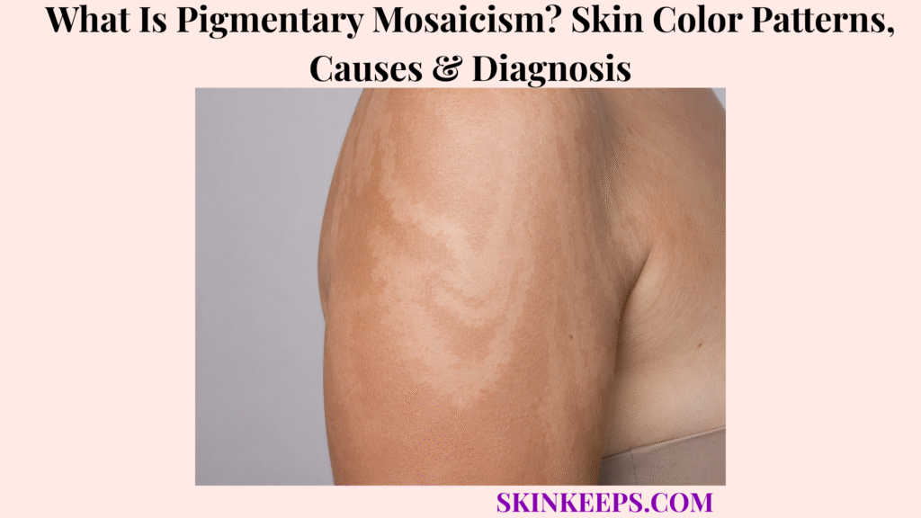
Pigmentary mosaicism is patterned lighter, darker or mixed skin colour caused by genetically different cell populations developing within the same person. The phenotype is usually flat, follows recognizable developmental geometry and may be visible at birth or become clearer during early childhood.
Pigmentary mosaicism can remain limited to the skin or reflect a broader mosaic disorder involving other tissues, so neurologic, skeletal, ocular, hearing, growth and developmental findings matter. Diagnosis begins with pattern recognition and whole-patient assessment, while molecular testing is selective and may require affected skin because a mosaic variant can be absent or extremely low-level in blood.
This article is for educational purposes only and does not constitute medical advice, diagnosis, or treatment. Patterned pigmentation accompanied by seizures, developmental or learning difficulties, abnormal muscle tone, limb or body asymmetry, skeletal abnormalities, visual problems, hearing abnormalities, unusual growth patterns or other congenital differences should be evaluated by a licensed dermatologist, pediatrician, geneticist or other qualified healthcare professional because mosaicism may involve tissues beyond the skin.
How Can You Recognize Pigmentary Mosaicism?
Pigmentary mosaicism causes flat areas of lighter, darker or mixed skin colour arranged in recognizable developmental patterns rather than random isolated colour changes.
Can Pigmentary Mosaicism Make Skin Lighter, Darker or Both?
Yes; pigmentary mosaicism can cause hypopigmentation, hyperpigmentation or mixed areas, so colour alone does not define the condition.
The defining clue is developmental pattern rather than whether melanin appears increased or reduced. Two people can therefore share a mosaic-pattern phenotype even when one has lighter skin areas and another has darker areas.
What Do Blaschko-Line Patterns Look Like?
Blaschkoid pigmentary mosaicism can form V-shaped or chevron patterns on the back, S-shaped whorls across the trunk and linear streaks along the limbs.
Blaschko lines reflect developmental pathways of epidermal cell migration and proliferation that are normally invisible but can become apparent when one cell clone differs genetically from neighbouring tissue.
These lines do not follow peripheral nerves or blood vessels.
Can Pigmentary Mosaicism Form Segmental or Phylloid Patterns?
Yes; mosaic pigmentation can also form block-like or checkerboard segments and larger leaf-shaped or phylloid areas rather than narrow Blaschko lines.
Segmental patterns can form broad blocks with clear midline influence, while phylloid patterns form larger leaf-like or oblong areas. Modern classification emphasizes the developmental pattern separately from whether the lesion is hypo- or hyperpigmented.
Are Pigmentary Mosaicism Lesions Flat and Present From Birth?
Classic pigmentary mosaicism is usually flat and developmental, becoming visible at birth or during early childhood even when subtle contrast is noticed later.
The colour difference is macular and cannot be felt. A raised, verrucous or nodular lesion shifts the differential toward another type of mosaic nevus or developmental skin disorder.
Figure 1. Pattern recognition comes before colour classification: Blaschkoid, segmental/checkerboard and phylloid forms can each be lighter, darker or mixed.
Why Does Pigmentary Mosaicism Develop?
Pigmentary mosaicism develops when a postzygotic genetic change creates a genetically distinct cell population that expands through the skin along developmental pathways.
What Does Genetic Mosaicism Mean?
Genetic mosaicism means that two or more genetically different cell populations arose from the same fertilized egg after a genetic change occurred during development.
A variant that appears after fertilization is postzygotic. Descendants of the altered cell can carry the variant while neighbouring cells derived from unaffected lineages do not.
Why Does Mosaicism Produce a Pattern Instead of Affecting All Skin?
Only descendants of the genetically altered developmental cell carry the mosaic change, so their expansion creates lines, segments or other patterns among unaffected neighbouring cells.
The sequence is postzygotic change → altered developmental clone → migration and expansion → visible pigment pattern. The geometry therefore reflects embryonic cell history rather than random adult spread.
Is There One Gene That Causes Pigmentary Mosaicism?
No; pigmentary mosaicism can result from many chromosomal and single-gene mosaic abnormalities, so there is no single “pigmentary mosaicism gene.”
Examples reported across mosaic pigmentation disorders include MTOR, RHOA, GNA13, TP63, TFE3 and chromosomal mosaicism, but the same visible phenotype can arise from different molecular causes.
Is Pigmentary Mosaicism Inherited, and Does Everyone Have a Syndrome?
Most postzygotic mosaic changes arise sporadically, and many people have isolated skin pigmentation without a broader syndrome, although reproductive risk and systemic involvement depend on the specific variant.
Whether a mosaic variant can be transmitted depends on the genotype and whether reproductive cells are involved. No universal recurrence percentage applies to pigmentary mosaicism as a broad phenotype.
Figure 2. A postzygotic variant creates an altered clone whose descendants spread through developmental pathways; systemic involvement depends on whether other tissues share that clone.
How Is Pigmentary Mosaicism Distinguished From Other Pigment Disorders?
Pigmentary mosaicism is distinguished by developmental pattern and early-life onset, while vitiligo, piebaldism and specific syndromes have different timing, morphology, biology or extracutaneous features.
How Is Pigmentary Mosaicism Different From Vitiligo and Piebaldism?
Pigmentary mosaicism follows developmental lines or segments and can be lighter or darker, while vitiligo is acquired immune-mediated depigmentation and piebaldism is a specific congenital stable melanocyte-development disorder often featuring a white forelock.
Vitiligo develops after previously normal pigmentation and can progress. Piebaldism is a specific congenital disorder, while pigmentary mosaicism is a broader phenotype with many possible genotypes.
Is Hypomelanosis of Ito the Same as Pigmentary Mosaicism?
Not exactly; hypomelanosis of Ito is a historical phenotype term for Blaschkoid hypopigmentation, while pigmentary mosaicism is the broader developmental and genetic concept.
Modern terminology works best when the pigment pattern is described first and a genotype is added when known rather than treating hypomelanosis of Ito as one genetically uniform disease.
How Do Segmental Pigmentation Disorder and Incontinentia Pigmenti Differ?
Segmental pigmentation disorder usually presents as a localized block-like colour difference, while incontinentia pigmenti is a specific X-linked disorder with evolving inflammatory skin stages and possible involvement of teeth, eyes and the nervous system.
Segmental pigmentation disorder often appears in infancy, is localized and frequently respects the midline. Incontinentia pigmenti can include blistering and verrucous stages before later pigmentation, so it is not a synonym for flat pigmentary mosaicism.
Which Findings Suggest a Broader Mosaic Syndrome?
Seizures, developmental delay, abnormal muscle tone, body asymmetry, skeletal abnormalities, eye problems, hearing abnormalities or other congenital findings make multisystem mosaic disease more likely.
Skin pattern alone can represent an isolated phenotype. Patterned pigmentation plus systemic findings should prompt broader genetic and specialist evaluation.
Figure 3. Isolated patterned pigmentation may need little investigation, while neurologic, developmental, skeletal, ocular, hearing or asymmetry findings raise concern for multisystem mosaic disease.
How Is Pigmentary Mosaicism Diagnosed?
Diagnosis begins with clinical mapping of the pigment pattern and systemic examination, while genetic testing is targeted to cases where identifying the mosaic genotype would change counseling, surveillance or management.
Can Pigmentary Mosaicism Usually Be Recognized Clinically?
Yes; the colour, lesion type and developmental distribution often establish a clinical pigmentary-mosaicism phenotype before molecular testing is considered.
Assessment records whether pigmentation is hypo-, hyper- or mixed, whether the lesion is flat, the developmental pattern, timing of appearance and whether systemic findings are present. Pattern comes first; genetics follows the clinical question.
What Does a Wood’s Lamp Add, and Does Every Child Need Extensive Testing?
Wood’s lamp examination can map subtle pigment contrast, but isolated pigmentary mosaicism without concerning history or examination findings does not automatically require extensive investigation.
Wood’s lamp helps show contrast and extent but does not identify genotype. Testing intensity should stay proportional to clinical findings.
Why Can Blood Genetic Testing Miss Pigmentary Mosaicism?
Blood testing can be negative because the mosaic variant may be concentrated in affected skin and absent or present at extremely low levels in circulating blood cells.
A postzygotic clone can have different variant allele fractions across tissues, so blood may not contain enough altered cells for standard sequencing to detect the variant.
A negative blood result therefore does not exclude mosaicism when the clinical phenotype remains convincing.
When Should Affected Skin Be Tested, and Does a Negative Test Exclude the Diagnosis?
Affected skin can provide a higher-yield sample when molecular confirmation matters, but even skin testing can miss low-level mosaic variants, so a negative result does not automatically exclude clinically convincing pigmentary mosaicism.
Affected-skin biopsy may enrich variant-containing cells. High-depth or mosaic-sensitive sequencing can be useful when the expected variant allele fraction is low.
A negative result may reflect low variant level, wrong tissue, assay limitations or an untested mechanism. Diagnosis therefore integrates clinical pattern, systemic phenotype and molecular findings.
Figure 4. Molecular diagnosis is phenotype-led and tissue-aware: blood can miss a tissue-limited variant, while affected skin plus sensitive sequencing may improve detection when genotyping matters.
Does Pigmentary Mosaicism Need Treatment or Long-Term Follow-Up?
Isolated pigmentary mosaicism usually needs no medical treatment, while follow-up is determined by extracutaneous findings or the risks associated with a confirmed underlying mosaic genotype.
Does Isolated Pigmentary Mosaicism Harm the Skin or Need Treatment?
Isolated skin-only pigmentary mosaicism is generally benign and usually does not require medical treatment simply because the colour difference is present.
The pigmentation can be permanent without representing active skin damage. Current pediatric dermatology guidance does not identify the colour difference itself as increasing skin-cancer risk or requiring routine skin checks solely because mosaic pigmentation is present.
Can Camouflage or Sun Protection Help?
Cosmetic camouflage can reduce visible contrast if desired, while consistent sun protection can reduce contrast created when surrounding skin tans more strongly.
Sunscreen and sun-protection measures can make the colour difference less noticeable in some people. Camouflage makeup remains optional and patient-directed.
Do Lasers Reliably Remove Pigmentary Mosaicism?
No; ordinary pigmentary mosaicism does not have a reliably effective laser treatment, so procedural normalization of skin colour should not be promised.
Response is variable, and appearance-directed intervention is optional. The Society for Pediatric Dermatology states that laser is typically not effective for these colour changes.
When Does Long-Term Follow-Up or Genetic Counseling Matter?
Long-term specialist follow-up becomes more important when the phenotype or genotype carries neurological, skeletal, ocular, growth or other organ risks, while genetic counseling is most informative after the molecular cause is known.
Surveillance should follow the actual phenotype and genotype rather than pigmentation alone. Genetic counseling can address possible reproductive-cell involvement and germline transmission when relevant; there is no universal recurrence percentage for the broad phenotype.
What Should You Remember About Pigmentary Mosaicism?
Pigmentary mosaicism is a developmental phenotype in which genetically different cell populations produce patterned lighter, darker or mixed skin colour, and its clinical importance depends on whether the finding is isolated or part of a broader mosaic disorder.
- Pigmentary mosaicism is a phenotype, not one disease.
- It can be hypopigmented, hyperpigmented or mixed.
- Developmental pattern matters more than colour.
- Blaschkoid, segmental/checkerboard and phylloid patterns can occur.
- Classic pigmentary-mosaicism lesions are flat.
- The causal genetic change is postzygotic.
- Altered cell clones create developmental skin geometry.
- Many chromosomal and single-gene abnormalities can produce similar appearances.
- No single gene defines the phenotype.
- Most postzygotic cases arise sporadically.
- Not every person has a multisystem syndrome.
- Vitiligo is acquired immune-mediated depigmentation.
- Piebaldism is a specific congenital melanocyte-development disorder.
- Hypomelanosis of Ito is better treated as a phenotype term than one uniform molecular disease.
- Neurologic, developmental, skeletal, ocular and hearing findings increase syndromic concern.
- Clinical pattern recognition comes before molecular testing.
- Wood’s lamp can map subtle pigment contrast.
- Not every isolated case needs extensive testing.
- Blood can miss tissue-limited mosaic variants.
- Affected skin may be a better molecular sample.
- High-depth or mosaic-sensitive sequencing may be needed.
- Negative testing does not always exclude a convincing mosaic phenotype.
- Isolated skin-only disease generally needs no medical treatment.
- Laser response is unreliable.
- Follow-up should be phenotype- and genotype-driven.
Recognize pattern → Assess whole patient → Distinguish mimics → Decide whether molecular diagnosis matters → Test appropriate tissue → Manage skin conservatively or follow genotype-specific systemic risks.
Frequently Asked Questions About Pigmentary Mosaicism
The most important pigmentary-mosaicism questions concern how genetically different skin-cell populations create the pattern, how modern terminology is used, whether other organs can be involved, why blood testing can miss mosaic variants and whether treatment or lifelong monitoring is necessary.
What Causes the Lighter or Darker Skin Patterns in Pigmentary Mosaicism?
The patterns develop when a postzygotic genetic change creates a genetically distinct cell population whose descendants spread through the skin along developmental pathways. Altered and unaffected neighbouring cells can then produce hypo-, hyper- or mixed pigmentation.
Is Pigmentary Mosaicism the Same as Hypomelanosis of Ito?
No; hypomelanosis of Ito is a historical term for a Blaschkoid hypopigmentation phenotype, while pigmentary mosaicism is the broader concept covering many mosaic genetic patterns and causes. Modern terminology works best when phenotype and genotype are described separately.
Can Pigmentary Mosaicism Affect Organs Other Than the Skin?
Yes in some mosaic disorders, but many people have isolated skin-only pigmentation without systemic disease. Neurologic, developmental, skeletal, ocular, hearing or asymmetry findings increase the need for broader assessment.
Why Can Blood Genetic Testing Miss Pigmentary Mosaicism?
A mosaic variant may be concentrated in affected skin and absent or too low-level in blood for standard testing to detect. Affected-skin sampling and mosaic-sensitive sequencing can improve diagnostic yield when identifying the genotype would change care.
Does Pigmentary Mosaicism Need Treatment or Lifelong Monitoring?
Isolated skin-only pigmentary mosaicism usually needs neither corrective treatment nor intensive lifelong surveillance, while broader follow-up depends on associated abnormalities or the confirmed genotype. Camouflage is optional, and surveillance should be phenotype- or genotype-directed.
Which Sources Support This Pigmentary Mosaicism Guidance?
British Journal of Dermatology — Mosaic Disorders Affecting Pigmentation, Part 1: How to Make a Clinical Diagnosis — Primary source for developmental pattern classification, lesion type, hypo/hyperpigmentation and phenotype-first assessment.
British Journal of Dermatology — Mosaic Disorders Affecting Pigmentation, Part 2: How to Make a Genetic Diagnosis — Primary source for tissue-aware sampling, variant allele fraction, sequencing sensitivity, genotype selection and transmission nuance.
DermNet — Blaschko Lines — Used for developmental epidermal migration patterns, V/S/linear morphology and the distinction from neural or vascular pathways.
DermNet — Hypomelanosis of Ito — Used narrowly for Blaschkoid hypopigmentation and neurologic, hearing, visual and orthopedic red flags.
Society for Pediatric Dermatology — Pigmentary Mosaicism Patient Perspective — Used for flat lighter/darker presentation, clinical diagnosis, Wood’s lamp, proportional testing, benign skin-only disease, sun protection, camouflage, laser limitations and follow-up.



