
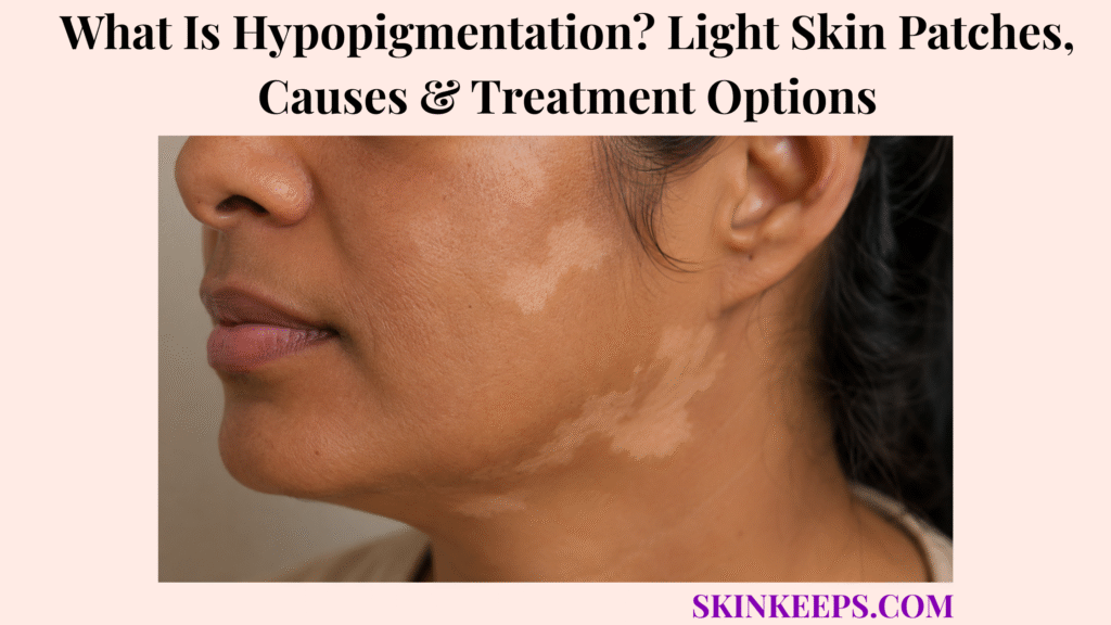
Hypopigmentation is an area of skin that has become lighter than the person’s surrounding natural skin colour. It may reflect reduced melanin, fewer functioning melanocytes or near-complete pigment loss, and it is a skin finding rather than one diagnosis.
Some light patches recover after inflammation or fungal infection resolves, while vitiligo, deep scars and inherited disorders can persist. Scale, texture, distribution, progression and sensation guide diagnosis, and numbness, weakness, ulceration or rapid spreading requires clinical assessment.
How Can You Recognize Hypopigmentation on the Skin?
Hypopigmentation is recognized when a patch is lighter than the surrounding skin, but not every pale patch has the same cause.
- Cream, pale-brown, pink-white or off-white colour.
- Milky-white or chalk-white depigmentation.
- Poorly defined or sharply outlined borders.
- One patch, several patches or a symmetrical distribution.
- Smooth skin or fine dryness and scale.
- Colour change after a healed rash, injury or procedure.
- Small white spots on sun-exposed limbs.
- Broad pale trunk patches.
- White hair within a light patch.
- Greater contrast after surrounding skin tans.
Ask about itching, scaling, previous redness, numbness, pain, tightening, hair loss, sweating change and spread.
Figure 1. Light patches are classified by remaining pigment, surface scale, border definition, sensation, body distribution and progression rather than by colour alone.
Where Can Light Skin Patches Develop?
Light patches can develop anywhere, but location often narrows the cause.
| Location | Pattern Clue |
|---|---|
| Child’s cheeks | Pityriasis alba |
| Chest, back, shoulders or neck with scale | Pityriasis versicolor |
| Hands, face, body openings or folds | Nonsegmental vitiligo |
| One-sided body segment | Segmental vitiligo or mosaic disorder |
| Sun-exposed shins and forearms | Idiopathic guttate hypomelanosis |
| Scar or burn site | Post-inflammatory or permanent scar loss |
| Exact product or workplace contact site | Chemical leukoderma |
| One numb pale patch | Nerve-affecting disease assessment |
How Are Hypopigmentation and Depigmentation Different?
What Does Partial Hypopigmentation Mean?
Partial hypopigmentation means some melanin remains, so the area is lighter but not completely white and may have indistinct borders.
What Does Complete Depigmentation Mean?
Complete or near-complete depigmentation produces milky-white or chalk-white skin, often with sharp borders and possible white hair.
Can Skin Look Pale Without Losing Melanin?
Yes. Reduced blood flow can create vascular pallor, which is not a melanocyte disorder and may behave differently with pressure or Wood-lamp examination.
| Pattern | Pigment Status | Common Clues |
|---|---|---|
| Hypopigmentation | Reduced but present | Cream or pale, often indistinct |
| Depigmentation | Nearly absent | Milky-white, sharper border |
| Vascular pallor | Melanin preserved | Blanching or blood-flow difference |
How Does the Skin Lose or Reduce Its Pigment?
Skin becomes lighter when melanocyte number, melanocyte function or melanin transfer is reduced.
- Inflammation temporarily suppresses melanocyte activity.
- Severe inflammation or scarring destroys melanocytes.
- Autoimmunity targets melanocytes directly.
- Fungal overgrowth interferes with normal pigmentation.
- Chemicals can be toxic to pigment-producing cells.
- Procedures can injure epidermal and follicular pigment reservoirs.
- Genetic differences reduce melanin production.
- Contrast increases when surrounding skin tans.
Figure 2. Inflammation may temporarily suppress pigment, fungus can disrupt colour while scale is active, vitiligo removes melanocytes through autoimmunity, and chemicals or scars may cause persistent pigment loss.
Which Main Conditions Can Cause Hypopigmentation?
The main cause groups are inflammatory, fungal, autoimmune, congenital, chemical, medicine-related, scar-related and vascular.
| Cause Group | Examples | Main Diagnostic Clue |
|---|---|---|
| Post-inflammatory | Eczema, psoriasis, burns or procedures | Prior rash or injury |
| Mild dermatitis | Pityriasis alba | Faint dry cheek patches |
| Fungal | Pityriasis versicolor | Fine scale on trunk |
| Autoimmune | Vitiligo | Smooth milky-white patches |
| Sun/age-associated | Idiopathic guttate hypomelanosis | Tiny white limb spots |
| Truncal pigment disorder | Progressive macular hypomelanosis | Smooth non-scaly midline patches |
| Chemical/medicine | Contact leukoderma or drug-induced vitiligo | Exposure or medicine timeline |
| Congenital | Albinism, piebaldism or naevus depigmentosus | Present from birth/childhood |
| Scarring | Deep burns, inflammation or procedures | Texture change and tissue loss |
What Is Post-Inflammatory Hypopigmentation?
Post-inflammatory hypopigmentation is a pale patch that appears where a rash, wound or procedure has healed.
A pale patch after dermatitis / eczema often means pigment production is temporarily reduced while the barrier recovers.
- Matches the shape of the original eruption.
- May be smooth or mildly dry.
- Can follow eczema, psoriasis, infection, burns or trauma.
- Temporary melanocyte dysfunction may recover.
- Deep tissue injury can create permanent loss.
- Contrast is often more visible in darker skin.
Which Skin Problems Commonly Leave Pale Patches After Healing?
Many inflammatory conditions can leave temporary or persistent pale patches after active disease settles.
A product reaction such as allergic contact dermatitis can leave pigment change and may continue while the trigger product remains in use.
- Atopic, seborrhoeic, contact or nummular dermatitis.
- Psoriasis and pityriasis rosea.
- Lichen planus or discoid lupus.
- Fungal infection or herpes zoster.
- Burns, cuts, abrasions and scratching.
- Chemical peels, cryotherapy or laser treatment.
- Radiation treatment.
What Is Pityriasis Alba?
Pityriasis alba is a mild dermatitis-related condition that causes faint pale, dry or finely scaly patches, especially on children’s cheeks.
- Poorly defined faint patches.
- Mild dryness or fine scale.
- Cheeks, chin, neck, shoulders or upper arms.
- Association with dry or atopic skin.
- More visible after tanning.
- Mild itch or no symptoms.
- Not fungal.
- Usually resolves gradually.
How Is Pityriasis Alba Different From Vitiligo?
Pityriasis alba is usually faint, dry and poorly defined, while vitiligo is generally smooth, sharply bordered and milky-white.
| Feature | Pityriasis Alba | Vitiligo |
|---|---|---|
| Colour | Pale or off-white | Milky or chalk-white |
| Border | Poorly defined | Sharper |
| Surface | Dry or finely scaly | Smooth |
| Common age/site | Children’s cheeks | Any age; face, hands, folds |
| Wood lamp | No strong enhancement | Bright accentuation |
| Course | Usually resolves | May spread or recur |
How Can Pityriasis Versicolor Cause Light Skin Patches?
Pityriasis versicolor can cause light patches because Malassezia yeast overgrowth disrupts normal pigmentation in the outer skin layer.
- Patches may be lighter, darker, pink or tan.
- Fine scale is usually present.
- Round patches can merge.
- Chest, upper back, shoulders and neck are common.
- Mild itch may occur.
- Warm humid conditions encourage recurrence.
- Colour can remain uneven after antifungal treatment.
How Is Pityriasis Versicolor Different From Vitiligo?
Pityriasis versicolor has scale and fungal involvement, while vitiligo is smooth autoimmune depigmentation.
| Feature | Pityriasis Versicolor | Vitiligo |
|---|---|---|
| Surface | Fine scale | Smooth |
| Distribution | Chest, back, shoulders, neck | Face, hands, folds, openings |
| Colour | Pale, tan, pink or brown | Milky-white |
| Testing | Fungal microscopy may be positive | Fungal testing negative |
| Treatment | Antifungal | Immune and repigmentation therapy |
| Colour return | Delayed after yeast clears | Depends on melanocyte response |
What Is Progressive Macular Hypomelanosis?
Progressive macular hypomelanosis causes smooth, poorly defined pale trunk patches without scale or a previous rash.
- Chest, abdomen or back involvement.
- Patches may merge near the midline.
- No preceding inflammation.
- No scale.
- Little or no itch.
- Often affects adolescents or young adults.
- May persist, progress or resolve.
What Are Idiopathic Guttate Hypomelanosis Spots?
Idiopathic guttate hypomelanosis causes small smooth white spots on sun-exposed skin, especially with ageing.
- Usually 2–5 mm in diameter.
- Common on forearms, shins and upper chest.
- Multiple separate spots.
- No scale, itch or pain.
- Associated with ageing, sun exposure and susceptibility.
- Benign and usually does not merge into large patches.
- Treatment is optional.
What Is Vitiligo—and Why Does It Cause White Patches?
Vitiligo is an acquired chronic depigmenting condition in which the immune system targets pigment-producing melanocytes.
- Chalk- or milky-white patches.
- Well-defined borders.
- Smooth surface without scale.
- White hair may appear within patches.
- New patches can follow friction or injury.
- Periods of spreading and stability.
- Perifollicular repigmentation may occur.
- Not contagious and not caused by poor hygiene.
How Do Nonsegmental and Segmental Vitiligo Differ?
What Is Nonsegmental Vitiligo?
Nonsegmental vitiligo usually affects both sides of the body and commonly involves the hands, face, openings and folds.
What Is Segmental Vitiligo?
Segmental vitiligo affects one side or anatomical segment, often begins younger, spreads early and may later stabilize.
What Is Mixed Vitiligo?
Mixed vitiligo combines segmental and nonsegmental patterns, and classification can change as new patches appear.
| Feature | Nonsegmental | Segmental |
|---|---|---|
| Distribution | Often bilateral/symmetrical | Usually one-sided |
| Course | Can recur over time | Early spread then stability |
| Autoimmune association | More typical | Less prominent |
| Surgery | Only stable selected disease | Stable localized disease may qualify |
Which Autoimmune Conditions Can Occur With Vitiligo?
Vitiligo can occur with other autoimmune conditions, especially autoimmune thyroid disease, but testing should be targeted.
Some people with vitiligo also have alopecia areata, so symptoms and family history should guide screening.
- Autoimmune thyroid disease.
- Type 1 diabetes.
- Pernicious anaemia.
- Addison disease.
- Alopecia areata.
- Other autoimmune syndromes in uncommon cases.
Fatigue, weight change, temperature intolerance, palpitations, constipation or neck swelling may justify thyroid evaluation.
Which Congenital or Inherited Conditions Can Cause Light Skin?
What Is Oculocutaneous Albinism?
Oculocutaneous albinism is an inherited reduction in melanin affecting skin, hair and eyes, with light sensitivity, visual problems and increased sun-related skin risk.
What Is Naevus Depigmentosus?
Naevus depigmentosus is a stable congenital or early-childhood light patch that usually grows proportionally with the person.
What Is Piebaldism?
Piebaldism causes congenital sharply defined depigmented areas, often with a white forelock and a stable distribution.
What Are Ash-Leaf Macules and Hypomelanosis of Ito?
Ash-leaf macules and Blaschko-line pigment patterns can signal mosaic or genetic disorders when multiple lesions occur with neurologic or developmental features.
| Condition | Pattern | Additional Clues |
|---|---|---|
| Albinism | Generalized reduced pigment | Eye involvement and sun sensitivity |
| Naevus depigmentosus | Stable localized pale patch | Present early |
| Piebaldism | Stable depigmentation | White forelock possible |
| Ash-leaf macules | Leaf-shaped pale macules | Tuberous-sclerosis assessment when patterned |
| Hypomelanosis of Ito | Linear/whorled pattern | Developmental or neurologic evaluation when indicated |
How Can Scarring or Skin Procedures Cause Permanent Pigment Loss?
Deep inflammation, burns, surgery, radiation, cryotherapy, peels or lasers can destroy melanocytes and follicular pigment reservoirs.
Scarring inflammatory disease such as discoid lupus erythematosus can leave light or dark patches with texture change, not simple pigment variation.
- Texture change suggests scar involvement.
- White hair suggests follicular pigment loss.
- Severe burns may remove repigmentation reservoirs.
- Repeated aggressive procedures can enlarge the pale area.
- Ordinary creams cannot restore absent melanocytes.
What Is Chemical Leukoderma?
Chemical leukoderma is vitiligo-like pigment loss caused by repeated contact with melanocyte-toxic chemicals.
- Sharply defined white macules.
- Confetti-like spots may appear.
- Pattern follows repeated contact.
- Possible sources include phenolic or catechol compounds, rubber, adhesives, hair dyes and industrial materials.
- Itch can occur, but dermatitis is not always present.
- Spread can continue after repeated exposure.
Which Medicines Can Trigger Vitiligo-Like Pigment Loss?
Selected medicines can trigger or reveal vitiligo-like depigmentation through immune activation or local melanocyte injury.
- Immune checkpoint inhibitors.
- Selected targeted cancer therapies.
- Imiquimod.
- Some cytokine or immune-modifying treatments.
- BRAF or tyrosine-kinase pathway medicines.
- Local medicine or patch exposure in rare patterns.
Do not stop essential cancer or immune treatment independently; the prescribing team should review the timeline and risks.
Why Does a Numb Pale Patch Need Prompt Assessment?
A numb pale or reddish patch needs prompt assessment because sensory loss can indicate leprosy or another peripheral nerve disorder.
- Reduced touch, pain or temperature sensation.
- Tingling or altered sweating.
- Thickened or tender peripheral nerve.
- Weak grip or foot weakness.
- Repeated painless burns or injuries.
- Slow-healing wound or ulcer.
Early diagnosis and treatment can prevent disability.
Which Other Disorders Can Resemble Ordinary Hypopigmentation?
Several inflammatory, scarring, vascular and tumour-related disorders can resemble ordinary pigment loss.
- Lichen sclerosus, especially genital white patches.
- Morphoea with hard or bound-down skin.
- Discoid lupus with scale, scarring or hair loss.
- Hypopigmented mycosis fungoides with persistent atypical patches.
- Halo nevus around a mole.
- Naevus anaemicus from vascular pallor.
- Healed injury or post-inflammatory contrast.
How Do Clinicians Diagnose the Cause of Hypopigmentation?
Diagnosis combines pigment degree, surface scale, sensation, texture, distribution, onset and progression.
- Present from birth or acquired later.
- Stable or spreading.
- Previous rash, infection or procedure.
- Fine scale or dryness.
- Normal or reduced sensation.
- White hair or hair loss.
- Chemical or occupational exposure.
- New medicine or immune treatment.
- Mucosal, genital, eye or neurologic symptoms.
- Family history and autoimmune clues.
How Does a Wood Lamp Help Diagnose Light Patches?
A Wood lamp can sharpen true pigment-loss borders and accentuate complete depigmentation, but it does not establish the diagnosis alone.
| Finding | Possible Meaning |
|---|---|
| Bright blue-white accentuation | Complete depigmentation such as vitiligo |
| No strong enhancement | Pityriasis alba or vascular pallor |
| Yellow-green/blue-green fluorescence | Possible pityriasis versicolor |
| Sharper subtle border | Reduced pigment in light skin |
| Unchanged vascular area | Pallor rather than melanin loss |
When Are Dermoscopy, Fungal Testing or Biopsy Needed?
What Can Dermoscopy Show?
Dermoscopy can show border activity, perifollicular repigmentation, scale, vascular change and melanocyte-related patterns.
When Is Fungal Scraping Useful?
Fungal scraping or potassium-hydroxide microscopy is useful when light patches have fine scale, especially on the trunk.
When Is a Skin Biopsy Needed?
Biopsy may be needed for persistent atypical lesions with hardening, atrophy, ulceration, scarring, unusual texture or concern for inflammatory disease or lymphoma.
Which Blood Tests May Be Used for Vitiligo or Systemic Clues?
Blood tests are targeted to symptoms, family history and examination rather than ordered as a universal panel.
| Clinical Clue | Possible Testing |
|---|---|
| Thyroid symptoms/family history | TSH and selected thyroid antibodies |
| Diabetes symptoms/risk | Blood glucose or HbA1c |
| Anaemia or neurologic symptoms | CBC and vitamin B12 |
| Fatigue, low pressure or salt craving | Adrenal evaluation |
| Broader autoimmune features | Cause-specific testing |
Does Every Light Skin Patch Require Treatment?
Not every patch needs treatment, but infection, active vitiligo, sensory loss, chemical exposure and atypical scarring require cause-specific care.
- Mild pityriasis alba can be observed.
- Post-inflammatory patches may recover with time.
- Idiopathic guttate hypomelanosis treatment is optional.
- Pityriasis versicolor needs antifungal treatment.
- Vitiligo treatment depends on activity and goals.
- Numb patches require prompt assessment.
- Congenital patterned lesions may need syndromic evaluation.
Why Must the Underlying Rash, Infection or Exposure Be Treated First?
Repigmentation is less likely while inflammation, fungus, chemical exposure or immune activity continues.
- Control eczema and scratching.
- Treat fungal infection.
- Stop melanocyte-toxic exposure.
- Review medicine-related pigment loss safely.
- Prevent repeated burns or procedures.
- Stabilize active vitiligo before surgery.
- Protect pale skin during recovery.
How Does Sun Protection Help Hypopigmented Skin?
Sun protection reduces sunburn risk in pigment-deficient skin and lowers contrast by preventing surrounding skin from tanning.
- Use broad-spectrum SPF 30 or higher.
- Apply to exposed pale and surrounding skin.
- Reapply during prolonged outdoor activity.
- Use hats, clothing and shade.
- Avoid deliberate sunburn.
- Do not substitute tanning beds for medical phototherapy.
How Is Post-Inflammatory Hypopigmentation Treated?
Post-inflammatory hypopigmentation is treated by controlling the original rash or injury and allowing melanocyte function to recover.
- Treat active eczema, psoriasis or infection.
- Use gentle moisturizer.
- Avoid scratching and abrasive exfoliation.
- Protect from sun contrast.
- Avoid repeated steroid or acid use on an undiagnosed patch.
- Expect gradual recovery over weeks or months.
- Refer texture-changing or scarred areas.
How Is Pityriasis Alba Treated?
Pityriasis alba usually improves with gentle barrier care and time.
- Use moisturizer for dryness.
- Use a mild cleanser.
- Apply sunscreen to reduce contrast.
- Use a limited mild topical corticosteroid when inflamed and advised.
- Tacrolimus or pimecrolimus may be selected for some patients.
- Avoid antifungal treatment unless testing supports fungus.
- Expect colour recovery over months and sometimes two or three years.
How Is Pityriasis Versicolor Treated?
Pityriasis versicolor is treated with antifungal medicine, but normal skin colour may return slowly after the yeast is cleared.
- Topical antifungal shampoo or cream is common first treatment.
- Oral antifungal medicine may be used for widespread or resistant disease.
- Scaling should settle before colour fully normalizes.
- Warm humid conditions can promote recurrence.
- Maintenance antifungal use may be recommended for frequent relapses.
- Persistent colour alone does not always mean active yeast remains.
Which Topical Medicines Can Treat Vitiligo?
Topical vitiligo treatment aims to reduce immune attack and stimulate repigmentation where melanocytes remain.
| Medicine | Typical Role | Main Boundary |
|---|---|---|
| Topical corticosteroid | Recent or localized disease | Limited duration; skin-thinning risk |
| Tacrolimus/pimecrolimus | Face, folds or sensitive areas | Burning or irritation possible |
| Ruxolitinib cream | Eligible nonsegmental vitiligo | Not for every cause of light patches |
| Supportive moisturizer | Barrier comfort | Does not restore absent melanocytes alone |
The FDA indication for ruxolitinib cream covers nonsegmental vitiligo in people aged 12 years and older, applied twice daily to eligible areas up to 10% body surface area; meaningful repigmentation may require more than 24 weeks.
How Does Phototherapy Restore Pigment in Vitiligo?
Narrowband UVB or targeted excimer treatment uses measured ultraviolet doses to stimulate remaining melanocytes and reduce immune activity.
- Treatment usually requires repeated sessions over months.
- Face and neck often respond better than fingertips or toes.
- Repigmentation commonly begins around hair follicles.
- Dose is adjusted for skin response.
- Burning and blistering indicate excessive exposure.
- Home units require medical supervision.
- Tanning beds are not a safe substitute.
When Are Oral Medicines Used for Vitiligo?
Oral treatment is reserved for selected rapidly spreading or extensive vitiligo under specialist direction.
- Short corticosteroid regimens may slow rapid spread.
- Systemic treatment requires infection and metabolic risk review.
- Long-term oral steroids are avoided when risk exceeds benefit.
- Other immune treatments may be considered in specialist care or research settings.
- Oral medicine does not guarantee complete repigmentation.
When Are Vitiligo Surgery or Pigment Grafting Considered?
Surgery or cell transplantation is considered only for stable, selected localized or segmental vitiligo.
- No recent new or enlarging patches.
- Cause and diagnosis confirmed.
- Recipient skin suitable for grafting.
- Donor pigment available.
- Realistic expectations about colour match.
- Risk of scarring, infection, uneven pigment or Koebner change reviewed.
- Active disease is not grafted.
Which Camouflage Options Can Cover Light Patches?
Camouflage changes appearance immediately without treating the pigment disorder.
- Colour-correcting makeup.
- Waterproof body camouflage.
- Dihydroxyacetone self-tanner.
- Clothing or accessories.
- Medical tattooing in carefully selected stable disease.
- Patch testing to avoid contact reactions.
How Is Chemical Leukoderma Managed?
Chemical leukoderma management starts by identifying and stopping the melanocyte-toxic exposure.
- Map the exact contact pattern.
- Review work materials, rubber, adhesives, cosmetics and hair dyes.
- Stop or substitute the suspected exposure safely.
- Use protective equipment when occupational contact cannot be avoided.
- Treat associated dermatitis.
- Monitor for spread beyond contact sites.
- Consider vitiligo-directed therapy when depigmentation persists.
Why Can Scar-Related White Skin Be Difficult to Repigment?
Scar-related pigment loss is difficult to reverse when melanocytes and hair-follicle reservoirs have been destroyed.
- Texture and flexibility may be altered.
- Hair may be absent or white.
- Topical medicine cannot replace destroyed cells.
- Phototherapy response may be limited.
- Camouflage is often first-line support.
- Micropigmentation or grafting is considered only in stable selected scars.
How Is Hypopigmentation Managed in Children?
Children need diagnosis before treatment because pityriasis alba, fungal infection, congenital marks and vitiligo require different care.
- Ask whether the patch was present from birth.
- Check for eczema, fine scale or previous injury.
- Test fungal trunk patches when uncertain.
- Assess sensation in every unusual pale patch.
- Use gentle moisturizer and SPF 30 or higher.
- Use steroids or immune medicines only with age-appropriate guidance.
- Assess multiple congenital patterned lesions for syndromic clues.
- Address bullying or stigma.
How Is Hypopigmentation Managed During Pregnancy?
Pregnancy management begins with diagnosis, photoprotection and the safest cause-specific topical care.
- Use moisturizer and broad-spectrum sunscreen.
- Treat proven fungal infection with pregnancy-compatible therapy.
- Use anti-inflammatory medicine only after clinician review.
- Avoid uncontrolled UV exposure.
- Defer elective grafting, tattooing or aggressive procedures.
- Review ruxolitinib and systemic therapy with the relevant specialist.
- Changing, painful or sensory lesions still require assessment.
Which Hypopigmentation Treatment Mistakes Should Be Avoided?
Treatment mistakes can cause burns, infection, skin thinning, further pigment loss or delayed diagnosis.
| Mistake | Possible Harm | Safer Direction |
|---|---|---|
| Assume every patch is vitiligo | Wrong treatment | Check scale, sensation and history |
| Use antifungal indefinitely | Irritation and delayed diagnosis | Confirm active fungus |
| Use potent steroid long-term | Atrophy and pigment change | Time-limited clinician plan |
| Deliberate sunburn/tanning bed | Burn and cancer risk | Medical phototherapy only |
| Use ruxolitinib for any pale patch | Wrong indication and risk | Eligible nonsegmental vitiligo only |
| Graft active vitiligo | New lesions or poor match | Wait for stability |
| Tattoo an uncertain lesion | Mask progression | Diagnose first |
| Ignore numbness | Delayed nerve treatment | Prompt assessment |
How Long Does Repigmentation Take?
Repigmentation timing depends on cause, depth of injury, body site and whether melanocyte reservoirs remain.
| Cause | Expected Course |
|---|---|
| Mild post-inflammatory change | Weeks to months |
| Pityriasis alba | Months to two or three years |
| Versicolor colour change | Weeks to months after yeast clears |
| Vitiligo topical/light therapy | Usually months |
| Scar-related loss | May be permanent |
| Congenital stable mark | Often persists |
Can Hypopigmentation Return After Treatment?
Light patches can recur when eczema, fungal overgrowth, vitiligo activity, chemical exposure or repeated injury returns.
- Pityriasis versicolor commonly recurs.
- Eczema flares can create new pale marks.
- Vitiligo can reactivate after stability.
- Chemical exposure can restart pigment loss.
- Procedures can trigger new injury.
- Maintenance care depends on the cause.
Can Hypopigmentation Be Permanently Cured?
Permanent recovery depends on the cause and whether pigment-producing cells survive.
- Temporary inflammatory suppression may fully recover.
- Pityriasis alba usually resolves.
- Fungal pigment change often normalizes after treatment.
- Vitiligo can repigment but may relapse.
- Congenital marks may remain stable.
- Deep scars may not repigment.
- No single treatment cures every cause.
How Can Light Skin Patches Affect Mental Health and Quality of Life?
Visible pigment difference can affect confidence, social comfort and treatment choices even when the condition is medically benign.
- Stigma or cultural misconceptions.
- Bullying in children.
- Avoidance of photographs or social settings.
- Time and cost of camouflage.
- Anxiety about spread.
- Frustration with slow repigmentation.
- Pressure to use unsafe remedies.
- Need for counselling or peer support.
When Should Light Skin Patches Be Checked by a Clinician?
Clinical assessment is recommended when the cause is uncertain, patches spread, scale persists, sensation changes or treatment could carry risk.
- Rapidly spreading or complete white patches.
- White hair within a new patch.
- Fine scale that does not respond as expected.
- Chemical or occupational exposure pattern.
- New pigment loss after medicine or cancer treatment.
- Genital white patches.
- Texture change, hardening, scarring or ulceration.
- Several congenital patterned patches.
- Eye symptoms or systemic autoimmune clues.
- Any numb pale patch.
Which Light Skin Changes Require Prompt or Urgent Assessment?
Prompt assessment is needed for sensory loss, weakness, rapid depigmentation, eye involvement, genital scarring, ulceration or a pale halo around a changing mole.
A pale ring around a changing mole may resemble a halo nevus, but the central mole still needs assessment when it is irregular, evolving or symptomatic.
- Definite numbness or reduced temperature/pain sensation.
- Weak grip, foot weakness or repeated painless injuries.
- Rapidly spreading white eruption.
- New pigment loss during immune or cancer treatment.
- Eye pain, redness or visual change.
- Genital pain, tearing, narrowing or scarring.
- Skin hardening, atrophy or ulceration.
- A halo around a changing, bleeding or irregular mole.
- Seizures or developmental change with congenital patterned pigment.
Prompt route: a numb pale patch, new weakness, eye symptoms, genital scarring or pigment loss around a changing mole should not be treated at home before medical assessment.
Figure 3. Diagnosis combines pigment degree, scale, sensation, distribution and exposure history, then treatment is matched to inflammation, fungus, vitiligo, chemical injury, scarring or congenital disease.
What Should You Remember About Hypopigmentation?
Hypopigmentation means skin is lighter than its surrounding baseline and is a finding rather than one diagnosis.
- Partial pigment reduction differs from complete depigmentation.
- Pale patches have many causes beyond vitiligo.
- Fine scale suggests dermatitis or fungal testing.
- Smooth milky-white patches can suggest vitiligo.
- A numb pale patch requires prompt assessment.
- Pityriasis alba usually resolves gradually.
- Versicolor colour recovery follows antifungal clearance.
- Scars may permanently remove pigment reservoirs.
- Vitiligo treatment is immune- and repigmentation-directed.
- Sunscreen prevents burns and contrast.
- Tanning beds are not medical phototherapy.
- Treatment must match the cause.
Frequently Asked Questions About Hypopigmentation?
Is hypopigmentation a disease or a skin finding?
Hypopigmentation is a skin finding, not one diagnosis. It means an area contains less pigment than the surrounding baseline skin.
What is the difference between hypopigmentation and depigmentation?
Hypopigmentation means some pigment remains, while depigmentation means epidermal melanin is absent or nearly absent.
Does every pale patch mean vitiligo?
No. Eczema, pityriasis alba, fungal infection, inflammation, chemical exposure, scars, vascular differences and congenital disorders can look similar.
What do hypopigmented patches look like?
They may be cream, pale-brown, pink-white, off-white or milky-white, with sharp or indistinct borders and either smooth skin or fine scale.
Why does skin become lighter after eczema?
Inflammation can temporarily reduce melanocyte activity, and scratching or deeper injury can prolong or permanently alter pigment.
How is pityriasis alba different from vitiligo?
Pityriasis alba is usually faint, dry, scaly and poorly defined, while vitiligo is generally smooth, sharply bordered and milky-white.
Is pityriasis alba a fungal infection?
No. Pityriasis alba is a mild dermatitis-related condition, and fungal tests are negative.
How long does pityriasis alba take to disappear?
Colour usually returns gradually over months, but complete recovery can take two or three years.
How does pityriasis versicolor cause light patches?
Malassezia yeast disrupts pigmentation and creates fine-scaled trunk patches; colour can remain uneven after the yeast is cleared.
How is pityriasis versicolor different from vitiligo?
Versicolor commonly has fine scale and positive fungal testing, while vitiligo is smooth autoimmune depigmentation with Wood-lamp accentuation.
What is progressive macular hypomelanosis?
It causes smooth, poorly defined pale trunk patches without scale or a preceding rash, often in adolescents or young adults.
What are idiopathic guttate hypomelanosis spots?
They are small benign white spots, usually 2–5 mm, on sun-exposed forearms, shins and other sites, especially with ageing.
What causes vitiligo?
Vitiligo is an autoimmune condition in which the immune system targets pigment-producing melanocytes.
Is vitiligo contagious?
No. Vitiligo is not contagious and is not caused by poor hygiene.
What is the difference between segmental and nonsegmental vitiligo?
Nonsegmental vitiligo is usually bilateral and can recur, while segmental vitiligo often affects one side and may stabilize after early spread.
Can vitiligo turn hair white?
Yes. Hair within a depigmented patch can lose pigment, a change called leukotrichia.
Which autoimmune diseases can occur with vitiligo?
Associations include autoimmune thyroid disease, type 1 diabetes, pernicious anaemia, Addison disease and alopecia areata, but testing is targeted.
Can a white patch be present from birth?
Yes. Naevus depigmentosus, piebaldism, albinism, hypomelanosis of Ito and ash-leaf macules are congenital or inherited possibilities.
Can scars permanently lose pigment?
Yes. Deep burns, inflammation or procedures can destroy melanocytes and hair-follicle reservoirs, limiting repigmentation.
What is chemical leukoderma?
Chemical leukoderma is vitiligo-like pigment loss after repeated contact with melanocyte-toxic chemicals, often in an exposure pattern.
Can medicine trigger pigment loss?
Yes. Immune checkpoint inhibitors, targeted therapies, imiquimod and other medicines can trigger vitiligo-like depigmentation.
Why is a numb pale patch concerning?
Definite sensory loss in a pale or reddish patch can indicate leprosy or another nerve-affecting disorder and needs prompt assessment.
What does a Wood lamp show in hypopigmentation?
It can sharpen true pigment-loss borders and make complete depigmentation appear bright blue-white; some infections may fluoresce.
When is fungal scraping needed?
Scraping is useful when a patch has fine scale, especially on the chest, back, shoulders or neck.
When is a biopsy needed for a light patch?
Biopsy may be needed for persistent atypical patches with texture change, hardening, atrophy, ulceration or uncertain diagnosis.
Does every light patch need treatment?
No. Stable birthmarks, idiopathic guttate hypomelanosis and mild post-inflammatory changes may be observed, while infection, vitiligo or sensory loss needs specific care.
How does sunscreen help hypopigmentation?
SPF 30 or higher protects pigment-deficient skin from sunburn and reduces contrast caused when surrounding skin tans.
Which medicines are used for vitiligo?
Options include limited-course topical corticosteroids, tacrolimus or pimecrolimus, and topical ruxolitinib for eligible nonsegmental vitiligo.
Who can use topical ruxolitinib for vitiligo?
The FDA indication is for nonsegmental vitiligo in adults and children aged 12 years or older, applied to eligible affected areas.
Can phototherapy restore pigment?
Narrowband UVB or targeted excimer therapy can stimulate remaining melanocytes over months, but response varies by body site.
Which light-patch changes require urgent assessment?
Prompt assessment is needed for numbness, weakness, rapidly spreading depigmentation, eye symptoms, genital pain or scarring, ulceration or a pale halo around a changing mole.
Which Sources Support This Hypopigmentation Guidance?
DermNet — Vitiligo — Autoimmune melanocyte loss, depigmented pattern, segmental and nonsegmental disease, investigations and treatment principles.
American Academy of Dermatology — Vitiligo Treatment — Topical corticosteroids, calcineurin inhibitors, phototherapy, oral treatment for rapid spread and stable-disease surgery.
DermNet — Pityriasis Alba — Childhood facial patches, Wood-lamp distinction, moisturizer, limited anti-inflammatory care and months-to-years recovery.
NHS — Pityriasis Versicolor — Fine-scaled trunk patches, antifungal shampoo or cream, delayed colour recovery and recurrence.
DermNet — Contact Leukoderma — Vitiligo-like chemical depigmentation, confetti macules, exposure pattern and diagnosis.
World Health Organization — Leprosy — Definite sensory loss in a pale or reddish patch as a cardinal diagnostic sign and the need for prompt treatment.
DermNet — Wood Lamp Examination — Wood-lamp accentuation of pigment loss, vascular distinction and infection fluorescence.
U.S. FDA — Opzelura for Nonsegmental Vitiligo — Ruxolitinib indication from age 12, twice-daily application to up to 10% body surface area and possible treatment beyond 24 weeks.
This SkinKeeps article is educational and does not diagnose or replace dermatology, infectious-disease, neurologic, pediatric, pregnancy, oncology, eye or emergency care. Seek prompt assessment for a numb pale patch, weakness, repeated painless injury, rapid depigmentation, eye symptoms, genital pain or scarring, ulceration, new pigment loss during immune or cancer treatment, or a pale halo around a changing mole. Do not use prolonged potent steroids, indefinite antifungals, tanning beds, deliberate sunburn, ruxolitinib on undiagnosed patches, tattooing or grafting before diagnosis and stability are confirmed.



