
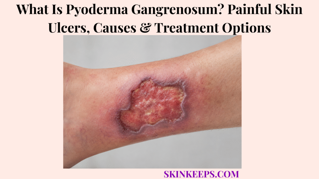
Pyoderma gangrenosum is a rare neutrophilic inflammatory skin disease that can cause extremely painful ulcers which enlarge rapidly and may be mistaken for serious infection or gangrene. PG is driven by abnormal inflammatory activity rather than ordinary bacterial infection or loss of blood supply, although an open ulcer can still become secondarily infected.
PG can worsen after trauma through pathergy and may occur with inflammatory bowel disease, inflammatory arthritis or selected hematologic disorders. Diagnosis requires careful exclusion of important mimics, while treatment combines inflammation control with atraumatic wound care, pain management and coordination of associated disease when relevant.
This article is for educational purposes only. A rapidly enlarging, very painful or systemically unwell skin ulcer requires prompt medical evaluation.
What Is Pyoderma Gangrenosum and What Does It Look and Feel Like?
Pyoderma gangrenosum is a neutrophilic inflammatory skin disease that can begin as a tender bump, pustule or small break in the skin and rapidly progress into an extremely painful ulcer.
How Does Abnormal Inflammation Cause PG Ulcers?
PG appears to involve dysregulated innate immunity and excessive neutrophilic inflammation that injures skin tissue and drives painful ulcer formation.
The useful mechanism is immune dysregulation → neutrophilic inflammation → tissue injury → ulceration. The complete disease pathway is not fully understood, so neutrophilic inflammation should be treated as a central feature rather than a complete explanation of every PG case.
Why Is PG Not the Same as Infection or Gangrene?
PG is primarily an inflammatory disease rather than bacterial infection or ordinary gangrene, although an open PG ulcer can still develop secondary infection.
Despite its name, PG is not the same disease process as gangrene, which refers to tissue death from a different mechanism. Bacterial conditions such as cellulitis also belong to a different disease category, although infection can mimic PG or coexist with an open PG wound.
How Does a Typical PG Ulcer Progress?
Classic PG can evolve rapidly from a painful inflammatory lesion into an enlarging ulcer with irregular undermined borders and dusky red, blue or violaceous edges.
The ulcer may have an exudative or necrotic-looking base and severe pain that seems disproportionate to its early size. Colour alone is not diagnostic; rapid progression, border shape, pain, history and exclusion of dangerous alternatives matter more than one visual feature.
Where Does PG Occur, and Are There Different Forms?
PG commonly affects the lower legs but can occur elsewhere, including surgical or peristomal sites, and several variants can produce ulcerative, bullous, pustular or more superficial patterns.
Classic ulcerative PG is the best-known pattern, while bullous, pustular, superficial granulomatous and peristomal variants broaden the clinical spectrum. These variants support recognition but should not replace careful diagnostic exclusion when an ulcer is rapidly progressive or atypical.
Figure 1. PG recognition is a progression problem: a tender inflammatory lesion may enlarge rapidly into a severely painful ulcer with an undermined violaceous edge, but dangerous infectious and vascular mimics still need exclusion.
What Causes Pyoderma Gangrenosum, and Which Conditions Are Associated With It?
PG has no single external cause; it reflects immune dysregulation and may occur alone or alongside inflammatory bowel disease, inflammatory arthritis or selected hematologic disorders.
Is There One Specific Cause of PG?
No; PG is linked to immune dysregulation, and some people develop it without any identifiable associated disease.
PG should not be reduced to infection, one gene or one trigger. The disease is best understood as an inflammatory neutrophilic process whose exact pathogenesis remains incomplete and whose associated conditions differ from person to person.
How Is Inflammatory Bowel Disease Associated With PG?
PG can occur in people with inflammatory bowel disease such as ulcerative colitis or Crohn disease, but neither condition is present in every PG patient.
The relationship is an association rather than a guarantee of causation. A PG diagnosis may prompt review for inflammatory bowel symptoms when clinically appropriate, but this page remains centered on the skin disease rather than IBD management.
Which Arthritis or Blood Disorders Can Be Associated With PG?
PG can also occur with inflammatory arthritis and selected hematologic disorders such as monoclonal gammopathy or myelodysplastic disease.
Inflammatory arthritis, including rheumatoid arthritis, and selected blood disorders are relevant because they can shape broader assessment and treatment coordination. Their presence is not required for PG, and associated disease should not be mistaken for the direct cause of every ulcer.
What Is Pathergy, and Can Surgery Trigger PG?
Pathergy is an exaggerated inflammatory reaction to minor trauma, meaning surgery, needle injury or wound manipulation can sometimes trigger a new PG lesion or worsen an existing ulcer.
The sequence is trauma or procedure → inflammatory amplification → new or worsening lesion. Pathergy is a trigger phenomenon, not the root cause of PG, and it does not mean that every wound-care procedure or every operation is forbidden.
Figure 2. PG is easiest to understand when three ideas stay separate: immune/neutrophilic dysregulation is the disease mechanism, inflammatory or hematologic conditions may be associated, and trauma or surgery can act as a pathergy trigger.
How Is Pyoderma Gangrenosum Diagnosed and Distinguished From Dangerous Mimics?
PG is diagnosed from a combination of clinical pattern, biopsy and exclusion of alternative ulcer causes because no single blood test or microscopic finding proves the disease by itself.
Why Can PG Be Difficult to Diagnose?
PG can resemble bacterial infection, necrotizing infection, vascular ulcers, vasculitis, malignancy and other inflammatory ulcerative disorders.
Rapidly progressive deep infection such as necrotizing fasciitis is a safety-critical mimic that must not be dismissed when PG is being considered. The diagnostic task is therefore two-sided: avoid missing dangerous infection while also avoiding unnecessary trauma to active PG.
Why Are Wound Cultures and Other Tests Sometimes Needed?
Wound cultures and targeted investigations can help identify infection, exclude alternative ulcer causes, assess associated disease and prepare for safe immunosuppressive treatment.
Testing should answer specific clinical questions rather than follow one fixed universal panel. A negative culture does not prove PG, and positive culture results must be interpreted in context because secondary infection can occur in an open ulcer.
What Does a Skin Biopsy Show in Suspected PG?
Biopsy can support PG and exclude important mimics, but its histology is not uniquely diagnostic and may show neutrophilic inflammation depending on lesion stage and biopsy site.
Biopsy is most useful when combined with the clinical pattern and differential diagnosis. Neutrophils can support the diagnosis, but they do not uniquely establish PG on their own.
Why Can Aggressive Debridement Be Risky Before PG Is Controlled?
Extensive trauma to active PG can worsen ulceration through pathergy, so aggressive debridement should not be undertaken casually before the diagnosis and inflammatory disease activity are properly assessed.
This is a caution against unnecessary or aggressive trauma during active disease, not a rule that debridement is never appropriate. When serious infection or another necrotic process remains possible, urgent specialist evaluation and appropriate treatment of that dangerous alternative take priority.
Figure 3. PG diagnosis is a safety pathway: first address dangerous infectious, necrotizing and vascular alternatives, then integrate targeted testing and biopsy with the clinical pattern while minimizing unnecessary trauma if active PG remains likely.
How Is Pyoderma Gangrenosum Treated?
PG treatment primarily suppresses the abnormal inflammatory process, using local therapy for limited disease and systemic corticosteroids, cyclosporine or other specialist immunomodulatory approaches for more severe or refractory disease.
How Are Corticosteroids Used in PG?
Corticosteroids can be used locally or systemically to suppress PG inflammation, with systemic treatment commonly considered when ulcers are rapidly progressive, extensive or severe.
Local anti-inflammatory treatment may be appropriate for selected limited disease, while systemic corticosteroid treatment is clinician-directed when inflammatory burden is greater. Dose and taper decisions depend on the individual case and should not be treated as a universal schedule.
When Is Cyclosporine Used?
Cyclosporine is another established systemic treatment option for active PG and should be presented as an alternative therapeutic strategy rather than universally superior to corticosteroids.
Corticosteroids and cyclosporine have the strongest conventional evidence base among systemic approaches, but treatment choice depends on ulcer severity, patient factors, treatment risks and associated disease rather than a claim that one drug is always best.
When Are Biologic or Other Immunomodulating Treatments Considered?
Biologic or other steroid-sparing immunomodulatory treatments may be considered for refractory, complex or associated inflammatory disease when conventional approaches are insufficient or unsuitable.
TNF-targeted therapy and other biologic or steroid-sparing strategies may be used by specialists in selected cases, but PG is rare and the quality of evidence varies across treatments. The goal is targeted inflammation control, not a long drug catalogue.
Why Must Associated Disease Be Considered During PG Treatment?
Associated inflammatory bowel, arthritic or hematologic disease can influence treatment selection, but controlling the associated condition does not guarantee that every PG ulcer will heal automatically.
PG treatment and associated-disease treatment can overlap without being identical. Coordinated care may help align systemic therapy and monitoring when a patient has both PG and an inflammatory or hematologic condition.
Figure 4. PG management works through parallel tracks: suppress inflammation, protect the ulcer with atraumatic wound care, treat pain and coordinate associated disease when relevant; wound dressing alone is not a substitute for controlling PG inflammation.
How Should PG Ulcers Be Protected, and When Is Urgent Medical Evaluation Needed?
PG ulcers require gentle atraumatic wound care, adequate pain management and continued inflammation control, while rapidly worsening ulcers or systemic illness require prompt reassessment to exclude serious infection and other dangerous mimics.
How Can Dressings Protect a PG Ulcer?
PG wound care should protect the ulcer, manage moisture and exudate and minimize traumatic dressing changes while anti-inflammatory therapy treats the underlying disease.
The practical goals are gentle handling, moisture balance, exudate control when needed and protection of surrounding skin. Dressing care is adjunctive: it can create a safer wound environment, but it does not replace treatment of the underlying inflammatory process.
How Should Pain Be Managed?
PG pain should be treated as a major disease burden through inflammation control, appropriate wound care and individualized pain management.
Severe pain is part of the clinical burden, not a cosmetic issue or something to ignore because ulcer pain is expected. Pain relief may improve as inflammation comes under control, while wound handling and dressing changes should also be planned with comfort in mind.
Why Should Unnecessary Trauma Be Avoided During Healing?
Repeated trauma can amplify PG inflammation through pathergy and may enlarge an active ulcer, so wound handling should be as atraumatic as practical.
The mechanism is trauma → pathergy → inflammatory worsening. That principle supports gentle dressing changes and careful procedural planning without turning pathergy into a blanket prohibition against all interventions.
When Does PG Need Urgent Reassessment?
Urgent medical assessment is warranted for a very painful rapidly enlarging ulcer, fever or systemic illness, unexpected postoperative deterioration, suspected serious infection, increasing tissue involvement or worsening despite treatment.
Prompt reassessment is also important when secondary infection is suspected, rapid skin breakdown occurs or the diagnosis remains uncertain before major surgery or debridement. PG can mimic serious infection, and serious infection can mimic or coexist with PG, so rapidly progressive ulcers require medical assessment rather than visual self-diagnosis. Healing can be slow, scars may remain and recurrence can occur, which makes follow-up important even after improvement.
What Should You Remember About Pyoderma Gangrenosum?
Pyoderma gangrenosum is a painful neutrophilic inflammatory ulcerative disease that can resemble serious infection but requires careful diagnostic exclusion, inflammation control and atraumatic wound management.
- PG is an inflammatory neutrophilic dermatosis.
- The exact mechanism remains incompletely understood.
- PG is not primarily a bacterial infection.
- PG is not ordinary gangrene.
- Secondary infection can occur in an open PG ulcer.
- PG may start as a papule, pustule, blister or small skin break.
- Ulcers can enlarge quickly and become extremely painful.
- Irregular undermined violaceous borders are useful clinical clues.
- The lower legs are common sites.
- Surgical and peristomal PG can occur.
- Ulcerative, bullous, pustular and superficial variants exist.
- No single external cause explains every PG case.
- Inflammatory bowel disease can be associated with PG.
- Inflammatory arthritis can be associated with PG.
- Selected hematologic disorders can be associated with PG.
- Associated disease is not automatically the direct cause of PG.
- Pathergy can worsen PG after trauma or procedures.
- Trauma is a possible trigger, not the underlying cause.
- Diagnosis requires exclusion of important mimics.
- No single blood test proves PG.
- Cultures can help investigate infection.
- Biopsy supports diagnosis and excludes mimics but is not uniquely diagnostic.
- Aggressive trauma can worsen active PG through pathergy.
- Corticosteroids are established anti-inflammatory treatment.
- Cyclosporine is another established systemic option.
- Selected biologic or immunomodulatory treatments may be used by specialists.
- Wound care should be gentle and atraumatic.
- Pain needs active management.
- Healing may leave scars.
- PG can recur.
- Rapid worsening or systemic illness requires urgent assessment.
Recognize the inflammatory ulcer pattern → exclude infection and other dangerous mimics → minimize unnecessary trauma → suppress inflammation → protect the wound → control pain → reassess rapid progression.
Frequently Asked Questions About Pyoderma Gangrenosum
The main PG questions concern contagion, infection, healing, pathergy after injury or surgery and recurrence after treatment.
Is Pyoderma Gangrenosum Contagious?
No; PG is an inflammatory skin disease and is not transmitted from one person to another through ordinary contact.
Is Pyoderma Gangrenosum a Bacterial Infection?
No; PG is primarily a neutrophilic inflammatory disease, although an open PG ulcer can become secondarily infected.
Can Pyoderma Gangrenosum Heal Completely?
PG ulcers can heal with effective inflammation control and wound care, although healing may take time and can leave scars.
Can Surgery or Skin Injury Make PG Worse?
Yes; some people with PG develop pathergy, in which trauma or surgery can trigger a new lesion or worsen active ulceration.
Can Pyoderma Gangrenosum Come Back After Treatment?
Yes; PG can recur after healing, so follow-up remains important when new painful inflammatory lesions appear.
Which Sources Support This Pyoderma Gangrenosum Guidance?
DermNet — Pyoderma Gangrenosum — Used for PG definition, painful ulcer progression, pathergy, lesion appearance, locations, variants, treatment, secondary infection, debridement caution, wound care, pain and healing context.
British Association of Dermatologists — Pyoderma Gangrenosum — Used for neutrophilic-disease framing, the distinction from gangrene, non-contagious status, no-single-cause framing and inflammatory, bowel, arthritic and hematologic association context.
JAMA Dermatology — Delphi Consensus Diagnostic Criteria of Ulcerative Pyoderma Gangrenosum — Used for diagnosis as an integrated clinical-pathologic framework rather than a single-test diagnosis.
Updated Review — Established and Emerging Pharmacological Treatments for Pyoderma Gangrenosum — Used for corticosteroid, cyclosporine and specialist immunomodulatory treatment context and for the limits of the evidence base.
Systematic Review — Local Wound Care Management for Pyoderma Gangrenosum — Used for atraumatic wound-care principles and the lack of one standardized evidence-based wound-care regimen.



