Langerhans cells recognize foreign antigens in the epidermis by surveying the local skin environment with dendritic extensions, detecting foreign or abnormal material through immune-recognition systems, and then capturing and processing that material into an immune signal. This recognition step allows the epidermis to respond to pathogens and allergens before they spread deeper.
This process is one of the key reasons the epidermis acts as an immune organ, not just a physical barrier. Foreign-antigen recognition at the skin surface allows local defense to begin quickly while also preparing information for broader immune activation.
Understanding how Langerhans cells recognize foreign antigens in the epidermis makes it easier to explain how epidermal surveillance works, how local immune detection becomes a coordinated response, and what goes wrong when recognition becomes weak, excessive, or unstable.
Why is early foreign-antigen recognition by Langerhans cells important in the epidermis?
Early foreign-antigen recognition by Langerhans cells is important because the epidermis is the first tissue layer exposed to many pathogens and allergens, so danger has to be identified locally before deeper invasion or escalation occurs.
Recognition at the epidermal level allows defense to start before foreign material reaches deeper tissue compartments, such as the dermis. This “front-gate” detection is essential for maintaining epidermal structural integrity and minimizing overall damage.
Fast local immune detection improves downstream organization because the response begins where exposure actually happens. By identifying threats early, the skin can coordinate a targeted removal process that preserves overall cutaneous stability.
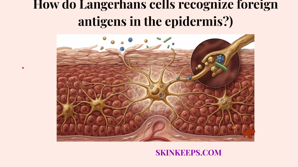
Where are Langerhans cells positioned to recognize foreign antigens in the epidermis?
Langerhans cells are positioned to recognize foreign antigens mainly in the suprabasal epidermis—the epidermal layers located above the basal layer—especially the stratum spinosum, the main suprabasal layer where these cells are most concentrated.
Their dendritic shape allows them to extend dendritic extensions, branching cellular projections used for surveillance, between neighboring keratinocytes—the primary epithelial cells of the epidermis. These processes survey the epidermal environment continuously without needing the cell body to move.
This arrangement places them at the overlap between environmental exposure and epidermal immune surveillance. A strong review describes Langerhans cells as occupying the stratum spinosum and extending dendrites through tight-junction-associated spaces toward outer layers without disrupting the barrier (Deckers et al., 2018).
How do Langerhans cells detect foreign antigens in the epidermis?
Langerhans cells detect foreign antigens in the epidermis by actively surveying the local tissue environment and encountering material that appears non-self, abnormal, or danger-associated rather than waiting passively for deeper inflammatory recruitment.
Detection begins through active environmental sampling, not static waiting. The cells use their dendritic processes to constantly “taste” the extracellular fluid between keratinocytes, looking for molecular signatures of invasion.
This first step is not yet full immune activation; it is the critical point at which suspicious material is encountered and interpreted as potentially meaningful. This encounter prepares the cell for the subsequent step of engaging its specialized receptor systems.
| Detection Phase | Action | Immune Significance |
|---|---|---|
| Outer Exposure | Foreign material contacts skin | Threat enters surveillance zone |
| Active Encounter | Dendrites meet the material | Transition from sampling to detection |
| Recognition Initiation | Systems begin to engage | Threat interpreted as “non-self” |
How do recognition receptors help Langerhans cells recognize foreign antigens?
Recognition receptors—immune-sensing molecules that detect danger or non-self material—help Langerhans cells recognize foreign antigens by translating environmental exposure into a biologically actionable immune signal.
Recognition depends on both surface and intracellular immune-sensing systems, specifically PRRs (pattern-recognition receptors) that detect conserved danger signals, such as TLRs (Toll-like receptors).
Langerhans cells use innate-sensing systems including C-type lectin pathways. Reviews note that epidermal Langerhans cells participate in TLR-linked sensing, while Langerin (CD207) is a characteristic C-type lectin receptor involved in antigen uptake (Stoitzner, 2011).
How do Langerhans cells capture foreign antigens after recognizing them?
Langerhans cells capture foreign antigens after recognizing them by internalizing suspicious material into the cell, allowing the recognition event to move beyond surface detection and into direct immune handling.
Capture is necessary because recognition alone does not yet preserve the material in a form the immune system can use. Antigen capture (internalization) is the physical uptake of recognized material into the cell’s interior.
This step converts environmental exposure into an intracellular immune event. Once internalized, the cell can begin the chemical transformation of the material into information that other immune cells can understand.
How does antigen capture begin after recognition?
Antigen capture begins after recognition when suspicious material is internalized by the Langerhans cell, turning a detection event into direct handling of the foreign signal. A focused review describes langerin/CD207 as a characteristic uptake receptor involved in pathogen and antigen internalization (Stoitzner, 2011).
Why is antigen capture necessary for immune coordination?
Antigen capture is necessary for immune coordination because the immune system cannot respond in a targeted way unless the foreign material is first preserved, handled, and prepared for interpretation. A review of antigen presentation explains that uptake and intracellular handling are prerequisites for later immune functions (Igyártó et al., 2012).
How do Langerhans cells process foreign antigens after capture?
Langerhans cells process foreign antigens after capture by breaking the internalized material down into smaller immunologically meaningful components that can later be used for structured immune communication.
Antigen processing is the intracellular breakdown of antigens into immune-relevant fragments. Raw foreign material must be transformed into these “molecular snapshots” before it can be presented to T-cells.
Processing is what makes local recognition relevant beyond the epidermis. It prepares the threat information for presentation, ensuring that the broader immune system receives a clear message about what has breached the barrier (Igyártó et al., 2012).
How does foreign-antigen recognition activate Langerhans cells?
Foreign-antigen recognition activates Langerhans cells by shifting them from a resting surveillance state into a more responsive state marked by stronger signaling, antigen-handling commitment, and migration-related readiness.
An activated Langerhans cell is a recognition-engaged cell that has stopped its resting surveillance and shifted toward signaling and migration. This shift changes how the cell interacts with its microenvironment, especially during the process of keratinization in the surrounding tissue.
Activation should be understood as a functional shift, not simply as generalized inflammation. It is a biological pivot that prepares the cell to leave the epidermis and transport its captured information to the command centers of the immune system.
How do resting and activated Langerhans cells differ during foreign-antigen recognition?
Resting and activated Langerhans cells differ during foreign-antigen recognition because resting cells emphasize ongoing surveillance, while activated cells emphasize stronger antigen handling and downstream coordination.
Resting Langerhans cells are built for readiness and sampling. They maintain the dendritic network that makes the epidermis an “intelligent” barrier, similar to how keratinocytes maintain the physical structure.
Activated Langerhans cells are functionally changed by recognition. They downregulate their sampling dendrites and upregulate molecules that allow them to communicate with the specialized memory cells of the adaptive system.
| State | Main Behavior | Recognition Status | Immune Role |
|---|---|---|---|
| Resting | Ongoing surveillance | Monitoring for danger | Maintains readiness |
| Activated | Antigen handling | Foreign antigen recognized | Initiates coordination |
How does Langerhans cell foreign-antigen recognition connect to adaptive immunity?
Langerhans cell foreign-antigen recognition connects to adaptive immunity—the T-cell–linked immune phase that follows local detection—by converting local detection into processed information that can be carried outward.
This handoff requires CCR7, a migration-associated chemokine receptor linked to lymph-node trafficking. A migration model for Langerhans cells identifies CCR7-linked trafficking as part of the route from activated epidermal surveillance toward skin-draining lymph nodes (Villablanca et al., 2008).
How can you tell when Langerhans cell foreign-antigen recognition is weak or dysregulated?
You can tell when Langerhans cell foreign-antigen recognition is weak or dysregulated when the skin becomes less effective at controlled defense and more prone to recurrent infection or chronic inflammatory instability.
Warning Signs of Recognition Failure
What factors can weaken or dysregulate Langerhans cell foreign-antigen recognition?
Langerhans cell foreign-antigen recognition works best in a stable epidermal environment and becomes weaker or less balanced when the barrier is repeatedly stressed, UV-exposed, or damaged.
Excess UV exposure is especially relevant because UV radiation is known to alter Langerhans-cell number and morphology, which can weaken surveillance quality over time. This instability is a major factor that contributes to barrier disruption and increased water loss (Fukunaga et al., 2010).
Repeated inflammation and harsh routines can add persistent immune noise, making balanced recognition harder to maintain. When the background signaling is too high, the specific signal of a foreign antigen can be lost.
What habits help support healthy Langerhans cell foreign-antigen recognition?
Healthy Langerhans cell foreign-antigen recognition is best supported by barrier stability, lower chronic irritation, and protection from cumulative environmental immune stress.
How does barrier support help preserve foreign-antigen recognition?
Barrier support helps preserve foreign-antigen recognition by reducing background immune stress and creating a more stable epidermal surveillance environment. Human skin studies have linked barrier-related filaggrin defects to altered Langerhans-cell maturation markers (Leitch et al., 2016).
How does sun protection help preserve foreign-antigen recognition?
Sun protection helps preserve foreign-antigen recognition by reducing cumulative UV-related impairment of epidermal immune surveillance. Review literature describes UV exposure as capable of functionally disturbing these cells, which is why daily SPF is relevant to recognition quality (Fukunaga et al., 2010).
How do gentler routines help support healthy epidermal surveillance?
Gentler routines help support healthy epidermal surveillance by reducing unnecessary inflammatory noise and avoiding repeated destabilization of the environment. Because barrier instability is associated with altered Langerhans-cell behavior, gentler routines help maintain a more reliable surveillance state (Leitch et al., 2016).
Problem: Barrier stress and immune imbalance
Implication: Foreign-antigen recognition may weaken or become dysregulated
Solution: Protect the barrier, reduce chronic irritation, and support epidermal stability
What are the key takeaways about how Langerhans cells recognize foreign antigens in the epidermis?
The key takeaways center on the sequence of surveillance, receptor-supported detection, antigen capture, processing, activation, and adaptive immune handoff.
- ● Langerhans cells recognize antigens by surveying, detecting, and internalizing material.
- ● Recognition depends on both epidermal positioning and PRR/TLR mechanisms.
- ● Activation shifts the cell from resting surveillance to active coordination.
- ● Healthy recognition requires a stable epidermal microenvironment.
What daily steps can you take to support healthy foreign-antigen recognition in the epidermis?
You can support healthy foreign-antigen recognition in the epidermis by protecting the barrier, minimizing chronic irritation, and reducing cumulative environmental stress on immune surveillance.
Immune Surveillance Support Checklist
Healthy antigen recognition depends on more than immune cells being present. It depends on an epidermal environment that allows those cells to detect, process, and communicate without chronic disruption.
Build your routine around barrier stability and immune balance if your goal is healthier epidermal defense and more resilient skin immunity.