
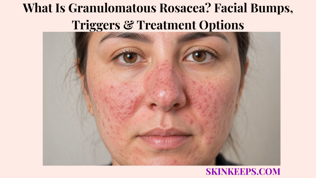
Granulomatous rosacea is an uncommon chronic inflammatory facial condition that produces firm, persistent papules or small nodules. The bumps may be skin-coloured, pink, red, yellow-brown, red-brown, or purple-brown, while flushing, diffuse redness, and visible vessels may be mild or absent.
It is not acne or contagious and cannot be diagnosed reliably from facial bumps alone. Care may involve biopsy, targeted exclusion of LMDF, sarcoidosis, or infection, gentle skin care, topical or oral treatment, eye assessment, and long-term relapse management.
How Can You Recognize Granulomatous Rosacea Facial Bumps?
Granulomatous rosacea often appears as persistent firm facial papules or small nodules that look relatively uniform, or monomorphic.
The lesions may remain for weeks or months and occur symmetrically on normal-looking skin or within mild redness. They are usually smooth and contain little or no pus.
Tenderness, burning, sensitivity, or swelling can occur. The absence of blackheads and whiteheads is an important clue, but it does not confirm the diagnosis by itself.
- Firm papules or small nodules.
- Relatively uniform lesion size and shape.
- Skin-coloured, pink, red, yellow-brown, red-brown, or purple-brown colour.
- Central or periocular facial distribution.
- Persistence for weeks or months.
- Little or no pus.
- No comedones.
- Redness may be mild or absent.
- Burning, tenderness, or sensitivity may occur.
Figure 1. Granulomatous rosacea often causes firm, persistent, relatively uniform papules on central or periocular facial skin, commonly without blackheads or whiteheads.
Where Does Granulomatous Rosacea Usually Appear?
Granulomatous rosacea usually appears on central and upper-central facial areas such as the cheeks, sides of the nose, eyelids, around the eyes, forehead, chin, and mouth area.
Periocular involvement matters because LMDF, sarcoidosis, periorificial dermatitis, and infections can affect similar sites.
Broader facial involvement is possible, but location supports rather than confirms the diagnosis.
| Facial Area | Common Pattern | Diagnostic Importance |
|---|---|---|
| Cheeks | Firm symmetric papules | Common central-face site |
| Eyelids and periocular skin | Small papules or nodules | Strong overlap with LMDF and ocular disease |
| Sides of nose | Yellow-brown or red-brown papules | Rosacea and sarcoidosis overlap |
| Forehead | Monomorphic papules | Seen in reported granulomatous rosacea cases |
| Around mouth | Papules with or without redness | Compare periorificial dermatitis |
| Chin | Persistent papules | Compare acne and periorificial disease |
| Broader face | Less typical spread | Consider expanded differential |
Is Granulomatous Rosacea a Separate Rosacea Type or a Phenotype?
Granulomatous rosacea has traditionally been described as a rare rosacea variant, while modern consensus increasingly uses phenotype-based diagnosis and treatment.
The phenotype approach identifies the visible and symptomatic features present rather than forcing every patient into one rigid subtype.
A person may have granulomatous papules together with flushing, persistent redness, visible vessels, ocular symptoms, or other rosacea features.
| Feature | Granulomatous Presentation | Papulopustular Presentation | Erythematous Presentation |
|---|---|---|---|
| Dominant lesion | Firm papules or nodules | Papules and pustules | Redness and flushing |
| Comedones | Absent | Absent | Absent |
| Background redness | May be mild or absent | Often present | Dominant |
| Visible vessels | May be absent | May occur | Common |
| Biopsy role | Often useful | Usually not needed | Usually not needed |
| Treatment logic | Feature-based and diagnosis-sensitive | Inflammatory lesion control | Vascular redness control |
How Does Granulomatous Rosacea Differ From More Typical Rosacea?
Firm yellow-brown or red-brown papules may dominate granulomatous rosacea while classic flushing, diffuse redness, or visible vessels remain subtle.
More typical rosacea may emphasise recurrent flushing, persistent central redness, telangiectasia, softer inflammatory papules or pustules, burning, eye symptoms, or skin thickening.
The presentations can overlap. Granulomatous bumps do not replace other phenotypes; they may coexist with them.
How Does Granulomatous Rosacea Develop Within the Skin?
Granulomatous rosacea develops when clusters of inflammatory cells form within the dermis, creating firm papules or nodules.
Rosacea involves dysregulated immune and neurovascular responses. In granulomatous disease, histiocytes, lymphocytes, epithelioid cells, and multinucleated giant cells may form granulomas around follicles and sebaceous structures.
Follicular disruption, keratin material, and Demodex may contribute in selected cases, but no single pathway explains every patient.
Figure 2. Granulomatous rosacea involves dermal inflammatory clusters, often around follicular structures, producing firm papules while immune, follicular, microbial, genetic, and environmental contributors remain incompletely defined.
What Causes Granulomatous Rosacea?
The exact cause of granulomatous rosacea remains unknown.
It is not caused by poor hygiene, a contagious bacterial or fungal infection, or the same comedonal process that defines acne.
Immune dysregulation, abnormal inflammatory responses, follicular factors, Demodex in selected patients, genetic susceptibility, and environmental influences may contribute.
| Proposed Factor | Possible Role | Universal Cause? | Care Implication |
|---|---|---|---|
| Immune dysregulation | Persistent dermal inflammation | No | Anti-inflammatory treatment |
| Follicular disruption | May initiate granuloma formation | No | Biopsy and morphology context |
| Demodex | May contribute in selected patients | No | Targeted ivermectin only when clinically suitable |
| Genetic susceptibility | May influence rosacea tendency | No | Not diagnostic |
| Environmental aggravators | May worsen rosacea features | No | Individual trigger record |
| Poor hygiene | Myth | No | Gentle care, not scrubbing |
Which Triggers May Worsen Granulomatous Rosacea?
Rosacea triggers may worsen redness, burning, sensitivity, or inflammation, but they are not proven to create every granulomatous papule.
Possible aggravators include sunlight, hot weather, cold wind, hot showers, spicy foods, alcohol, hot drinks, emotional stress, strenuous exercise, friction, irritating cosmetics, harsh cleansers, topical corticosteroids, and medicines that promote flushing.
Evidence is stronger for flushing and general rosacea flares than for granulomatous papules specifically. A personal trigger record is more useful than avoiding every published trigger.
| Exposure | Possible Change | Record | Interpretation Boundary |
|---|---|---|---|
| Sun or heat | Redness, burning, sensitivity | Timing and duration | Aggravator, not proven cause |
| Cold or wind | Barrier irritation | Weather and skin response | Individual pattern |
| Spicy food, alcohol, hot drinks | Flushing | Repeatability | May not change papules |
| Exercise or stress | Heat and flushing | Intensity and recovery | Context-dependent |
| Cosmetics or harsh cleanser | Stinging and dermatitis | Product and application | May mimic or worsen disease |
| Topical steroid | Rosacea-like rebound | Strength and duration | Requires medical review |
Does Granulomatous Rosacea Look Different Across Skin Tones?
Granulomatous rosacea may be harder to recognize on brown or black skin because background redness can be subtle.
Inflammation may appear purple, brown, grey-brown, yellow-brown, or darker than nearby skin. Firm texture, distribution, burning, swelling, and sensitivity can be more informative than visible redness.
After papules flatten, post-inflammatory hyperpigmentation may remain longer than the active inflammation.
- Feel for firm uniform papules.
- Assess central and periocular distribution.
- Look for purple, brown, grey-brown, or yellow-brown change.
- Ask about burning, sensitivity, and swelling.
- Use good lighting or dermoscopy when needed.
- Separate residual pigment from active raised lesions.
How Is Granulomatous Rosacea Different From Acne?
Granulomatous rosacea usually lacks blackheads and whiteheads, while acne vulgaris commonly includes comedones.
The absence of comedones argues against acne, but it does not prove granulomatous rosacea by itself.
Granulomatous rosacea tends to create firmer, more uniform central or periocular papules. Acne often combines comedones, papules, pustules, nodules, or cysts and may affect the jaw, chest, or back.
Strong acne acids or rough exfoliation can worsen facial sensitivity when the diagnosis is wrong.
| Feature | Granulomatous Rosacea | Acne Vulgaris |
|---|---|---|
| Comedones | Absent | Common |
| Lesion uniformity | Relatively uniform | Mixed lesion types |
| Main location | Central or periocular face | Face, jaw, chest, back |
| Pus | Usually little | Pustules possible |
| Skin sensitivity | Common | Variable |
| Biopsy role | Often useful when uncertain | Usually unnecessary |
| Harsh acne products | May irritate | May help selected true acne |
How Is Granulomatous Rosacea Different From Periorificial Dermatitis?
Granulomatous rosacea can affect the cheeks, eyelids, nose, forehead, and mouth area, while periorificial dermatitis clusters around the mouth, nose, or eyes.
Perioral dermatitis often spares a narrow area beside the lip margin and may follow topical corticosteroid exposure.
Granulomatous periorificial dermatitis can occur in children. Biopsy may be needed when morphology overlaps or treatment fails.
| Condition | Mouth Border | Eyelids | Central Cheeks | Flushing | Biopsy Role |
|---|---|---|---|---|---|
| Granulomatous rosacea | May involve | May involve | Common | May occur or be mild | Useful if uncertain |
| Periorificial dermatitis | Often lip-margin sparing | May involve | Less dominant | Usually not dominant | Atypical or resistant cases |
| Granulomatous periorificial dermatitis | Often perioral in children | Possible | Variable | Not core | Specialist assessment |
How Is Granulomatous Rosacea Different From LMDF?
LMDF can closely resemble granulomatous rosacea because both may cause red-brown or yellow-brown facial papules with granulomatous inflammation.
LMDF often involves the eyelids strongly, may show caseation or necrosis on biopsy, and is more likely to leave pitted scars.
Granulomatous rosacea more often overlaps with other rosacea features and may show noncaseating perifollicular granulomas. Historical terminology is inconsistent, so clinical and microscopic findings must be interpreted together.
| Feature | Granulomatous Rosacea | LMDF |
|---|---|---|
| Distribution | Central and periocular face | Strong eyelid and central-face pattern |
| Rosacea features | May coexist | Usually less prominent |
| Biopsy | Often noncaseating or perifollicular granulomas | Caseation or necrosis may occur |
| Scarring | Less typical | Pitted scarring more likely |
| Treatment logic | Rosacea-directed options | Separate specialist plan |
| Certainty | Clinical and biopsy correlation | Clinical and biopsy correlation |
How Is Granulomatous Rosacea Different From Sarcoidosis?
Sarcoidosis can resemble granulomatous rosacea on the face, but sarcoidosis may also involve the lungs, eyes, lymph nodes, heart, scars, and other skin sites.
Both conditions can produce red-brown, purple, yellow-brown, or skin-coloured papules. Sarcoidosis may show smooth translucent lesions and relatively sparse inflammation around granulomas.
Biopsy patterns overlap. Respiratory symptoms, eye symptoms, swollen nodes, scar infiltration, or suspicious histology may justify chest imaging and targeted systemic evaluation.
| Feature | Granulomatous Rosacea | Sarcoidosis |
|---|---|---|
| Skin distribution | Central or periocular face | Face, scars, nose, other sites |
| Systemic disease | Not a core feature | Lungs, eyes, nodes, heart possible |
| Biopsy | Granulomatous inflammation | Granulomas with possible sparse rim |
| Extra testing | Targeted if atypical | Systemic work-up when suspected |
| Treatment risk | Rosacea-directed | Do not miss systemic disease |
Which Other Conditions Can Resemble Granulomatous Rosacea?
Several inflammatory, infectious, granulomatous, and tumour-like conditions can resemble granulomatous rosacea.
Follicular pustules or infection-like facial bumps can resemble folliculitis, while other key mimics include demodicosis, cutaneous tuberculosis, lupus vulgaris, leishmaniasis, fungal disease, foreign-body granuloma, granulomatous contact dermatitis, lupus, sebaceous hyperplasia, and benign tumours.
Ulceration, scarring, drainage, systemic symptoms, travel, procedure history, or unusual spread should widen the differential.
| Condition | Defining Clue | Systemic Relevance | Helpful Test |
|---|---|---|---|
| Demodicosis | Follicular roughness and mite burden | Usually skin-limited | Microscopy or clinical response |
| Cutaneous TB or lupus vulgaris | Chronic plaque and exposure risk | Important infection | Biopsy, stain, culture, PCR |
| Leishmaniasis | Travel or vector exposure, ulcerating papule | Infection | Smear, biopsy, PCR |
| Fungal disease | Scale or pustules | Usually skin-limited | KOH, culture, stain |
| Foreign-body granuloma | Procedure or trauma history | Local | Biopsy |
| Granulomatous contact dermatitis | Exposure pattern | Usually skin-limited | Patch testing and biopsy |
| Systemic lupus | Photosensitive or systemic clues | Systemic possible | Labs and biopsy |
| Sebaceous hyperplasia or tumour | Persistent focal growth | Usually local | Dermoscopy or biopsy |
How Do Clinicians Diagnose Granulomatous Rosacea?
Clinicians diagnose granulomatous rosacea by combining facial morphology, rosacea phenotypes, history, biopsy, and exclusion of meaningful alternatives.
Assessment includes distribution, symmetry, colour, firmness, uniformity, comedones, flushing, persistent erythema, telangiectasia, eye symptoms, skin-care and cosmetic history, topical steroid exposure, medicines, travel, infection risk, systemic symptoms, and previous treatment response.
Other skin sites are examined when sarcoidosis, infection, or another granulomatous disorder is possible. There is no single blood test that confirms granulomatous rosacea.
- Facial distribution and symmetry.
- Colour, firmness, and uniformity.
- Presence or absence of comedones.
- Flushing, redness, and visible vessels.
- Eye and eyelid symptoms.
- Skin-care, cosmetic, and steroid history.
- Medication and treatment-response history.
- Travel, infection, procedure, and systemic-disease clues.
- Examination beyond the face when indicated.
- Biopsy and targeted exclusion tests when needed.
Why Is a Skin Biopsy Often Needed for Granulomatous Rosacea?
A skin biopsy is often useful because several granulomatous facial diseases can look similar.
Biopsy becomes more important with firm monomorphic papules, periocular or unusual distribution, minimal classic rosacea features, scarring, ulceration, treatment resistance, or concern for LMDF, sarcoidosis, infection, or another granulomatous disease.
Possible findings include noncaseating or perifollicular granulomas, epithelioid histiocytes, multinucleated giant cells, dilated vessels, follicular disruption, and lymphocytic inflammation.
Biopsy supports the diagnosis but may not settle it alone because several conditions share microscopic features.
| Histologic Finding | May Support | Does Not Exclude | Possible Follow-Up |
|---|---|---|---|
| Noncaseating granulomas | GR or sarcoidosis pattern | Sarcoidosis or infection | Clinical and systemic correlation |
| Perifollicular granulomas | GR pattern | LMDF or infection | Special stains if indicated |
| Giant cells and histiocytes | Granulomatous inflammation | Several diseases | Biopsy correlation |
| Follicular disruption | Rosacea-like reaction | Foreign body or infection | Culture or stains if suspicious |
| Caseation or necrosis | LMDF or infection concern | TB or leishmaniasis | Microbiology or PCR |
| Dilated superficial vessels | Rosacea context | Other inflammation | Phenotype assessment |
Which Tests May Be Needed to Exclude Other Granulomatous Diseases?
Testing for granulomatous rosacea mimics should be targeted by biopsy findings, exposure history, systemic symptoms, and treatment risk.
Possible investigations include bacterial, fungal, or mycobacterial stains; tissue cultures; PCR; tuberculosis testing; leishmaniasis testing; chest imaging; symptom-guided blood tests; ophthalmology assessment; or patch testing.
Not every patient needs every test. Infection exclusion is especially important before systemic immune suppression.
| Suspicion | Test | Purpose | Management Change |
|---|---|---|---|
| TB or lupus vulgaris | Stain, culture, PCR, TB testing | Identify infection | Antimicrobial pathway |
| Leishmaniasis | Smear, biopsy, PCR | Confirm parasite | Antiparasitic treatment |
| Fungal disease | KOH, culture, stain | Exclude fungus | Antifungal treatment |
| Sarcoidosis | Chest imaging and targeted labs | Assess systemic disease | Systemic evaluation |
| Ocular disease | Ophthalmology exam | Protect eye surface and vision | Eye-directed treatment |
| Contact dermatitis | Patch testing | Identify allergen | Avoidance plan |
Does Granulomatous Rosacea Require Treatment?
Granulomatous rosacea often requires treatment when papules persist, spread, cause symptoms, affect the eyes, or create psychosocial distress.
Mild stable disease may be monitored while diagnosis is confirmed, but firm lesions can persist longer than ordinary rosacea papules.
No universally accepted granulomatous-rosacea-specific regimen exists. Several treatments are adapted from rosacea care or supported by case reports, so response is variable and rapid clearance cannot be promised.
| Situation | Direction | Reason |
|---|---|---|
| Few stable lesions | Confirm and discuss | Avoid overtreatment |
| Burning or tender lesions | Active treatment | Symptom control |
| Spreading lesions | Dermatology treatment | Persistent inflammatory course |
| Periocular lesions | Assess eye and eyelid involvement | Ocular risk |
| Refractory disease | Reassess and escalate carefully | Exclude mimics first |
| Ulceration, scarring, or systemic clues | Reconsider diagnosis | LMDF, infection, or sarcoidosis |
How Should Facial Skin Be Cared for During Granulomatous Rosacea Treatment?
Gentle skin care and sun protection support treatment by reducing irritation and background rosacea flares.
- Wash with a mild fragrance-free cleanser.
- Use fingertips rather than brushes or scrubs.
- Rinse with lukewarm rather than hot water.
- Pat dry gently.
- Apply a non-irritating moisturizer.
- Use broad-spectrum sunscreen regularly.
- Choose sensitive-skin products.
- Introduce one new product at a time.
- Avoid astringents, rough exfoliants, and aggressive treatments.
- Keep the routine stable long enough to assess response.
- Use skin care as support rather than a replacement for prescribed treatment.
Which Topical Treatments May Help Granulomatous Rosacea?
Topical treatment is individualized and often adapted from rosacea medicines used for inflammatory facial lesions.
How May Metronidazole Help Granulomatous Rosacea?
Metronidazole is used mainly for anti-inflammatory activity rather than because granulomatous rosacea is a bacterial infection.
Improvement is gradual, and dryness, irritation, or stinging may occur. Evidence specific to the granulomatous presentation is limited.
How May Azelaic Acid Reduce Facial Inflammation?
Azelaic acid has anti-inflammatory and keratinization-modifying effects and may help papules, redness, or residual pigmentation.
Burning or tingling can occur, so sensitive skin may need a cautious introduction.
When May Topical Ivermectin Be Considered?
Ivermectin has anti-inflammatory and anti-Demodex activity and may be considered when a follicular or mite-associated pattern fits.
Evidence is stronger for papulopustular rosacea than for granulomatous rosacea, and temporary irritation can occur.
When Are Pimecrolimus or Tacrolimus Used?
Calcineurin inhibitors have been reported in selected granulomatous cases when other treatments are unsuitable.
Evidence is limited to small reports, burning may occur, and infection should be reasonably excluded first.
| Option | Primary Action | Best-Fit Feature | Limitation | Evidence |
|---|---|---|---|---|
| Metronidazole | Anti-inflammatory | Inflammatory facial lesions | Dryness or stinging | Rosacea evidence; limited GR data |
| Azelaic acid | Anti-inflammatory and keratinization effects | Papules, redness, pigmentation | Burning or tingling | Rosacea evidence; individualized |
| Ivermectin | Anti-inflammatory and anti-Demodex | Follicular or mite-associated pattern | Irritation | Stronger for papulopustular rosacea |
| Pimecrolimus or tacrolimus | Calcineurin inhibition | Selected resistant cases | Burning; exclude infection | Small reports |
Which Oral Treatments May Control Granulomatous Rosacea?
Oral treatment may be considered for persistent, spreading, or refractory disease after the diagnosis and major mimics have been reviewed.
How Are Oral Tetracyclines Used?
Doxycycline, minocycline, or tetracycline may help through anti-inflammatory effects rather than treatment of a proven bacterial cause.
Improvement often takes weeks. Photosensitivity, gastrointestinal upset, oesophageal irritation, pregnancy, age, and drug interactions affect suitability, and relapse can occur after stopping.
When Is Oral Isotretinoin Considered?
Isotretinoin may be considered for severe or refractory disease under specialist supervision.
Dryness, lipids, liver function, pregnancy prevention, and relapse require monitoring.
Are Other Systemic Medicines Used?
Dapsone, macrolides, antiparasitic treatment, or selected immune-modifying medicines have been reported in difficult cases.
They are not interchangeable first-line options. Selection depends on biopsy findings, comorbidities, infection exclusion, and medicine-specific risks.
| Medicine | Role | Expected Timing | Monitoring | Major Restriction |
|---|---|---|---|---|
| Tetracyclines | Anti-inflammatory control | Weeks | Side effects and interactions | Pregnancy and age limitations |
| Isotretinoin | Refractory or severe disease | Gradual | Lipids, liver, dryness | Pregnancy prevention |
| Dapsone | Selected specialist option | Variable | Blood and liver risks | Not routine |
| Macrolides | Alternative in selected cases | Variable | Interactions and GI effects | Not universal |
| Antiparasitic treatment | Diagnosis-dependent | Variable | Targeted assessment | Avoid blanket Demodex assumption |
| Immune-modifying medicines | Highly selected | Variable | Infection and systemic risks | Exclude infection first |
Can Lasers or Light Treatments Remove Granulomatous Rosacea Bumps?
Lasers and light treatments may help redness or visible vessels, but they do not reliably remove underlying dermal granulomas.
Vascular lasers and intense pulsed light target erythema and telangiectasia more directly than firm papules.
Active inflammation can become irritated, and pigment change is possible, especially in darker skin tones. Procedural treatment should target a defined phenotype and be performed by an experienced clinician.
| Procedure | Redness | Vessels | Granulomatous Papules | Boundary |
|---|---|---|---|---|
| Vascular laser | May help | May help | Not reliable | Targets vascular phenotype |
| Intense pulsed light | May help | May help | Limited | Skin-tone caution |
| Photodynamic treatment | Uncertain | Variable | Case-level evidence | Specialist use |
| Home laser device | Avoid | Unsafe | Unsafe | Burn and pigment risk |
Are Newer Treatments Proven for Granulomatous Rosacea?
Newer treatments remain emerging possibilities for highly refractory disease, not routine standards.
Case reports describe improvement with topical ruxolitinib, tapinarof, oral JAK inhibitors, or biologic immune-modifying treatment.
Reports may involve one or a few patients, spontaneous fluctuation can complicate interpretation, long-term safety and relapse remain uncertain, and systemic infection risks may be significant.
Evidence ladder: established rosacea therapy → small case series → individual case report → experimental possibility. Case reports do not establish routine care.
Which Granulomatous Rosacea Treatment Mistakes Should Be Avoided?
Treatment mistakes often occur when firm facial papules are treated repeatedly as acne, Demodex alone, or infection without diagnostic reassessment.
| Unsafe Action | Why It Fails | Possible Harm | Safer Response |
|---|---|---|---|
| Squeezing or piercing | Papules are not removable plugs | Scarring and infection | Dermatology assessment |
| Aggressive scrubbing | Does not remove dermal granulomas | Barrier damage | Gentle cleanser |
| Strong acne acids | Wrong pathway if acne is absent | Burning and dermatitis | Confirm diagnosis |
| Unsupervised topical steroids | Can worsen rosacea-like disease | Rebound and thinning | Clinician plan |
| Steroid-antifungal combination | May mask the diagnosis | Worsening and atrophy | Avoid unless prescribed |
| Indefinite antibiotics | No reassessment | Side effects and resistance | Review diagnosis |
| Treating every case as Demodex | Oversimplifies cause | Delayed diagnosis | Targeted therapy |
| Antifungals without evidence | Wrong target | Irritation and delay | Test if infection suspected |
| Immune suppression before infection exclusion | Can worsen TB or leishmaniasis | Disseminated infection | Targeted exclusion testing |
| Harsh peels or home lasers | Irritates active disease | Burn and pigment change | Specialist procedures |
| Automatic escalation after failure | Diagnosis may be wrong | Higher treatment risk | Re-biopsy or reassess |
How Long Does Granulomatous Rosacea Last—and Can It Return?
Granulomatous rosacea is usually chronic, and individual firm papules may persist for weeks or months.
Improvement may require prolonged therapy, and lesions can recur after apparent clearance. Trigger control may reduce flushing without preventing every granulomatous papule.
Residual red, brown, or purple pigmentation may remain after bumps flatten. Scarring is not typical and should prompt reconsideration of LMDF or another diagnosis.
Management aims for control and longer remission rather than a guaranteed permanent cure.
Course: persistent papules → gradual treatment response → fading pigmentation → remission → possible recurrence.
Can Granulomatous Rosacea Affect the Eyes?
Granulomatous rosacea can coexist with eye or eyelid symptoms, and eye complaints may not match the severity of facial bumps.
- Dry or burning eyes.
- Gritty sensation.
- Watery eyes.
- Red eyelid margins.
- Recurrent styes.
- Crusting around eyelashes.
- Light sensitivity.
- Blurred vision.
- Eye pain.
Urgent eye care: eye pain, marked light sensitivity, blurred vision, or a very red eye may indicate corneal inflammation and needs prompt ophthalmic assessment.
When Should Persistent Facial Bumps Be Checked by a Dermatologist?
Persistent firm facial papules should be checked when they last for several weeks, cluster around the eyes, lack comedones, or fail acne treatment.
- Firm papules persist for several weeks.
- Bumps concentrate around the eyes.
- Acne treatment repeatedly fails.
- No comedones are present.
- The diagnosis remains uncertain.
- Lesions spread despite gentle care.
- Facial changes cause substantial distress.
- Eye or eyelid symptoms develop.
- Oral therapy or biopsy is being considered.
Prompt reassessment: scarring, ulceration, bleeding, pain, pus, unusual spread, fever, cough, breathing symptoms, swollen lymph nodes, TB or leishmaniasis exposure, worsening during immune suppression, or treatment failure should trigger diagnostic reconsideration.
Figure 3. Granulomatous rosacea care combines phenotype assessment, biopsy when needed, targeted exclusion of infection and systemic mimics, gentle skin care, individualized medication, and diagnostic reassessment when disease behaves atypically.
What Should You Remember About Granulomatous Rosacea?
Granulomatous rosacea is an uncommon chronic facial disorder characterized by firm granulomatous papules or nodules.
- Papules may be skin-coloured, pink, red, yellow-brown, red-brown, or purple-brown.
- Flushing and diffuse redness may be mild or absent.
- The exact cause remains unknown.
- Demodex may contribute but is not the sole proven cause.
- The absence of comedones helps separate the condition from acne.
- LMDF, sarcoidosis, periorificial dermatitis, and infection can closely resemble it.
- Biopsy often helps when lesions are atypical or resistant.
- Biopsy alone may not settle the diagnosis.
- Gentle skin care and sunscreen support medical treatment.
- Topical rosacea medicines and oral tetracyclines are commonly considered.
- Isotretinoin and other systemic options require specialist supervision.
- Lasers treat vascular features more reliably than dermal granulomas.
- Emerging case reports are not established routine care.
- Eye pain, blurred vision, scarring, ulceration, or systemic clues require reassessment.
What Questions Do People Ask About Granulomatous Rosacea?
Is granulomatous rosacea a true type of rosacea?
It has traditionally been described as a rare rosacea variant, but modern guidance increasingly uses a phenotype-based approach. Clinicians identify and treat the features present, such as granulomatous papules, redness, flushing, visible vessels, or ocular symptoms.
What do granulomatous rosacea bumps look like?
They are often firm, relatively uniform papules or small nodules that may be skin-coloured, pink, red, yellow-brown, red-brown, or purple-brown on central or periocular facial skin.
Can granulomatous rosacea occur without facial redness?
Yes. Flushing, diffuse redness, and visible vessels may be mild or absent, making persistent firm papules the main clue.
Is granulomatous rosacea contagious?
No. It does not spread through touch, towels, kissing, or ordinary contact.
Is granulomatous rosacea caused by Demodex mites?
Demodex may contribute in selected patients, but it is not the proven sole cause. The condition is likely multifactorial.
How is granulomatous rosacea different from acne?
Granulomatous rosacea usually lacks blackheads and whiteheads, has firmer and more uniform facial papules, and often affects central or periocular areas. Acne commonly includes comedones and mixed lesion types.
How is granulomatous rosacea different from LMDF?
LMDF may show strong eyelid involvement, caseation or necrosis on biopsy, and a greater risk of pitted scarring. Clinical correlation and biopsy are often needed because the two conditions overlap.
Can granulomatous rosacea be mistaken for sarcoidosis?
Yes. Both can cause granulomatous facial papules. Sarcoidosis may involve lungs, eyes, lymph nodes, heart, scars, or other skin sites, so systemic clues may require targeted assessment.
Does granulomatous rosacea require a skin biopsy?
Biopsy is often useful for firm monomorphic or periocular papules, atypical or treatment-resistant disease, scarring, ulceration, or concern for LMDF, sarcoidosis, infection, or another granulomatous condition.
Can sun exposure worsen granulomatous rosacea?
Sun exposure is a common rosacea aggravator and may worsen redness or sensitivity. Its role in forming granulomatous papules specifically is less certain.
Do topical steroids make granulomatous rosacea worse?
Repeated unsupervised facial corticosteroid use can provoke or worsen rosacea-like and periorificial eruptions and may cause rebound inflammation or skin thinning.
Which creams treat granulomatous rosacea?
Clinician-directed options may include metronidazole, azelaic acid, ivermectin, or selected calcineurin inhibitors. Evidence specific to granulomatous rosacea is limited, so treatment is individualized.
Can oral doxycycline clear granulomatous rosacea?
Oral tetracyclines such as doxycycline may help through anti-inflammatory effects, but improvement usually takes weeks and recurrence can occur after treatment stops.
When is isotretinoin considered?
Isotretinoin may be considered for severe or refractory disease under specialist supervision, with pregnancy prevention and appropriate laboratory monitoring.
Can granulomatous rosacea return after treatment?
Yes. It is chronic, individual papules may persist for weeks or months, lesions can recur, and residual pigmentation may remain after inflammation settles.
When do granulomatous facial bumps require urgent reassessment?
Prompt reassessment is needed for scarring, ulceration, bleeding, pain, pus, unusual spread, systemic symptoms, infection exposure risk, worsening during immune-suppressing therapy, eye pain, marked light sensitivity, blurred vision, or a very red eye.
Which Sources Support This Granulomatous Rosacea Guidance?
ROSCO 2019 Consensus — Phenotype-Based Rosacea Guidance — Transition from rigid subtypes to diagnosis and management based on each patient’s visible rosacea features.
American Academy of Dermatology — Rosacea Diagnosis and Treatment — Clinical diagnosis, testing to exclude mimics, and biopsy when granulomatous rosacea is suspected.
DermNet — Rosacea — Triggers, skin care, sunscreen, topical metronidazole, azelaic acid, ivermectin, oral treatment, vascular procedures, and phenotype management.
Facial Granulomatous Rosacea Case Report — Firm monomorphic facial papules, perifollicular granulomas, giant cells, biopsy role, and overlap with sarcoidosis and LMDF.
LMDF Versus Granulomatous Rosacea — Clinical overlap, eyelid distribution, histologic distinctions, caseation, and scarring risk.
StatPearls — Perioral Dermatitis — Periorificial distribution, corticosteroid association, clinical diagnosis, and biopsy for atypical or resistant disease.
DermNet — Ocular Rosacea — Eye and eyelid symptoms, ocular treatment boundaries, and ophthalmic care.
Topical Ruxolitinib Case Report — Emerging case-level evidence for a topical JAK inhibitor in treatment-refractory granulomatous rosacea.
This SkinKeeps article is educational and does not diagnose or replace dermatology, ophthalmology, infectious-disease, rheumatology, pathology, or emergency care. Have persistent, periocular, scarring, ulcerated, painful, draining, spreading, treatment-resistant, or systemically associated facial bumps examined; seek urgent eye care for pain, marked light sensitivity, blurred vision, or a very red eye, and do not squeeze lesions, use unsupervised facial steroids, or begin immune suppression before infection is excluded.



