
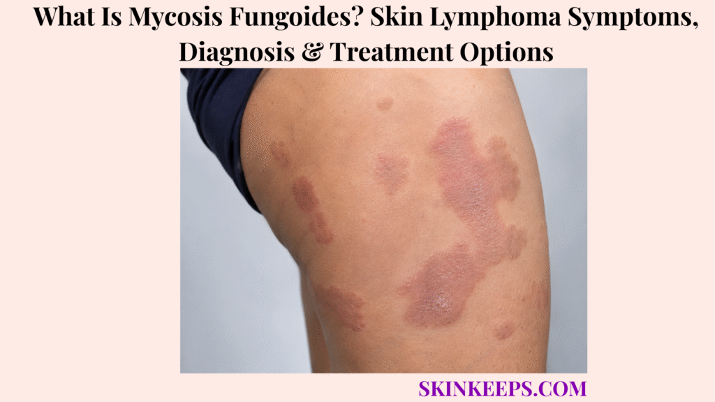
Mycosis fungoides is a cutaneous T-cell lymphoma in which malignant T lymphocytes preferentially accumulate in the skin, often beginning as persistent patches that resemble eczema or psoriasis. Despite the word “mycosis,” MF is not a fungal infection, and its cutaneous spectrum ranges from patches and plaques to tumours in a subset of patients rather than following one inevitable progression.
Early MF can remain difficult to confirm because inflammatory-looking patches may wax and wane and one biopsy may not contain enough diagnostic malignant cells. Treatment and prognosis depend strongly on stage, with early skin-limited disease usually managed with skin-directed therapy and more advanced or extracutaneous disease increasingly requiring systemic treatment.
This article is for educational purposes only and does not constitute medical advice, diagnosis, or treatment. A persistent unexplained rash that does not improve as expected with appropriate treatment, develops thicker plaques or tumours, ulcerates, becomes extensively red, or occurs with enlarged lymph nodes, unexplained fever, weight loss, or other persistent unusual symptoms should be evaluated by a licensed dermatologist or qualified healthcare professional.
How Can You Recognize Mycosis Fungoides?
Mycosis fungoides most often begins as persistent irregular patches that may resemble eczema or psoriasis and can gradually become thicker plaques or, in some patients, skin tumours.
What Does Early Patch-Stage Mycosis Fungoides Look Like?
Early patch-stage MF commonly causes persistent irregular, finely scaly patches that may be red, brownish, violaceous or subtly discoloured.
The patches may show fine scale, wrinkling or mild atrophy and can itch or cause little discomfort. Buttocks, thighs and the lower trunk are common sites, particularly relatively sun-protected skin.
Persistence and unusual behaviour matter more than one exact colour.
Why Can Early MF Look Like Eczema or Psoriasis?
Early MF can resemble eczema or psoriasis because it may cause chronic scaling, itching and inflammatory-looking patches.
Atopic dermatitis usually follows a recognizable inflammatory pattern, while MF becomes more concerning when lesions remain fixed in unusual sites or progressively change despite appropriate management.
Psoriasis can also form scaly plaques, but lesion-to-lesion variation, atrophy, unusual pigment change or structural progression can justify biopsy.
Unexpected treatment response raises suspicion but does not by itself prove lymphoma.
What Happens When MF Reaches the Plaque Stage?
Plaque-stage MF produces thicker, more infiltrated and better-defined lesions than patch-stage disease.
Plaques may be red, violaceous or brown and can become scaly, annular, arc-shaped or irregular. Some enlarge with central regression, and not every plaque can be traced to a known earlier patch.
What Do Tumour-Stage MF Lesions Look Like?
Tumour-stage MF forms raised nodules or masses that may become shiny, ulcerated or secondarily infected.
These lesions can be red or violaceous and may arise within or near previous plaques. Tumour-stage skin disease corresponds to the T3 skin category but does not automatically mean distant metastatic disease.
What Is Hypopigmented Mycosis Fungoides?
Hypopigmented MF causes persistent lighter-than-surrounding patches, often with fine scale, and is particularly important in younger patients and darker skin tones.
It can resemble vitiligo, pityriasis alba or post-inflammatory pigment loss, although hypopigmented MF is not limited exclusively to darker skin.
What Is Folliculotropic Mycosis Fungoides?
Folliculotropic MF preferentially infiltrates hair follicles and can cause follicular papules, acne-like lesions, plaques, tumours or hair loss.
The head and neck are common sites. Because malignant cells extend around follicles, this variant can cause alopecia and may behave differently from superficial patch-stage disease.
Can Mycosis Fungoides Cause Generalized Red Skin?
Yes; advanced MF can cause erythroderma, but erythroderma with substantial circulating malignant T cells raises the separate diagnosis of Sézary syndrome.
Blood assessment becomes more important when generalized redness develops. Sézary syndrome is a related leukemic CTCL and is not the inevitable final stage of MF.
Figure 1. Classic MF can progress from patches to plaques and tumours in some patients, while hypopigmented and folliculotropic forms follow different visual patterns and Sézary syndrome remains a related but distinct CTCL entity.
Why Does Mycosis Fungoides Develop, and How Can It Progress?
Mycosis fungoides develops from a malignant clone of skin-homing T lymphocytes, although the original cause of that malignant transformation remains unknown.
What Causes Mycosis Fungoides?
The exact cause of mycosis fungoides is unknown, and no single infection, exposure or lifestyle factor has been established as the cause of most cases.
Research has explored chronic antigen stimulation, genetic susceptibility and environmental exposures, but none is a proven universal cause. MF is not caused by fungal infection, poor hygiene or one known dietary factor.
What Happens to T Cells in Mycosis Fungoides?
MF develops when a clonal population of malignant T lymphocytes preferentially migrates into and accumulates within the skin.
These cells can infiltrate the epidermis and superficial dermis, producing patches initially and denser plaques or tumours in some patients. The process is malignant even when the visible rash looks inflammatory.
Does Everyone With MF Progress From Patches to Tumours?
No; many patients remain in patch or plaque stages for long periods, and progression to tumour or extracutaneous disease is not inevitable.
Early MF often behaves indolently for years. A subset develops thicker skin disease, extracutaneous involvement or other higher-risk features.
How Is Skin Extent Used to Describe Mycosis Fungoides?
The skin component of MF staging classifies limited patches or plaques, more extensive skin involvement, cutaneous tumours and generalized erythroderma as progressively different T categories.
T1 describes patches or plaques involving less than 10% of body surface area. T2 describes patches or plaques involving at least 10%.
T3 means one or more cutaneous tumours, while T4 means generalized erythroderma. The T category alone is not the final stage.
Can Mycosis Fungoides Spread Outside the Skin?
Yes; advanced MF can involve regional lymph nodes, blood and, less commonly, internal organs.
Regional lymph nodes are a major extracutaneous assessment site. Blood and visceral involvement shift MF into a more advanced staging and treatment context.
What Is Large-Cell Transformation in MF?
Large-cell transformation occurs when MF develops a population of larger, more aggressive malignant cells and is associated with a less favourable prognosis.
This affects a minority of cases and can alter treatment planning. CD30 expression may become relevant, but tumour-stage MF and large-cell transformation are not interchangeable.
Figure 2. MF begins with a malignant skin-homing T-cell clone; denser skin infiltration can develop in some patients, while nodal, blood or visceral involvement belongs to more advanced disease and is not inevitable.
How Is Mycosis Fungoides Distinguished, Diagnosed and Staged?
MF diagnosis depends on clinicopathologic correlation using skin biopsy, histology and immunophenotyping, with repeat biopsies and staging studies used when the diagnosis or extent remains uncertain.
How Is Mycosis Fungoides Different From Eczema?
MF becomes more suspicious than ordinary eczema when patches remain chronically fixed, recur in the same unusual sites, progressively infiltrate or fail to behave as expected despite appropriate management.
Eczema generally follows a recognizable inflammatory distribution and treatment pattern. Persistent clinicopathologic mismatch or progressive structural change is more important than treatment resistance alone.
How Is Mycosis Fungoides Different From Psoriasis?
MF can resemble psoriasis, but greater lesion-to-lesion variation, persistent patches, atrophy, unusual pigment change, follicular abnormalities or unexpected treatment response can justify further investigation.
Scaling and plaque morphology overlap substantially. Biopsy is often needed when clinical behaviour does not fit a straightforward inflammatory pattern.
Why Can Early MF Be Difficult to Diagnose?
Early MF can be difficult to diagnose because malignant cells may be sparse and the biopsy can resemble benign inflammatory dermatitis.
The disease can wax and wane, and subtle histologic findings may not be diagnostic in one sample. A nondiagnostic early biopsy therefore does not always exclude MF when clinical suspicion remains high.
How Should a Skin Biopsy Be Planned for Suspected MF?
A biopsy for suspected MF should target a representative active lesion, and more than one site may be sampled when lesions differ in morphology.
Heavily excoriated, infected or treatment-altered areas can be less informative where alternatives exist. The dermatologist coordinates lesion selection, biopsy timing and whether multiple morphologies should be sampled.
What Does the Pathologist Look For in Mycosis Fungoides?
Pathology looks for an atypical T-cell infiltrate with epidermotropism and supportive immunophenotypic abnormalities consistent with MF.
Epidermotropism means atypical lymphocytes migrate into the epidermis. Cytologic atypia, tissue architecture, immunohistochemistry and T-cell phenotype are integrated with the clinical picture.
T-cell receptor clonality can support the diagnosis but does not establish MF by itself.
When Are Blood Tests Used in MF?
Blood testing becomes more important when MF is erythrodermic or advanced, or when circulating malignant T cells are suspected.
Testing can include general laboratory assessment and flow cytometry to characterize abnormal T-cell populations and assign the TNMB blood category. Extensive blood staging is not automatically required for every limited patch-stage patient.
When Are Lymph-Node Biopsy or Imaging Needed?
Lymph-node biopsy and imaging are used when enlarged nodes, tumours, erythroderma or other features raise concern for advanced or extracutaneous MF.
CT or PET/CT can help define disease extent in selected higher-risk cases, and abnormal lymph nodes may require biopsy. Routine PET/CT is not necessary for every patient with limited patch-stage disease.
How Is the Final Mycosis Fungoides Stage Determined?
The final MF stage combines skin involvement, lymph-node status, visceral disease and blood involvement through the TNMB system.
T describes skin extent and morphology, N lymph nodes, M visceral involvement and B blood involvement. Skin appearance alone is therefore not the final MF stage.
Figure 3. A representative biopsy starts the tissue diagnosis, repeat biopsy can be appropriate when clinic and pathology remain mismatched, and confirmed MF is staged using skin, lymph-node, visceral and blood information.
How Is Mycosis Fungoides Treated?
Mycosis fungoides treatment is stage-adapted, with early disease usually managed using skin-directed therapy and advanced, refractory or extracutaneous disease increasingly requiring systemic treatment.
Does Every Patient With MF Need Aggressive Cancer Treatment?
No; early-stage MF can often be controlled with skin-directed treatment, and selected very limited stable disease may be carefully observed.
Treatment intensity is balanced against disease extent, symptoms and toxicity. A lymphoma diagnosis does not automatically mean multi-agent chemotherapy.
How Are Topical Corticosteroids Used?
Topical corticosteroids can reduce inflammation, scale and itching and can improve limited patch-stage MF.
They are commonly used for early localized disease and can accompany other skin-directed treatments. Potency and regimen depend on clinical context.
What Is Topical Mechlorethamine?
Topical mechlorethamine is a skin-directed antineoplastic treatment used particularly for early cutaneous MF.
Also known as nitrogen mustard, it can produce regression of cutaneous lesions but may cause local irritation or dermatitis.
When Are Narrowband UVB and PUVA Used?
Narrowband UVB is particularly useful for superficial patch-stage MF, while PUVA penetrates more deeply and may be preferred for thicker plaques or more extensive skin disease.
PUVA combines psoralen with UVA, whereas NB-UVB is a more superficial phototherapy. Relapse can occur after either treatment.
How Is Radiation Therapy Used in Mycosis Fungoides?
MF is highly radiosensitive, so radiation can treat isolated lesions, symptomatic tumours or widespread cutaneous disease depending on the technique used.
Localized electron or photon radiation can target individual lesions, while total-skin electron-beam therapy is reserved for selected widespread cutaneous disease.
When Are Systemic Treatments Needed?
Systemic treatment becomes increasingly appropriate when MF is widespread, repeatedly refractory, tumour-stage, node-positive, blood-involved or extracutaneous.
Systemic options include bexarotene, interferon-based approaches and selected targeted or biologic therapies. Sequential biologic or targeted strategies are often preferred before reflexive aggressive chemotherapy when clinically appropriate.
When Are Brentuximab or Mogamulizumab Used?
Brentuximab vedotin and mogamulizumab are selected systemic therapies used according to tumour markers, prior treatment and disease distribution rather than routine first-line treatment for early MF.
Brentuximab targets CD30 and is used in appropriately CD30-expressing disease. Mogamulizumab targets CCR4 and is used in previously treated MF or Sézary syndrome, with blood involvement an especially relevant clinical context.
What Roles Do Pembrolizumab, Chemotherapy and Stem-Cell Transplant Have?
Pembrolizumab, conventional chemotherapy and allogeneic transplantation are reserved for selected advanced, relapsed or refractory MF rather than routine early-stage disease.
Pembrolizumab has activity in selected relapsed or refractory advanced disease. Conventional chemotherapy can produce responses but these are often less durable and treatment toxicity is greater.
Allogeneic stem-cell transplantation offers the strongest potential for durable disease eradication but carries major risks and is restricted to carefully selected advanced or refractory patients.
| Disease Context | Main Treatment Direction |
|---|---|
| Very limited stable early disease | Observation in selected cases |
| Patch-stage / limited skin disease | Topical corticosteroid or mechlorethamine |
| More extensive superficial disease | NB-UVB / PUVA |
| Localized symptomatic lesion or tumour | Local radiation |
| Widespread / refractory / tumour disease | Systemic biologic or targeted strategy |
| Node / blood / extracutaneous disease | Multidisciplinary systemic treatment |
| Selected aggressive refractory disease | Consider allogeneic transplant |
What Is the Outlook for Mycosis Fungoides, and Which Changes Need Follow-Up?
Mycosis fungoides prognosis depends strongly on stage, with limited early disease often behaving indolently and tumour, nodal, blood or visceral involvement carrying substantially greater risk.
Is Mycosis Fungoides Curable?
MF is usually considered controllable but chronically relapsing rather than reliably curable with conventional treatment.
Complete skin responses can occur and early disease can remain controlled for long periods, but recurrence is common. Selected allogeneic-transplant patients represent a different high-risk treatment context.
What Is the Outlook for Early Stage MF?
Early limited MF generally has a favourable long-term outlook, and some patients with very limited Stage IA disease can have survival similar to matched people without MF.
NCI notes that survival of more than eight years is common in early-stage disease and Stage IA outcomes can approach age- and sex-matched controls. This contrasts with the substantially higher-risk biology of advanced disease.
Which Findings Make MF Prognosis Less Favourable?
MF prognosis becomes less favourable with tumour-stage skin disease, involved lymph nodes, significant blood disease, visceral spread, higher overall stage or large-cell transformation.
Stage remains the central prognostic framework. Other factors such as age and selected laboratory findings can refine risk assessment.
Why Can Skin Infection Become Important in Advanced MF?
Advanced MF can disrupt the skin barrier through tumours, ulceration or erythroderma, increasing the risk of clinically significant secondary infection.
Bacterial, viral and fungal infections can complicate damaged skin. A secondary fungal infection is unrelated to the misleading historical word “mycosis” in the disease name.
Can Mycosis Fungoides Return After Successful Treatment?
Yes; MF commonly relapses after skin-directed or systemic treatment, either at previously affected sites or as new lesions elsewhere.
Recurrence does not automatically mean extracutaneous progression. A change in morphology or disease distribution can still justify renewed biopsy or staging assessment.
Which Changes Should Prompt Earlier Reassessment?
Earlier reassessment is warranted when previously flat MF becomes thick or nodular, lesions ulcerate, generalized redness develops, lymph nodes enlarge or unexplained systemic symptoms appear.
New tumours, erythroderma, enlarged nodes, unexplained fever or weight loss can justify repeat biopsy, infection evaluation or restaging according to context.
Why Does Long-Term Follow-Up Matter Even in Early MF?
Long-term follow-up matters because MF often behaves over years, can relapse after treatment and can progress in a minority of patients.
Follow-up tracks body-surface involvement, lesion thickness, new tumours, photographs, lymph nodes, symptoms and treatment response. Longitudinal change is part of MF assessment.
What Should You Remember About Mycosis Fungoides?
Mycosis fungoides is a cutaneous T-cell lymphoma that often begins as persistent inflammatory-looking patches and requires clinicopathologic correlation, stage-based treatment and long-term follow-up.
- MF is a cutaneous T-cell lymphoma.
- MF is not fungal.
- Early disease can resemble eczema or psoriasis.
- Patch-stage disease often affects protected skin.
- MF may progress to plaques or tumours, but progression is not inevitable.
- Hypopigmented and folliculotropic variants matter.
- Sézary syndrome is related but distinct.
- Diagnosis may require multiple biopsies.
- Histology and immunophenotype are central.
- TCR clonality is supportive, not independently diagnostic.
- TNMB staging includes skin, nodes, viscera and blood.
- Early disease is mainly skin-directed.
- Advanced or refractory disease may require systemic therapy.
- Aggressive chemotherapy is not automatic.
- Prognosis depends strongly on stage.
- Relapse is common.
- New tumours, nodes, blood or systemic changes can trigger restaging.
Recognize persistence → biopsy and correlate → repeat when necessary → TNMB stage → treat by extent → monitor relapse and progression.
Frequently Asked Questions About Mycosis Fungoides
The most important MF questions concern its early appearance, lymphoma classification, inflammatory-rash mimicry, repeated biopsies and long-term disease control.
What Does Early Mycosis Fungoides Look Like?
Early MF usually appears as persistent irregular scaly patches that may be red, brownish, violaceous or lighter than surrounding skin. Protected sites and long-term persistence are useful clues, but appearance alone cannot confirm lymphoma.
Is Mycosis Fungoides a Type of Skin Cancer or Lymphoma?
Mycosis fungoides is a cutaneous T-cell lymphoma, meaning it is a malignant T-cell lymphoma that primarily begins in the skin. It is not a fungal infection.
Why Can Mycosis Fungoides Be Mistaken for Eczema or Psoriasis?
MF can resemble eczema or psoriasis because early lesions may be scaly, itchy and inflammatory-looking for long periods. Persistence, unusual distribution and clinicopathologic findings help distinguish it.
Why Are Several Skin Biopsies Sometimes Needed to Diagnose Mycosis Fungoides?
Several biopsies may be needed because early MF can contain too few diagnostic malignant cells for one tissue sample to provide a conclusive result. Representative active lesions, histology, immunophenotype and longitudinal clinical correlation work together.
Can Mycosis Fungoides Be Cured or Controlled Long Term?
Many patients, especially with early-stage MF, can achieve long periods of disease control, although relapse after treatment is common and conventional therapy is not reliably curative for most patients. Prognosis depends strongly on stage.
Which Sources Support This Mycosis Fungoides Guidance?
National Cancer Institute — Mycosis Fungoides and Other Cutaneous T-Cell Lymphomas Treatment (PDQ®) — Primary evidence source for CTCL classification, T1–T4 skin categories, TNMB staging, prognosis, early versus advanced treatment, mechlorethamine, phototherapy, radiation, systemic therapy, chemotherapy, checkpoint inhibition and allogeneic transplantation.
National Cancer Institute — Mycosis Fungoides Treatment, Patient Version — Used for patient-facing lymphoma classification, staging and treatment explanations.
DermNet — Mycosis Fungoides — Used for patch, plaque and tumour morphology; sun-protected distribution; hypopigmented and folliculotropic MF; erythroderma; repeated biopsy; staging and relapse context.
DermNet — Mycosis Fungoides Pathology — Used for epidermotropism, immunophenotype, loss of T-cell markers, clonality studies and the need for multiple biopsies with clinical correlation.
FDA — ADCETRIS (Brentuximab Vedotin) Prescribing Information — Used specifically for CD30-directed systemic therapy in appropriate previously treated primary cutaneous anaplastic large-cell lymphoma or CD30-expressing MF.
FDA — POTELIGEO (Mogamulizumab-kpkc) Prescribing Information — Used specifically for CCR4-directed treatment of relapsed or refractory MF or Sézary syndrome after prior systemic therapy.



