
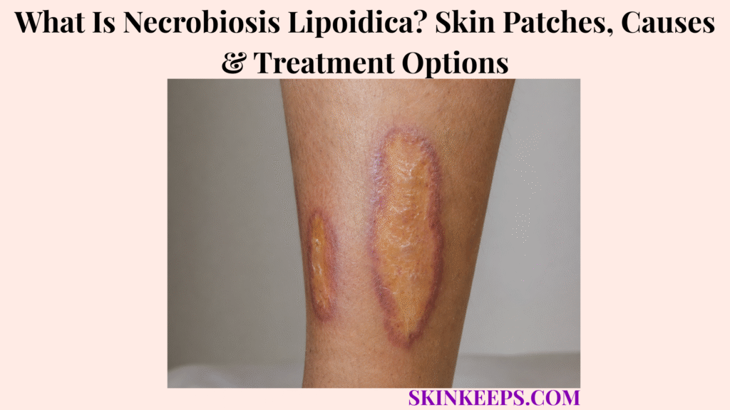
Necrobiosis lipoidica is a rare chronic granulomatous skin disease that most often affects the shins and produces enlarging yellow-brown atrophic plaques with an inflammatory red-purple border. Granulomatous inflammation, collagen degeneration and vascular change drive the transition from early red-brown lesions to thin shiny mature plaques.
Necrobiosis lipoidica is strongly associated with diabetes but can occur in people without diabetes, so the older term necrobiosis lipoidica diabeticorum is no longer the preferred disease name. The course is chronic, minor trauma can trigger ulceration, and treatment decisions depend on separating active inflammation from established atrophy.
This article is for educational purposes only and does not constitute medical advice, diagnosis, or treatment. A necrobiosis-lipoidica-like plaque that develops a new or non-healing ulcer, persistent crusting, increasing pain or redness, repeated bleeding, a rapidly enlarging nodule or thickened area, signs of wound infection, or another significant change from its usual appearance should be evaluated by a licensed dermatologist or qualified healthcare professional.
How Can You Recognize Necrobiosis Lipoidica?
Necrobiosis lipoidica typically begins as red-brown or violaceous lesions on the shins that gradually enlarge into yellow-brown, shiny, thinned plaques with a persistent inflammatory border.
What Does Early Necrobiosis Lipoidica Look Like?
Early NL usually begins as one or more well-defined red-brown or violaceous papules or small plaques on the lower legs.
These lesions represent the inflammatory phase and can enlarge gradually or merge with nearby lesions. Recognition should begin with this earlier phase rather than only the final yellow atrophic appearance.
What Does an Established Necrobiosis Lipoidica Plaque Look Like?
Established NL typically has a yellow-brown, shiny, atrophic centre surrounded by a red-brown or violaceous inflammatory edge.
Central thinning makes surface blood vessels, or telangiectasias, more visible. The plaque is often sharply defined and can become slightly depressed as dermal tissue is lost.
Where Does Necrobiosis Lipoidica Usually Develop?
Necrobiosis lipoidica most commonly develops over the pretibial shins and often affects both lower legs.
Anterior lower-leg involvement is the classic distribution. Other sites can occur, but a chronic yellow-brown atrophic shin plaque remains a particularly useful recognition clue rather than a diagnosis by itself.
Can Necrobiosis Lipoidica Itch or Hurt?
Necrobiosis lipoidica can be asymptomatic or cause itching, pain, tingling or reduced sensation depending on lesion activity and tissue damage.
Some plaques show reduced sweating or reduced hair growth as deeper skin structures are damaged. Absence of symptoms does not exclude NL.
Why Does the Centre of an NL Plaque Become Thin?
The centre becomes thin because chronic granulomatous inflammation and collagen degeneration progressively damage the dermis and produce atrophy.
This tissue loss creates the shiny or slightly sunken appearance of established disease. Atrophy can persist after inflammatory activity has decreased.
What Does Ulcerated Necrobiosis Lipoidica Look Like?
Ulcerated NL develops an open wound within the fragile atrophic plaque, often after relatively minor trauma.
The ulcer can be painful and slow to heal, and a prolonged open wound can become secondarily infected. Ulceration is an important complication but does not automatically mean cancer.
Figure 1. NL can progress from an inflammatory red-brown lesion to a yellow atrophic plaque with a persistent active border; fragile established tissue can later ulcerate after trauma.
Why Does Necrobiosis Lipoidica Develop, and How Is It Related to Diabetes?
The exact cause of necrobiosis lipoidica is unknown, but inflammatory, autoimmune and microvascular mechanisms appear important, and diabetes is a strong association rather than a universal cause.
What Causes Necrobiosis Lipoidica?
The exact cause of NL remains uncertain, with current evidence implicating microvascular abnormalities, immune dysregulation, granulomatous inflammation and abnormal collagen metabolism.
A useful simplified pathway is inflammatory or vascular susceptibility → granulomatous inflammation → collagen degeneration → tissue atrophy. No single mechanism has been proven to explain every case.
Why Is Diabetes Associated With Necrobiosis Lipoidica?
Diabetes occurs frequently among people with NL, and diabetes-related microvascular changes may contribute to disease susceptibility.
The association is especially recognized with type 1 diabetes, but it remains incomplete because most people with diabetes never develop NL and some people with NL do not have diabetes.
Does Poor Blood Sugar Control Cause Necrobiosis Lipoidica?
No simple direct relationship has been established between poor glycaemic control and the development of necrobiosis lipoidica.
Good diabetes care remains important for overall health and wound healing, but better glucose control alone should not be promised to clear an established plaque.
Can Someone Without Diabetes Develop Necrobiosis Lipoidica?
Yes; necrobiosis lipoidica can occur without diabetes, which is why the older term “necrobiosis lipoidica diabeticorum” is no longer preferred.
The broader name accurately includes people with and without diabetes and prevents the disease from being reduced to a diabetic-only skin complication.
Which Other Conditions Are Associated With NL?
NL has also been reported alongside thyroid disease, hypertension, obesity, hyperlipidaemia and selected inflammatory or autoimmune disorders.
These associations do not justify universal screening for every condition. Evaluation should follow the person’s medical history, symptoms, examination and metabolic risk profile.
Why Can Minor Trauma Make Necrobiosis Lipoidica Worse?
Minor trauma can trigger ulceration because established NL plaques contain thin, fragile and poorly resilient skin.
Knocks, scratching and repeated pressure can break down an atrophic plaque, so protecting the shins from avoidable trauma is an important practical strategy.
Figure 2. Diabetes may add vascular and metabolic susceptibility, but NL also occurs without diabetes and its exact pathogenesis remains multifactorial.
How Is Necrobiosis Lipoidica Distinguished and Diagnosed?
Typical necrobiosis lipoidica can often be diagnosed clinically and dermoscopically, while atypical, ulcerated or structurally changing lesions require biopsy.
How Is Necrobiosis Lipoidica Different From Diabetic Dermopathy?
Diabetic dermopathy usually causes small flat brown shin spots, while NL produces larger inflammatory-atrophic plaques with yellow centres, raised borders and visible vessels.
Diabetic dermopathy is typically macular and much less structurally destructive. NL develops true plaque-level inflammation, atrophy and telangiectasia.
How Is Necrobiosis Lipoidica Different From Granuloma Annulare?
NL more commonly develops yellow-brown central atrophy and prominent telangiectasia, whereas granuloma annulare usually lacks the characteristic thin yellow atrophic centre.
Both are granulomatous disorders and annular morphology can overlap. Histology can help when the clinical pattern is uncertain.
Which Other Conditions Can Resemble NL?
Sarcoidosis, necrobiotic xanthogranuloma and lipodermatosclerosis can resemble NL when lower-leg plaques lack the classic colour, atrophy or distribution.
These entities matter mainly when morphology stops being typical enough for confident clinical diagnosis and tissue confirmation becomes more useful.
Can Necrobiosis Lipoidica Be Diagnosed Clinically?
Yes; classic pretibial NL can often be diagnosed from its characteristic clinical and dermoscopic pattern.
Pretibial distribution, yellow atrophy, a violaceous border and prominent telangiectasia are strong supporting features. Clinical examination is not always sufficient when morphology is atypical or complications develop.
When Is a Skin Biopsy Needed?
Skin biopsy is recommended when NL has an unclear appearance, is ulcerated or develops features suspicious for malignant transformation.
Biopsy can distinguish NL from other granulomatous diseases and is especially important when a persistent nodule, progressive thickening, repeated bleeding or a non-healing ulcer changes the lesion’s structure.
What Does an NL Biopsy Show?
NL histopathology typically shows granulomatous inflammation surrounding areas of degenerated collagen.
Findings can include necrobiotic collagen, histiocytes, multinucleated giant cells, layered or palisading inflammation and vascular-wall or endothelial change.
When Are Vascular Tests Needed?
Vascular assessment becomes relevant when examination suggests venous or arterial insufficiency, particularly when an NL ulcer heals poorly.
Leg oedema, signs of venous disease, arterial concerns or unexpectedly slow ulcer healing can justify targeted circulation assessment. Vascular testing is not routine for every uncomplicated plaque.
Figure 3. Classic stable NL can often be recognized clinically, while atypical, ulcerated or changing lesions move toward biopsy and poorly healing legs with vascular signs move toward circulation assessment.
How Is Necrobiosis Lipoidica Treated?
Necrobiosis lipoidica treatment depends on whether the lesion is actively inflamed, stable and atrophic, ulcerated or resistant to initial therapy.
Does Every Necrobiosis Lipoidica Plaque Need Treatment?
No; stable asymptomatic NL without active inflammation or ulceration can sometimes be monitored rather than actively treated.
Treatment becomes more useful when plaques are expanding, inflamed, symptomatic or threatened by ulceration. Observation is a deliberate management choice rather than neglect.
How Are Active Early NL Lesions Treated?
Potent topical corticosteroids are commonly used for active inflammatory NL, while intralesional corticosteroids can be directed into selected active borders.
The inflammatory edge is the treatment target. The already thinned centre should not automatically receive the same treatment intensity as an active expanding border.
Why Should Strong Steroids Be Used Carefully on Atrophic NL Skin?
Strong corticosteroids should be used cautiously over an already thin inactive NL centre because additional steroid-induced atrophy can increase skin fragility.
Inflammatory edge and atrophic centre represent different tissue states. Persistent colour or thinness alone does not prove ongoing inflammation.
When Is Topical Tacrolimus Considered?
Topical tacrolimus can be considered as a steroid-sparing treatment when active NL requires local anti-inflammatory therapy near already thinned skin.
Tacrolimus avoids steroid-induced skin atrophy and has supportive case-series and case-level evidence, but it should not be presented as universally superior or curative.
Do Phototherapy or Photodynamic Therapy Help NL?
Light-based treatments such as PUVA can reduce inflammation in selected NL cases, but response is inconsistent and established atrophy is unlikely to reverse.
These options belong mainly to selected inflammatory disease. No single light-based treatment reliably works for all NL.
When Are Systemic Treatments Considered?
Systemic treatment is generally reserved for more extensive, ulcerated, refractory or otherwise difficult NL under specialist supervision.
Reported options include cyclosporine, fumarates, antimalarials, mycophenolate, pentoxifylline and selected biologic therapies. Evidence quality varies considerably, so no unsupported rank order should be assumed.
What Roles Do TNF-Alpha and JAK Inhibitors Have?
TNF-alpha inhibitors are specialist options for selected severe refractory or ulcerative NL, while JAK inhibition remains an emerging rather than established universal treatment approach.
Infliximab and adalimumab have been reported in severe or ulcerative disease. Topical ruxolitinib has prospective phase-2 evidence, but the study was small and does not make JAK inhibition a routine first-line standard.
| Disease State | Main Management Logic |
|---|---|
| Stable, inactive, asymptomatic plaque | Observation / protection |
| Active inflammatory border | Topical or selected intralesional anti-inflammatory therapy |
| Thin atrophic centre | Avoid treating as active inflammation |
| Persistent / refractory disease | Specialist-directed treatment |
| Severe ulcerative / refractory disease | Selected systemic or biologic options |
| Emerging JAK therapy | Developing evidence only |
How Are Ulcers and Long-Term Complications of Necrobiosis Lipoidica Managed?
NL ulcers require wound and contributing-vascular care, while persistent structural changes within longstanding ulcerated plaques must be assessed for infection or rare squamous cell carcinoma.
How Common Is Ulceration in Necrobiosis Lipoidica?
Ulceration is a significant NL complication and has occurred in roughly one-third of patients in some clinical series.
This figure is not a universal prevalence estimate. Minor trauma commonly precedes ulceration, making protection of fragile lower-leg plaques a practical priority.
How Should an NL Ulcer Be Treated?
An NL ulcer requires appropriate wound care together with assessment of infection, pain, oedema and relevant vascular disease.
Management can include appropriate dressings, pain care, infection assessment and treatment of contributing oedema. Compression can help when venous insufficiency or lymphoedema contributes, but adequate arterial circulation must be established first.
Can Necrobiosis Lipoidica Ulcers Become Infected?
Yes; prolonged open NL ulcers can develop secondary infection.
Increasing pain, spreading redness, purulent drainage or systemic illness can indicate infection. Redness around an ulcer should not automatically be attributed to active NL inflammation.
Can Necrobiosis Lipoidica Become Skin Cancer?
Rarely, squamous cell carcinoma can develop within longstanding NL, especially in chronically ulcerated or repeatedly traumatized lesions.
This is an uncommon complication and usually belongs to a long-duration disease context rather than ordinary stable NL.
Which Changes Should Raise Concern for Squamous Cell Carcinoma?
A persistent new nodule, progressive thickening, recurrent bleeding, enlarging crusted area or non-healing ulcer within NL deserves prompt reassessment and possible biopsy.
Meaningful structural change is more important than pain alone when deciding whether malignancy must be excluded.
Can Necrobiosis Lipoidica Heal Completely?
Active NL can improve or become inactive, but established atrophy, scarring and pigmentation may persist even after inflammation is controlled.
Ulcers can heal, and disease can reactivate or recur. Complete cosmetic restoration of tissue that has already atrophied cannot be guaranteed.
How Can Future Ulceration Risk Be Reduced?
Protecting fragile NL plaques from repeated trauma and managing wound-healing risks can reduce the likelihood of future ulceration.
Protective clothing, avoiding repeated knocks and scratching, managing leg oedema, addressing vascular disease when relevant, smoking cessation where applicable, good diabetes care and prompt wound attention can all reduce risk without guaranteeing prevention.
What Should You Remember About Necrobiosis Lipoidica?
Necrobiosis lipoidica is a chronic granulomatous skin disease that most often affects the shins, and management depends on distinguishing active inflammation from established atrophy and ulcer-related complications.
- NL is a rare chronic granulomatous disorder.
- The shins are the classic location.
- Early lesions are red-brown or violaceous.
- Mature plaques become yellow-brown and atrophic.
- Telangiectasia becomes more visible as skin thins.
- Diabetes is strongly associated but not required.
- Poor glucose control is not a proven direct cause.
- People without diabetes can develop NL.
- Trauma increases ulcer risk.
- Typical lesions can often be diagnosed clinically.
- Biopsy is appropriate when disease is unclear, ulcerated or structurally suspicious.
- Active inflammation and permanent atrophy must be separated.
- Local corticosteroid treatment targets active disease.
- Steroids require caution on thin inactive centres.
- More resistant disease may require specialist therapy.
- Ulcers need wound and vascular assessment.
- Secondary infection can occur.
- SCC is a rare chronic-lesion complication.
- Persistent nodules or non-healing ulcers need reassessment.
- Complete reversal of established atrophy is not guaranteed.
Recognize → determine activity → understand diabetes context → confirm when needed → treat active disease → protect fragile skin → manage ulcers → biopsy persistent change.
Frequently Asked Questions About Necrobiosis Lipoidica
The most important NL questions concern its typical appearance, diabetes association, ulcer healing, active-disease treatment and rare skin-cancer risk.
What Do Necrobiosis Lipoidica Skin Patches Look Like?
NL usually develops as enlarging shin plaques with a yellow-brown shiny atrophic centre and a red-brown or violaceous inflammatory border. Early lesions can begin as smaller red-brown papules, and telangiectasias often become visible as the centre thins.
Does Necrobiosis Lipoidica Always Mean You Have Diabetes?
No; diabetes is strongly associated with necrobiosis lipoidica, but people without diabetes can also develop the condition. That is why the older diabetes-specific name is no longer preferred.
Can Necrobiosis Lipoidica Ulcers Heal?
Yes; NL ulcers can heal, but healing may be slow and requires appropriate wound care and management of infection, oedema or vascular problems when present. Fragile underlying skin means recurrence can still occur.
Which Treatments Are Used for Active Necrobiosis Lipoidica?
Active NL is commonly treated with local anti-inflammatory therapy, while persistent, extensive or ulcerative disease may require specialist-directed systemic treatment. Topical corticosteroids, selected intralesional therapy and steroid-sparing topical options are used according to disease state.
Can Necrobiosis Lipoidica Turn Into Skin Cancer?
Rarely, squamous cell carcinoma can develop within longstanding NL, particularly in chronically ulcerated or repeatedly traumatized plaques. A new nodule, repeated bleeding, progressive thickening or non-healing ulcer deserves reassessment.
Which Sources Support This Necrobiosis Lipoidica Guidance?
2026 S1 Guideline — Diagnosis and Therapy of Necrobiosis Lipoidica — Highest-priority source for terminology, diagnostic framework, biopsy indications, clinical/dermoscopic recognition and current treatment strategy.
StatPearls / NCBI Bookshelf — Necrobiosis Lipoidica — Used for classic pretibial morphology, symptoms, diabetes association, ulceration, pathogenesis, histopathology, vascular assessment, wound care and treatment options.
Journal of Investigative Dermatology — Topical Ruxolitinib Phase 2 Study — Used specifically to frame topical JAK inhibition as emerging evidence from a small prospective study rather than established first-line therapy.
Dermatologic Therapy — Necrobiosis Lipoidica Treated With Topical Tacrolimus — Used specifically for selected steroid-sparing topical tacrolimus context.
Dermatologic Clinics — Necrobiosis Lipoidica Review — Used for chronic atrophic plaque morphology, therapeutic limitations and the ulceration/SCC complication context.
Journal of Cutaneous Medicine and Surgery — 2026 Systematic Review of Malignancies Arising From NL — Used specifically for the rare long-term malignant-transformation surveillance context.
Anais Brasileiros de Dermatologia — Squamous Cell Carcinoma Superimposed on Necrobiosis Lipoidica — Used specifically to support biopsy vigilance for persistent structural change in longstanding NL.



