
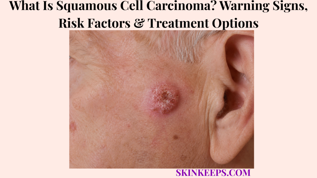
Cutaneous squamous cell carcinoma (cSCC) is a skin cancer that develops when squamous keratinocytes in the epidermis acquire malignant changes and grow without normal control. It often appears as a persistent rough or scaly patch, firm or dome-shaped growth, wart-like lesion, or sore that repeatedly crusts, bleeds, or fails to heal.
Most localized cSCCs are highly treatable, but some tumors can invade deeply, involve nerves, recur, spread to lymph nodes, or become metastatic. Risk assessment therefore goes beyond appearance: suspicious lesions need biopsy, confirmed cancers need tumor-risk assessment, and treatment is selected according to location, depth, recurrence, immune status and other high-risk features.
This article is for educational purposes only. Persistent, rapidly growing, bleeding, non-healing or recurrent skin lesions should be professionally evaluated.
What Is Squamous Cell Carcinoma and What Warning Signs Can It Cause?
Cutaneous squamous cell carcinoma is a malignant keratinocyte tumor that can appear as a persistent rough, scaly, crusted, firm, bleeding or non-healing lesion of the skin.
Which Skin Cells Become Cancerous in Cutaneous SCC?
Cutaneous SCC develops from squamous keratinocytes, the epidermal cells that normally mature toward the skin surface.
UV radiation and other cancer-promoting influences can damage cellular DNA and regulatory pathways. When abnormal keratinocytes escape normal growth controls, they can form a malignant tumor that initially remains in the epidermis or later invades deeper tissue.
What Is the Difference Between SCC in Situ and Invasive SCC?
SCC in situ remains confined to the epidermis, whereas invasive cSCC has crossed the basement-membrane boundary and entered deeper skin tissue.
This distinction matters because in situ disease and invasive disease do not carry the same biological risk. Invasive tumors can extend into dermis and, in higher-risk cases, reach fat, nerves, muscle, bone or regional lymph nodes.
How Is Actinic Keratosis Related to Squamous Cell Carcinoma?
Actinic keratosis is a UV-damage-associated precancerous keratinocyte lesion that can sometimes progress to cSCC, but it is not automatically squamous cell carcinoma.
Actinic keratosis is best treated as a related precursor condition rather than an interchangeable diagnosis. Not every AK progresses, and not every cSCC follows a visible AK → in situ → invasive sequence.
What Does Early Squamous Cell Carcinoma Look Like?
Early cSCC can look like a persistent rough or scaly patch, a firm raised bump, a dome-shaped growth, a wart-like lesion, or a sore that does not heal normally.
Color is variable. A lesion may be pink, red, skin-colored, brown, black or mixed depending on skin tone and tumor features. Persistent behavior—growth, crusting, repeated reopening or bleeding—is often more useful than memorizing one color or shape.
Can SCC Bleed, Crust or Fail to Heal?
Yes; repeated bleeding, crusting, ulceration, enlargement, or a sore that heals temporarily and then reopens are important cSCC warning patterns.
Pain, tenderness, burning, numbness or pins-and-needles sensations can also occur. New sensory symptoms near a known or suspected cSCC deserve attention because selected tumors can track along nerves.
What Causes Squamous Cell Carcinoma and Which Factors Increase the Risk?
Cumulative ultraviolet damage is a major driver of cutaneous SCC, while immune suppression, previous skin cancer, chronically damaged skin and selected environmental or inherited factors can further increase risk.
How Does UV Radiation Increase SCC Risk?
Repeated UV exposure increases cSCC risk by damaging DNA and allowing cancer-promoting changes in epidermal keratinocytes to accumulate over time.
Both sunlight and artificial UV from tanning devices contribute to this damage. Risk reflects cumulative exposure, individual susceptibility and other host factors rather than a single episode alone.
Who Has a Higher Risk of Developing SCC?
cSCC risk is higher with substantial cumulative UV exposure, increasing age, previous keratinocyte cancer, immune suppression and selected exposures such as prior radiation or arsenic.
People whose skin burns easily carry greater UV-related risk, but cSCC is not restricted to fair skin. A person can develop the disease even without every classic risk factor, so an actual changing lesion should be evaluated on its behavior rather than dismissed because background risk seems low.
Can People With Darker Skin Develop Squamous Cell Carcinoma?
Yes; people of every skin tone can develop cutaneous SCC, and the lesion may be skin-colored, brown, black, pink, red or mixed rather than fitting one “red scaly spot” stereotype.
In darker skin, cSCC can also occur on sites with little obvious sun exposure. Chronic scars, non-healing wounds and other long-standing areas of inflammation therefore remain important clinical contexts across skin tones.
Why Does Immune Suppression Increase SCC Risk?
Immune suppression increases cSCC risk because reduced immune surveillance makes abnormal keratinocytes harder to control and can also make some tumors behave more aggressively.
Organ-transplant recipients and people receiving significant immunosuppressive treatment are important higher-risk groups. In this setting, new or changing lesions warrant a lower threshold for professional assessment and follow-up.
Can SCC Develop in Scars or Chronic Wounds?
Yes; cSCC can arise in long-standing wounds, old burn scars and chronically inflamed or repeatedly damaged skin.
This does not mean an ordinary scar will become cancer. The concern is a scar or wound that develops a new persistent growth, bleeding, ulceration, foul change or failure to heal. The broader skin cancer page explains how persistent lesion behavior fits into whole-skin surveillance.
How Is Squamous Cell Carcinoma Diagnosed and Its Risk of Spreading Assessed?
Cutaneous SCC is confirmed by biopsy, after which tumor depth, site, recurrence status, nerve involvement, immune status and microscopic features help determine whether the cancer is lower or higher risk.
How Does a Dermatologist Evaluate a Suspicious SCC?
A dermatologist evaluates a suspected cSCC by reviewing how the lesion has changed and examining its size, surface, border, site and surrounding skin before deciding on biopsy.
The examination may also include palpation for firmness or fixation and assessment of nearby lymph nodes when the clinical situation makes regional spread more relevant. Visual inspection raises suspicion; it does not establish the diagnosis by itself.
Why Is a Skin Biopsy Needed to Confirm SCC?
Biopsy is needed because microscopic examination of tissue establishes whether malignant squamous cells are actually present.
Pathology can distinguish cSCC from benign mimics and from related keratinocyte lesions. It also provides information that can influence treatment, such as tumor differentiation, depth and other microscopic features.
Which Features Can Make an SCC Higher Risk?
A cSCC becomes more concerning when tumor or patient features increase the chance of recurrence, deep invasion or spread.
Important factors include greater size or depth, recurrence, certain anatomical sites, immune suppression, perineural involvement, extension into deeper tissue and aggressive microscopic characteristics. Risk assessment is multidimensional; one feature does not automatically determine the entire treatment plan.
When Are Lymph-Node Tests or Imaging Needed?
Lymph-node assessment or imaging is reserved for selected cSCCs with features suggesting meaningful risk of regional or distant spread rather than used routinely for every small localized tumor.
Deep invasion, concerning nerve symptoms, recurrence, enlarged regional nodes or other high-risk findings can increase the value of ultrasound or cross-sectional imaging. A routine low-risk cSCC does not automatically require CT, MRI or PET scanning.
What Skin Conditions Can Be Mistaken for SCC?
Actinic keratosis, basal cell carcinoma, irritated seborrheic keratosis, warts, keratoacanthoma and chronic inflammatory or infectious lesions can sometimes resemble cSCC.
Basal cell carcinoma is another keratinocyte skin cancer, but it usually has different biology and a much lower tendency to metastasize. Persistent suspicious lesions still require tissue diagnosis rather than visual guessing.
An irritated seborrheic keratosis can become crusted or inflamed enough to resemble malignant keratinocyte disease, so a changing “stuck-on” lesion should not be destroyed solely from appearance.
Warts can have a rough or wart-like surface, but cSCC can also look verrucous. A wart-like shape therefore does not prove that HPV is the cause.
How Is Squamous Cell Carcinoma Treated?
Cutaneous SCC treatment is risk-based: most localized tumors are treated surgically, while recurrent, high-risk or advanced disease may require stronger margin control, radiation or systemic therapy.
How Is Standard Surgical Excision Used?
Standard excision removes the cSCC together with an appropriate surrounding margin so the specimen can be examined for complete removal.
This is a common treatment for localized disease. The exact margin and surgical plan depend on tumor risk and anatomy, so one fixed measurement should not be applied to every lesion.
When Is Mohs Surgery Used?
Mohs surgery is particularly useful for selected high-risk, recurrent or anatomically sensitive cSCCs where detailed margin control and tissue preservation matter.
Mohs removes the visible tumor and thin layers of surrounding tissue, examines the margins during the procedure, and continues only where cancer remains. It is a powerful margin-control technique, not a requirement for every cSCC.
When Are Curettage, Electrosurgery or Other Local Treatments Used?
Selected superficial or lower-risk cSCCs may be treated with less extensive local methods, while invasive or high-risk tumors generally need treatment with more reliable margin control.
Curettage with electrodesiccation can be appropriate for carefully selected low-risk tumors. Cryosurgery or photodynamic therapy have narrower roles, particularly when disease is superficial or in situ and more definitive surgery is not appropriate. These choices require clinician selection rather than self-treatment.
When Is Radiation Therapy Used?
Radiation therapy may be used when surgery is unsuitable, when additional local control is needed, or when cSCC has become regionally or otherwise advanced.
Radiation can be definitive in selected patients or combined with surgery for higher-risk disease. The choice depends on tumor location, prior treatment, pathology, patient health and the likelihood of controlling the cancer while preserving function.
How Is Advanced Squamous Cell Carcinoma Treated?
Advanced cSCC may require multidisciplinary treatment with surgery, radiation and systemic immunotherapy when disease invades major structures, cannot be controlled with routine local treatment, or spreads beyond the primary site.
Checkpoint immunotherapy is an established systemic option for selected locally advanced or metastatic cSCC. Drug choice and sequencing belong to oncology care and should not be reduced to a single regimen in a general skin-disease article.
How Can SCC Recurrence Be Reduced, and When Should a Lesion Be Reassessed?
Long-term cSCC care combines UV-risk reduction, surveillance of actinic and chronically damaged skin, follow-up after treatment and prompt reassessment of recurrent or rapidly changing lesions.
How Does UV Protection Reduce Future SCC Risk?
Reducing intense and cumulative UV exposure lowers avoidable skin-cell damage that contributes to future cSCC formation.
A combined strategy is more useful than relying on sunscreen alone: seek shade when practical, use protective clothing and hats, use appropriate broad-spectrum sun protection on exposed skin, and avoid unnecessary intense UV exposure.
Why Should Indoor Tanning Be Avoided?
Indoor tanning should be avoided because tanning beds and sunlamps deliver carcinogenic ultraviolet radiation and add to cumulative skin damage.
A tan is evidence of UV injury rather than protection. Artificial UV exposure is therefore not a safe substitute for outdoor tanning or a preventive “base tan.”
Why Should Actinic Keratoses and Chronically Damaged Skin Be Monitored?
Actinic keratoses and chronically damaged skin deserve surveillance because they identify areas in which abnormal keratinocyte changes or long-standing tissue injury may persist.
Monitoring does not mean every actinic keratosis or scar will become cSCC. The goal is to recognize new thickening, growth, bleeding, pain or non-healing change early enough for diagnosis.
Why Is Follow-Up Important After SCC Treatment?
Follow-up matters because a previous cSCC increases the risk of recurrence and of developing additional skin cancers later.
Follow-up intensity depends on the original tumor’s risk and the patient’s broader risk profile. The key principle is that successful treatment of one tumor does not erase future risk, so the treated site and the rest of the skin remain part of surveillance.
Can Squamous Cell Carcinoma Come Back?
Yes; cSCC can recur after treatment, and recurrence risk depends on tumor biology, depth, location, immune status, margin control and whether the lesion had already recurred before.
Recurrence can appear as a new lump, sore, crusted area or change in or near the treatment scar. A new enlarged regional lymph node can also matter in higher-risk disease.
When Should a Suspected SCC Be Checked Promptly?
A persistent rough, scaly, growing, bleeding, crusted or non-healing lesion should be professionally assessed rather than repeatedly treated at home without a diagnosis.
Prompt assessment is particularly important for rapid growth, a lesion arising in a chronic wound or old burn scar, recurrent bleeding, or a suspicious lesion in someone who is significantly immunosuppressed.
Which Features Make an Existing SCC More Urgent to Reassess?
Rapid enlargement, increasing pain, numbness, tingling, fixation to deeper tissue, enlarged nearby lymph nodes or recurrence after treatment raise concern for more aggressive cSCC.
These findings can signal deeper invasion, nerve involvement or regional spread and therefore change the urgency and staging task. Persistent + growing + bleeding/crusting + non-healing = dermatologist assessment rather than prolonged home treatment.
What Should You Remember About Squamous Cell Carcinoma?
Cutaneous SCC is a malignant keratinocyte cancer that is often highly treatable when localized, but biopsy and high-risk assessment are essential because some tumors can invade deeply, recur or spread.
- Cutaneous SCC is a skin cancer arising from squamous keratinocytes.
- Actinic keratosis, SCC in situ and invasive SCC are related but different diagnoses.
- Persistent rough or scaly patches, firm or dome-shaped growths, wart-like lesions and non-healing sores can all be warning patterns.
- Repeated bleeding, crusting, ulceration or reopening deserves assessment.
- cSCC can occur in people of every skin tone and is not limited to sun-exposed sites.
- Cumulative UV exposure and indoor tanning increase avoidable risk.
- Immune suppression materially increases cSCC risk and can increase tumor aggressiveness.
- Long-standing wounds and burn scars are recognized higher-risk settings.
- Appearance raises suspicion; biopsy and histopathology confirm the diagnosis.
- Depth, site, recurrence, nerve involvement, immune status and microscopic features influence risk.
- Not every cSCC requires imaging or extensive staging.
- Standard excision is common; Mohs is selected for appropriate higher-risk or sensitive-site tumors.
- Selected superficial lower-risk lesions can use other local treatments, but these are not universal substitutes for surgery.
- Radiation has selected roles, and advanced cSCC may require systemic immunotherapy.
- After treatment, recurrence and additional skin cancers remain possible.
- UV-risk reduction, avoidance of intentional tanning and ongoing skin surveillance support long-term care.
- Rapid growth, new nerve symptoms or enlarged nearby lymph nodes increase urgency.
Frequently Asked Questions About Squamous Cell Carcinoma
The main cSCC questions concern danger, spread, early appearance, actinic keratosis and recurrence after treatment.
Is Squamous Cell Carcinoma a Dangerous Skin Cancer?
Most localized cutaneous squamous cell carcinomas are highly treatable, but high-risk tumors can invade deeply and sometimes spread to lymph nodes or distant sites.
Can Squamous Cell Carcinoma Spread to Other Parts of the Body?
Yes. Spread is uncommon in many routine localized cSCCs, but the risk becomes more important when high-risk tumor or patient features are present.
What Does Early Squamous Cell Carcinoma Usually Look Like?
Early cSCC may appear as a persistent rough or scaly patch, firm or dome-shaped growth, wart-like lesion, or a sore that repeatedly crusts, bleeds, or fails to heal.
Is Actinic Keratosis the Same as Squamous Cell Carcinoma?
No. Actinic keratosis is a UV-damage-associated precancerous keratinocyte lesion that can sometimes progress to cSCC, but it is not the same diagnosis as invasive squamous cell carcinoma.
Can Squamous Cell Carcinoma Come Back After Removal?
Yes. Recurrence is possible, especially when tumor, location, immune, microscopic, or treatment factors place the cSCC in a higher-risk category.
American Academy of Dermatology — Squamous cell carcinoma: From symptoms to treatments. Supports cSCC appearance, skin-tone variability, chronic-wound and scar context, biopsy-centered diagnosis, treatment selection, high-risk behavior and follow-up.
American Academy of Dermatology — Cutaneous squamous cell carcinoma clinical guideline. Supports risk stratification, biopsy principles, surgery as the mainstay of treatment, selected nonsurgical options and follow-up after cSCC.
National Cancer Institute — Skin Cancer Treatment (Patient Version). Supports biopsy confirmation, treatment selection by cancer type/stage/site, localized cSCC treatment options and immunotherapy for advanced disease.
National Cancer Institute — Skin Cancer Treatment (Health Professional Version). Supports cSCC risk factors, immune suppression, chronic inflammation, localized treatment approaches and advanced-disease treatment evidence.
CDC — Reducing Risk for Skin Cancer. Supports UV-risk reduction, protective clothing and shade, broad-spectrum sunscreen as one component of protection, and avoidance of tanning beds and sunlamps.



