
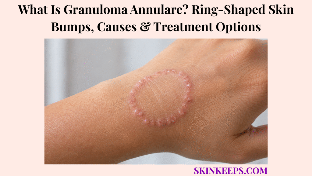
Granuloma annulare is a benign inflammatory skin condition that often creates smooth, firm bumps arranged in circular or semicircular patterns. The bumps may be skin-coloured, pink, red, reddish-brown, purple, or darker than nearby skin and usually have little or no surface scale.
It is not fungal, contagious, or skin cancer. Localized granuloma annulare often clears without treatment, while generalized, subcutaneous, perforating, and patch forms may persist longer and sometimes need biopsy or specialist-directed care.
How Can You Recognize Granuloma Annulare Skin Bumps?
Granuloma annulare usually appears as small, firm, smooth papules that group into a complete ring, partial ring, or curved arc.
The active border may expand outward while the centre remains normal-looking or slightly depressed. One ring, several rings, or many scattered lesions may develop.
Typical lesions have little or no surface scale and cause little pain. Mild itching or tenderness can occur, especially in generalized or perforating variants.
Colour contrast differs across skin tones, and the ring may be easier to feel than see.
- Smooth, firm individual papules.
- Complete ring, partial ring, or arc.
- Skin-coloured, pink, red, red-brown, purple, or darker colour.
- Quiet or slightly depressed centre.
- Little or no surface scale.
- Usually minimal pain or itching.
- One localized ring or several lesions.
- Uncertainty check before treating every ring as fungal.
Figure 1. Typical granuloma annulare forms smooth firm papules around a complete or partial ring, with little scale and a quiet centre, most often on hands, feet, wrists, or ankles.
Where Does Granuloma Annulare Usually Appear?
Granuloma annulare commonly appears on the hands, fingers, knuckles, feet, wrists, and ankles, but different variants can affect wider or deeper areas.
Localized rings also occur around elbows, forearms, and lower legs. Generalized disease may affect the trunk, arms, legs, and skin folds.
Subcutaneous granuloma annulare can form firm nodules around scalp margins, fingertips, hands, or shins, especially in children.
| Pattern | Common Locations | Typical Depth | Recognition Clue |
|---|---|---|---|
| Localized | Hands, fingers, knuckles, feet, wrists, ankles, elbows | Dermal | One or several smooth papular rings |
| Generalized | Trunk, arms, legs, skin folds | Dermal and widespread | Numerous papules or patches |
| Subcutaneous | Scalp margins, hands, fingertips, shins | Deep dermis or subcutis | Firm rubbery lump without a visible ring |
| Perforating | Hands common; other sites possible | Dermal with surface elimination | Crusted or indented papule |
| Patch | Variable | Dermal | Flat discoloured patch with subtle border |
How Does Granuloma Annulare Develop Within the Skin?
Granuloma annulare develops mainly in the dermis, where histiocytes and other immune cells collect around altered collagen and form firm papules.
Mucin may accumulate between collagen bundles, creating a granulomatous inflammatory pattern below the surface.
Because the process is deeper than the epidermis, typical lesions feel firm while showing little scale.
As papules remain active around the outer edge and central inflammation settles, the lesions form an expanding ring or arc.
Figure 2. Granuloma annulare develops mainly in the dermis as inflammatory cells gather around altered collagen and mucin, forming firm papules that can join around an outward-growing ring.
What Causes Granuloma Annulare—and Which Factors May Trigger It?
The exact cause of granuloma annulare remains unknown.
It appears to involve a localized immune reaction in the dermis. It is not caused by fungus, bacteria, poor hygiene, a universal food allergy, a vitamin deficiency, or one lifestyle behaviour.
Minor injury, insect or animal bites, sun exposure, infections, vaccinations, skin testing, and certain medicines have preceded lesions in some reports, but timing does not prove direct causation.
| Reported Event | Relationship | Universal Cause? | Reasonable Response |
|---|---|---|---|
| Minor skin injury | Sometimes precedes lesions | No | Record timing without assuming proof |
| Insect or animal bite | Possible preceding event | No | Reassess if painful, warm, or infected-looking |
| Sun exposure | Reported in selected cases | No | Use standard sun protection |
| Infection | Reported association | No | Treat only a confirmed infection |
| Vaccination or skin test | Rare temporal reports | No | Avoid unsupported causation claims |
| Medication | Uncommon reports | No | Review timing with a clinician |
| Food, allergy, or vitamin deficiency | Not a universal explanation | No | Avoid restrictive diets or unnecessary supplements |
Is Granuloma Annulare Associated With Diabetes or Thyroid Disease?
Studies have reported associations with diabetes, thyroid disease, and other immune-related conditions, but findings are inconsistent and a few localized rings do not prove systemic disease.
Targeted testing may be considered when granuloma annulare is widespread, recurrent, unusually persistent, or accompanied by relevant symptoms or medical history.
Excessive thirst, frequent urination, unexplained weight change, or slow healing may justify glucose assessment. Thyroid testing should follow fatigue, cold intolerance, neck swelling, or other clinical clues rather than the rash alone.
| Situation | Routine Extensive Testing? | Reasonable Direction |
|---|---|---|
| Few localized rings and otherwise well | Usually no | Confirm the diagnosis and observe |
| Widespread disease | More reasonable | Targeted medical assessment |
| Persistent or recurrent disease | Context-dependent | Review symptoms and risk factors |
| Thirst, frequent urination, or weight change | Targeted testing reasonable | Blood glucose assessment |
| Cold intolerance, fatigue, or thyroid history | Targeted testing reasonable | Thyroid assessment |
| Rash alone without other concern | Usually no | Avoid indiscriminate screening |
How Do the Different Types of Granuloma Annulare Appear?
Granuloma annulare variants differ by distribution, depth, surface behaviour, symptoms, and persistence.
What Does Localized Granuloma Annulare Look Like?
Localized granuloma annulare is the most common form and usually causes one or several rings of smooth firm papules on hands, feet, wrists, or ankles.
Symptoms are usually minimal, spontaneous clearance is common, and recurrence can occur after the skin clears.
How Does Generalized Granuloma Annulare Affect the Skin?
Generalized granuloma annulare causes numerous papules or broad patches across the trunk, arms, legs, or skin folds.
Rings may be less obvious, itching is more likely, and the condition can persist for years and resist treatment.
What Is Subcutaneous Granuloma Annulare?
Subcutaneous granuloma annulare forms firm rubbery nodules beneath normal-looking skin, most often in children.
Nodules around the scalp margins, fingertips, hands, or shins can resemble rheumatoid nodules or an epidermal cyst. They do not automatically indicate rheumatoid arthritis.
What Is Perforating Granuloma Annulare?
Perforating granuloma annulare produces papules with central crusting or indentation as altered collagen passes through the skin surface.
The hands are commonly affected, and itching, tenderness, pigment change, or scarring can occur.
What Does Patch Granuloma Annulare Look Like?
Patch granuloma annulare appears as flat or slightly raised pink, red-brown, purple, or discoloured patches without the classic raised papular ring.
Because it can resemble eczema, morphea, or inflammatory pigmentation, biopsy may be needed more often.
| Type | Lesion Shape | Usual Location | Symptoms | Persistence | Biopsy Need |
|---|---|---|---|---|---|
| Localized | Smooth papular ring | Hands, feet, wrists, ankles | Usually few | Often clears | Low if classic |
| Generalized | Numerous papules or patches | Trunk, arms, legs | Itch more likely | More persistent | Moderate if atypical |
| Subcutaneous | Deep rubbery nodules | Scalp margin, hands, shins | Usually minimal | Can recur | Higher if uncertain |
| Perforating | Crusted or indented papules | Hands common | Itch or tenderness possible | May scar | Higher |
| Patch | Flat discoloured patches | Variable | Usually mild | Can persist | Higher than classic ring |
Is Granuloma Annulare Contagious, Infectious, or Cancerous?
Granuloma annulare is not contagious, not a fungal infection, and not a form of skin cancer.
It does not spread through touch, kissing, towels, shared objects, animals, or sexual contact. Antifungal medicine does not treat confirmed granuloma annulare.
Typical granuloma annulare does not transform into cancer. Atypical or changing growths still deserve evaluation because another condition can imitate it.
- Fungal: no.
- Bacterial: no.
- Contagious: no.
- Cancerous: no.
- Inflammatory: yes.
- Atypical lesions still need evaluation: yes.
How Is Granuloma Annulare Different From Ringworm?
Granuloma annulare usually forms a ring from separate smooth firm papules with little scale, while ringworm forms a continuous scaly advancing border caused by dermatophyte fungi.
Ringworm is often itchy and may follow contact with an infected person, animal, or object. Granuloma annulare is noninfectious and commonly affects hands, feet, wrists, or ankles.
Ring shape alone cannot diagnose either condition. Scale, symptoms, exposure history, and fungal testing may be needed.
| Feature | Granuloma Annulare | Ringworm |
|---|---|---|
| Border | Separate smooth firm papules | Continuous scaly advancing edge |
| Surface scale | Little or none | Common |
| Symptoms | Often none or mild | Itch more common |
| Cause | Inflammatory and noninfectious | Dermatophyte fungus |
| Contagious | No | Yes |
| Common sites | Hands, feet, wrists, ankles | Any exposed skin; exposure-linked |
| Treatment | Observation or anti-inflammatory options | Antifungal treatment |
How Is Granuloma Annulare Different From Other Ring-Shaped Skin Conditions?
Other inflammatory, infectious, and granulomatous conditions can form rings, plaques, or nodules, so the surface and border pattern matter.
How Does Granuloma Annulare Differ From Nummular Eczema?
Granuloma annulare forms smooth grouped papules, while eczema can form coin-shaped itchy plaques with dryness, crusting, or weeping.
How Does Granuloma Annulare Differ From Psoriasis?
Granuloma annulare is usually smoother than psoriasis, which often forms sharply defined plaques with visible scale.
How Does Granuloma Annulare Differ From Erythema Annulare Centrifugum?
Erythema annulare centrifugum often forms expanding red rings with trailing scale, while granuloma annulare has a firmer papular border and little scale.
How Does Granuloma Annulare Differ From Necrobiosis Lipoidica?
Necrobiosis lipoidica usually affects the shins and may form yellow-brown shiny thinned plaques with visible blood vessels and occasional ulceration.
Granuloma annulare more often forms smooth papules without central skin thinning.
How Does Granuloma Annulare Differ From Sarcoidosis?
Cutaneous sarcoidosis can produce red-brown, purple, or skin-coloured papules and plaques and may involve internal organs.
Biopsy helps separate overlapping granulomatous patterns.
How Does Subcutaneous Granuloma Annulare Differ From a Cyst or Rheumatoid Nodule?
Subcutaneous granuloma annulare forms inflammatory nodules, while cysts contain keratin or fluid and rheumatoid nodules arise in a different clinical context.
Imaging or biopsy may be needed when a deep lump enlarges, persists, or remains uncertain.
| Condition | Surface Scale | Border or Texture | Central Change | Typical Symptom | Helpful Test |
|---|---|---|---|---|---|
| Granuloma annulare | Little or none | Smooth firm papules | Normal or slightly depressed | Usually mild | Clinical exam ± biopsy |
| Ringworm | Common | Scaly advancing edge | Central clearing common | Itchy | Fungal test if uncertain |
| Nummular eczema | Dry, crusted, or weeping | Eczematous plaque | No papular ring | Often itchy | Clinical exam |
| Psoriasis | Visible scale | Sharp plaque edge | Persistent plaque | Itch possible | Clinical exam ± biopsy |
| EAC | Trailing scale possible | Expanding red patch | Expanding ring | Variable | Clinical exam ± biopsy |
| Necrobiosis lipoidica | Shiny and thinned | Plaque | Yellow-brown atrophy and vessels | May ulcerate | Clinical exam ± biopsy |
| Sarcoidosis | Usually little scale | Papules or plaques | Variable | Variable | Biopsy and systemic review |
| Cyst or rheumatoid nodule | No annular scale | Deep lump | No ring | Variable | Exam, imaging, or biopsy |
How Do Clinicians Diagnose Granuloma Annulare?
Clinicians often diagnose classic localized granuloma annulare by examining lesion shape, papular border, surface, colour, distribution, and symptoms.
The assessment checks for scale, crusting, ulceration, central clearing, itching, pain, duration, exposure to infected people or animals, medicines, and medical history.
A classic smooth papular ring with little scale is often diagnosed clinically. A scaly border or fungal exposure may justify a skin scraping or other fungal evaluation.
There is no single blood test that confirms granuloma annulare.
- Shape and completeness of the ring.
- Separate firm papules at the border.
- Amount and location of scale.
- Crusting, ulceration, drainage, or central breakdown.
- Pain, itching, and duration.
- Body distribution and variant pattern.
- Exposure and medication history.
- Fungal evaluation when ringworm remains possible.
- Biopsy when atypical or uncertain.
When Is a Skin Biopsy Needed for Granuloma Annulare?
A biopsy is not required for every classic localized case, but it can help when lesions are atypical, widespread, deep, crusted, painful, ulcerated, rapidly changing, or treatment-resistant.
Biopsy is also useful when fungal testing is negative but uncertainty remains, or when sarcoidosis, necrobiosis lipoidica, a tumour, or another granulomatous disorder is possible.
Microscopy commonly shows altered dermal collagen surrounded by inflammatory cells, often with increased mucin. The pattern supports the diagnosis but does not reveal one universal cause.
- Flat or non-classic lesion.
- Widespread disease.
- Deep subcutaneous nodule.
- Crusting or perforation.
- Pain, ulceration, bleeding, or rapid change.
- Negative fungal test with persistent uncertainty.
- Repeated treatment failure.
- Concern for tumour or another granulomatous disease.
Does Granuloma Annulare Require Treatment?
Most localized granuloma annulare does not require treatment when the diagnosis is secure and the lesions cause no significant symptoms.
Many lesions clear spontaneously within months or a few years. Treatment may accelerate improvement but cannot guarantee permanent clearance or prevent recurrence.
Treatment is more reasonable when lesions are highly visible, itchy, tender, widespread, deep, persistent, or emotionally distressing.
| Situation | Best Direction | Reason |
|---|---|---|
| Few asymptomatic localized rings | Observe | Benign and may clear |
| Visible or distressing localized lesions | Consider local therapy | Cosmetic or emotional burden |
| Itchy or tender lesions | Treat if needed | Symptom relief |
| Widespread persistent disease | Dermatology-directed therapy | Greater persistence |
| Deep uncertain nodules | Examine or biopsy | Confirm subtype |
| Atypical or ulcerated lesion | Reassess before treatment | Exclude another diagnosis |
Which Treatments Can Clear Localized Granuloma Annulare Faster?
Localized treatment aims to reduce inflammation or remove selected raised papules while balancing the risk of atrophy, pigment change, blistering, or scarring.
How Are Topical Corticosteroids Used for Granuloma Annulare?
Prescription-strength topical corticosteroids may reduce inflammation in selected localized lesions, sometimes under clinician-directed occlusion.
Possible harms include skin thinning, stretch marks, visible blood vessels, and pigment change, especially with excessive strength or duration.
How Do Steroid Injections Treat Granuloma Annulare?
Intralesional corticosteroid injections deliver medicine into active papules when a few lesions are persistent or prominent.
They can cause discomfort, indentation, skin thinning, or colour change.
When Is Cryotherapy Used for Granuloma Annulare?
Cryotherapy uses liquid nitrogen on selected raised lesions but may require repeated sessions.
Pain, blistering, scarring, and pigment loss are possible, which may outweigh benefit for large rings or deeply pigmented skin.
Can Laser or Other Local Treatments Help?
Selected laser procedures, topical calcineurin inhibitors, or imiquimod may be considered in uncommon specialist-directed cases.
Evidence is limited, and no local therapy works consistently for every lesion.
| Option | Best Use | Expected Role | Main Risk |
|---|---|---|---|
| Topical corticosteroid | Localized inflamed rings | Reduce inflammation | Atrophy and pigment change |
| Steroid under occlusion | Selected thicker lesions | Increase penetration | Greater steroid effects |
| Steroid injection | Few persistent papules | Strong local effect | Indentation and colour change |
| Cryotherapy | Selected raised lesions | Destroy or stimulate clearance | Blister, scar, pigment loss |
| Laser or local alternatives | Specialist refractory cases | Selected lesion control | Limited evidence and cosmetic risk |
How Is Widespread or Persistent Granuloma Annulare Treated?
Widespread or persistent granuloma annulare may justify phototherapy or systemic treatment when the burden is greater than the risks of treating a benign condition.
How Can Phototherapy Help Generalized Granuloma Annulare?
Phototherapy uses controlled ultraviolet exposure to treat many lesions at once.
Narrowband UVB, UVA1, or PUVA may be used in selected settings. Response varies, repeated visits are needed, recurrence can occur, and cumulative ultraviolet risk must be considered.
Which Oral Medicines May Be Considered for Persistent Granuloma Annulare?
Specialists may consider hydroxychloroquine, methotrexate, dapsone, oral retinoids, ciclosporin, or other immune-modifying medicines when widespread disease justifies systemic risk.
Evidence is limited or inconsistent, many uses are off-label, improvement can be delayed, and blood, liver, kidney, eye, infection, and pregnancy-related monitoring may be needed.
Are Biologic or JAK-Inhibitor Treatments Used for Granuloma Annulare?
Biologic and JAK-pathway medicines remain emerging or experimental options for difficult generalized disease rather than standard treatment for a few localized rings.
Case reports and small studies suggest benefit in selected patients, but stronger trials are needed and serious systemic risks require specialist selection and monitoring.
| Escalation Level | Example Direction | Who It May Suit | Key Boundary |
|---|---|---|---|
| Observation | No active therapy | Few asymptomatic localized rings | Often the safest option |
| Local treatment | Topical steroid, injection, cryotherapy | Bothersome localized lesions | Cosmetic risks remain |
| Phototherapy | NB-UVB, UVA1, or PUVA | Widespread disease | Repeated sessions and UV risk |
| Systemic medicine | Hydroxychloroquine, methotrexate, dapsone, retinoid, ciclosporin | Persistent generalized disease | Off-label use and monitoring |
| Emerging specialist therapy | Biologic or JAK pathway | Difficult refractory disease | Limited evidence and systemic risk |
Which Granuloma Annulare Treatment Mistakes Should Be Avoided?
Treatment mistakes occur when every ring is assumed to be fungal or when a benign dermal condition is scraped, cut, burned, or chemically treated.
| Mistake | Why It Fails | Possible Harm | Safer Action |
|---|---|---|---|
| Indefinite antifungal cream | Confirmed GA is not fungal | Delay and irritation | Confirm ringworm when scale or exposure fits |
| Repeated steroid-antifungal combination | Treats uncertainty poorly | Skin thinning and masked infection | Establish diagnosis first |
| Aggressive scraping or exfoliation | Papules are dermal inflammation | Irritation and pigment change | Gentle skin care |
| Cutting, piercing, or squeezing | There is no pus to remove | Infection and scarring | Clinician assessment |
| Wart remover or salicylic acid | Wrong condition and depth | Chemical burn and scar | Avoid destructive OTC treatment |
| Home freezing | Poor depth control | Blister, scar, pigment loss | Clinician cryotherapy if suitable |
| Bleach, vinegar, or essential oils | Irritant chemicals | Burns and dermatitis | Avoid home chemicals |
| Antibiotics without infection | GA is not bacterial | Side effects and resistance | Use only for confirmed infection |
| Diabetes or thyroid medicine from rash alone | Rash is not a systemic diagnosis | Medical harm | Test only when clinically indicated |
| Long unsupervised potent steroid use | Over-treatment | Atrophy and stretch marks | Review strength and duration |
| High-risk systemic therapy for mild disease | Risk exceeds benefit | Infection and organ toxicity | Observe or use local care |
How Long Does Granuloma Annulare Last—and Can It Return?
Granuloma annulare duration varies: localized lesions may clear within months or a few years, while generalized and atypical variants can persist much longer.
Many localized cases clear within approximately two years, but no fixed deadline applies to every lesion.
Subcutaneous nodules can resolve and return, perforating lesions may leave pigment change or scars, and recurrence can occur months or years later in the same location.
Treatment does not guarantee permanent clearance.
Outcome pathway: appearance → outward expansion → stable period → spontaneous or treatment-assisted clearance → possible recurrence.
When Should Ring-Shaped Bumps Be Checked by a Clinician?
Ring-shaped bumps should be checked when the diagnosis is uncertain, lesions persist, spread, become painful, develop scale or crust, form deep lumps, or fail expected treatment.
- A new ring-shaped rash persists for several weeks.
- The diagnosis remains uncertain.
- Lesions spread widely.
- Bumps are intensely itchy or painful.
- Deep lumps form beneath the skin.
- A child develops scalp nodules.
- Crusting, drainage, or central breakdown occurs.
- Antifungal treatment fails.
- Lesions cause substantial emotional distress.
- Symptoms suggest diabetes, thyroid disease, or another systemic condition.
Prompt reassessment: ulceration, repeated bleeding, rapid growth, hardness, fixation, warmth, swelling, pus, fever, systemic illness, subtype mismatch, or continued worsening during treatment may require biopsy or a different diagnosis.
Figure 3. Classic localized granuloma annulare can often be observed, while scale may prompt fungal testing and atypical, deep, ulcerated, or treatment-resistant lesions may require biopsy and a different care plan.
What Should You Remember About Granuloma Annulare?
Granuloma annulare is a benign inflammatory condition that commonly forms smooth firm papules in rings or arcs with little surface scale.
- It is benign and inflammatory.
- It is not fungal, contagious, or cancerous.
- The exact cause remains unknown.
- Localized, generalized, subcutaneous, perforating, and patch forms behave differently.
- Ringworm usually has a continuous scaly advancing edge.
- Diagnosis is often clinical when the pattern is classic.
- Fungal testing or biopsy may be needed when uncertain.
- Most localized cases can be observed without treatment.
- Local corticosteroids, injections, or cryotherapy may help selected lesions.
- Widespread disease may require phototherapy or carefully selected specialist therapy.
- Recurrence can occur after spontaneous or treatment-assisted clearance.
What Questions Do People Ask About Granuloma Annulare?
Is granuloma annulare a fungal infection?
No. Granuloma annulare is a benign inflammatory condition in the skin, and confirmed granuloma annulare does not respond to antifungal medicine.
How can granuloma annulare be distinguished from ringworm?
Granuloma annulare usually has smooth firm papules forming the border with little or no scale, while ringworm often has a continuous scaly advancing edge, itch, and fungal exposure clues. Fungal testing can help when uncertain.
Is granuloma annulare contagious?
No. It does not spread through touch, towels, kissing, sexual contact, animals, or shared objects.
Can granuloma annulare become skin cancer?
Typical granuloma annulare is benign and does not transform into skin cancer. Atypical, ulcerated, rapidly changing, hard, fixed, or treatment-resistant lesions still need reassessment because another condition may imitate it.
Does granuloma annulare always form a complete ring?
No. It can form complete rings, partial rings, arcs, scattered papules, flat patches, or deep nodules depending on the variant.
Can granuloma annulare itch or hurt?
It usually causes little or no discomfort, but mild itching or tenderness can occur. Intense pain, warmth, pus, ulceration, or rapidly worsening lesions need reassessment.
Is granuloma annulare associated with diabetes?
Some studies report associations, especially with widespread or persistent disease, but a few localized rings do not prove diabetes. Testing should follow symptoms and clinical context.
Can children develop granuloma annulare?
Yes. Children can develop localized disease and subcutaneous granuloma annulare, which may form firm lumps beneath normal-looking skin on scalp margins, hands, fingertips, or shins.
What causes hard granuloma annulare lumps under the skin?
Subcutaneous granuloma annulare forms deeper inflammatory nodules. These can resemble cysts or rheumatoid nodules, so examination or biopsy may be needed when the diagnosis is uncertain.
Does granuloma annulare clear without treatment?
Yes. Many localized cases clear without treatment, so observation is reasonable when the diagnosis is secure and the lesions are not bothersome.
How long does granuloma annulare usually last?
Duration varies. Localized disease may clear within months, many cases clear within approximately two years, and generalized or atypical variants can persist much longer.
Can granuloma annulare return after treatment?
Yes. Recurrence can occur months or years after clearance, and treatment does not guarantee permanent prevention.
Do antifungal creams treat granuloma annulare?
No. Antifungal creams treat fungal infections such as ringworm, not confirmed granuloma annulare. Fungal testing may help before continuing treatment when the diagnosis is uncertain.
When does granuloma annulare require a skin biopsy?
Biopsy may be needed when lesions are atypical, flat, widespread, deep, crusted, perforating, painful, ulcerated, rapidly changing, treatment-resistant, or another diagnosis remains possible.
Which Sources Support This Granuloma Annulare Guidance?
American Academy of Dermatology — Granuloma Annulare Overview — Definition, ring-shaped bumps, non-contagious status, benign framing, spontaneous clearance, and ringworm confusion.
DermNet — Granuloma Annulare — Variants, locations, dermal inflammation, histology pattern, differential diagnosis, biopsy, treatment, and outlook.
Mayo Clinic — Granuloma Annulare Symptoms and Causes — Symptoms, appearance, possible reported triggers, cause uncertainty, body sites, and non-contagious status.
American Academy of Dermatology — Granuloma Annulare Diagnosis and Treatment — Clinical diagnosis, biopsy, observation, corticosteroids, injections, cryotherapy, phototherapy, duration, and recurrence.
This SkinKeeps article is educational and does not diagnose or replace dermatology, primary-care, pediatric, infectious-disease, rheumatology, or pathology evaluation. Have persistent, widespread, deep, painful, crusted, draining, ulcerated, bleeding, rapidly changing, hard, fixed, warm, swollen, pus-producing, treatment-resistant, or uncertain lesions examined; do not scrape, cut, squeeze, freeze, burn, chemically treat, or use long-term potent steroids or systemic medicines without clinician guidance.



