
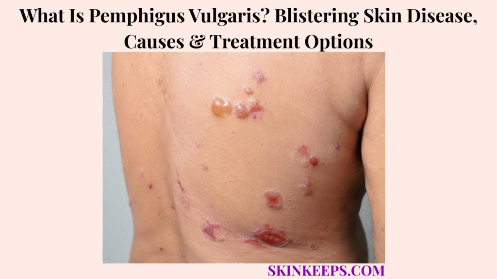
Pemphigus vulgaris is a serious autoimmune blistering disease that commonly begins with persistent painful mouth erosions and can later produce fragile skin blisters that rupture easily. The disease can affect both skin and mucous membranes, and the rapid collapse of thin blister roofs means many patients present mainly with raw erosions rather than numerous intact blisters.
PV develops when pathogenic IgG autoantibodies disrupt keratinocyte adhesion, especially through desmoglein 3 and sometimes desmoglein 1. Diagnosis requires immunopathologic confirmation with correctly selected biopsy specimens, and significant disease usually needs specialist systemic treatment, while infection prevention, wound care and nutritional support help reduce complications and modern therapy can produce prolonged disease control or remission.
This article is for educational purposes only and does not constitute medical advice, diagnosis, or treatment. Persistent unexplained mouth erosions, widespread or rapidly increasing blistering or skin loss, fever, pus or spreading infection, inability to eat or drink, painful swallowing, hoarseness, eye involvement, or severe genital mucosal disease should be evaluated promptly by a licensed dermatologist or qualified healthcare professional because pemphigus vulgaris can require urgent specialist systemic treatment.
How Can You Recognize Pemphigus Vulgaris?
Pemphigus vulgaris commonly causes persistent painful oral erosions followed by thin, flaccid skin blisters that rupture rapidly and leave raw or crusted erosions.
Why Does Pemphigus Vulgaris Often Begin in the Mouth?
Pemphigus vulgaris frequently begins with painful persistent oral erosions because mucosal epithelial adhesion is strongly affected by desmoglein-3-directed autoimmunity.
The inner cheeks, gums, palate and tongue are common sites, and oral disease can make eating, drinking and oral hygiene painful. Mouth disease may precede obvious skin lesions by weeks or months, and some patients remain predominantly mucosal.
What Do Pemphigus Vulgaris Skin Blisters Look Like?
PV skin blisters are thin-walled and flaccid, rupture easily and commonly leave painful moist or crusted erosions rather than remaining intact.
The scalp, face, upper chest and back are frequent sites. The superficial intraepidermal split creates a fragile blister roof, which explains why intact blisters can disappear quickly.
Which Other Mucosal or Skin Findings Support PV?
PV can affect the pharynx, larynx, nose, oesophagus, conjunctiva and genital or anal mucosa, while a positive Nikolsky sign can support—but not confirm—the diagnosis.
Painful swallowing, hoarseness, eye symptoms or genital erosions may indicate wider mucosal involvement. Nikolsky sign reflects superficial epidermal shearing and should be assessed clinically rather than deliberately reproduced at home.
Does Pemphigus Vulgaris Scar or Look Different on Darker Skin?
Typical PV erosions usually heal without deep scarring, while darker skin may show less obvious redness and more hyperpigmentation, haemorrhagic crusting or darker inflammatory change.
The ordinary blister split remains intraepidermal, so deep scarring is not typical. Post-inflammatory pigment may persist, while secondary infection or deeper injury can alter healing.
Figure 1. PV often begins with painful oral erosions; fragile skin blisters may appear later and rupture so quickly that erosions are more obvious than intact blisters.
Why Does Pemphigus Vulgaris Develop?
Pemphigus vulgaris develops when pathogenic IgG autoantibodies target desmosomal adhesion proteins—especially desmoglein 3 and sometimes desmoglein 1—causing keratinocytes to lose adhesion.
How Do Desmoglein Autoantibodies Produce Blisters?
Anti-desmoglein antibodies disrupt keratinocyte-to-keratinocyte adhesion, producing acantholysis and a fragile intraepidermal blister.
Desmosomes normally hold epidermal keratinocytes together. Autoantibody interference weakens that adhesion, creating suprabasal separation and the characteristic fragile blister.
Why Do Dsg3 and Dsg1 Affect the Clinical Pattern?
Dsg3-directed autoimmunity is strongly associated with mucosal disease, while additional Dsg1 involvement is more often seen when substantial skin blistering accompanies mucosal lesions.
This helps explain mucosal-predominant and mucocutaneous patterns, but antibody profiles are not absolute rules for every individual patient.
Is Pemphigus Vulgaris Inherited or Triggered by Medicines?
PV has genetic susceptibility but is not a simple inherited disorder, and selected medicines can trigger pemphigus in susceptible individuals.
HLA associations support inherited susceptibility rather than predictable parent-to-child transmission. Penicillamine, captopril and selected other drugs have been linked with drug-induced pemphigus, but a suspected medication should not be stopped without the prescribing clinician.
Can Infection, Trauma or Stress Cause Pemphigus Vulgaris?
Infection, trauma and environmental stressors may act as triggers in some susceptible people, but no single external trigger explains most PV and the disease itself is not contagious.
The primary mechanism remains autoimmune rather than bacterial, fungal or viral infection. Secondary infection can complicate open erosions later without being the cause of PV.
Figure 2. Pathogenic IgG against desmogleins disrupts desmosomal adhesion, producing acantholysis and a fragile intraepidermal blister.
How Is Pemphigus Vulgaris Distinguished and Diagnosed?
Pemphigus vulgaris is confirmed by combining the clinical pattern with histopathology and perilesional direct immunofluorescence, with desmoglein antibody testing providing additional diagnostic and monitoring information.
How Is Pemphigus Vulgaris Different From Other Blistering Diseases?
PV typically causes fragile flaccid blisters and prominent oral erosions, whereas bullous pemphigoid causes deeper tense itchy blisters and mucous membrane pemphigoid has a stronger tendency toward mucosal scarring.
Pemphigus foliaceus is more superficial and predominantly cutaneous, with characteristic oral erosions generally absent. Erosive lichen planus can also produce persistent oral erosions but follows a different inflammatory and histopathologic pattern.
Why Are Two Biopsy Samples Often Needed?
One biopsy is used to examine blister architecture by routine histology, while a separate perilesional biopsy is used for direct immunofluorescence.
The histology specimen should include a suitable active blister or erosion edge and typically shows suprabasal acantholysis. The DIF specimen is taken from relatively intact perilesional skin or mucosa because heavily eroded tissue can reduce diagnostic yield.
What Does Direct Immunofluorescence Show?
Direct immunofluorescence typically demonstrates intercellular IgG and/or complement deposition around epidermal keratinocytes, supporting the diagnosis of pemphigus.
DIF is performed on tissue, not blood, and provides a characteristic intercellular immunoreactant pattern that clinical morphology alone cannot replace.
What Do Desmoglein Blood Tests Show, and When Is a Broader Differential Needed?
Anti-Dsg1 and anti-Dsg3 serology supports PV diagnosis and can assist monitoring, while unusually severe or polymorphic disease may require consideration of alternative pemphigus syndromes such as paraneoplastic pemphigus.
ELISA titres can correlate with disease activity in some patients, but clinical activity remains central. Severe persistent stomatitis, polymorphic lesions or known neoplastic disease raises the value of a broader differential.
Figure 3. Histology and DIF usually require differently selected biopsy sites; serology supports the immunopathologic diagnosis rather than replacing it.
How Is Pemphigus Vulgaris Treated?
Pemphigus vulgaris requires specialist treatment that suppresses pathogenic autoantibody production, with rituximab-based systemic therapy playing a major role in appropriate moderate-to-severe disease while corticosteroid exposure is reduced as control develops.
What Is the Main Goal of PV Treatment?
Treatment aims to stop new blister formation, heal existing erosions, prevent infection and achieve sustained disease control while minimizing long-term medication toxicity.
Useful clinical endpoints include cessation of new blisters, healing erosions, improved oral comfort and intake, reduced corticosteroid exposure and durable disease control. Visible healing alone does not prove that autoimmune activity has completely disappeared.
When Are Rituximab and Corticosteroids Used?
Rituximab is a major first-line treatment for appropriate moderate-to-severe PV and is commonly combined initially with glucocorticoid therapy that is tapered as disease control improves.
Rituximab targets CD20-positive B cells and reduces the B-cell population that drives pathogenic autoantibody production. Corticosteroids can control active inflammation rapidly but carry important cumulative toxicity, so the modern strategy is to gain control while reducing prolonged steroid exposure.
No individual infusion schedule, corticosteroid dose or taper should be inferred from this general overview.
What Other Systemic or Local Treatments Can Be Used?
Azathioprine and mycophenolate remain established steroid-sparing options in selected patients, while topical corticosteroids and wound care can support localized lesions but do not replace systemic control of significant PV.
These steroid-sparing agents may be used when rituximab is unsuitable, unavailable or when another systemic strategy is needed. IVIG, immunoadsorption and other specialist therapies remain options for selected refractory disease rather than routine first-line equivalents.
What Safety Checks Are Needed Before and During Systemic Treatment?
Major systemic PV therapy requires pretreatment infection and laboratory assessment plus ongoing monitoring for immunosuppression-related complications.
Rituximab safety assessment includes hepatitis B screening, baseline blood counts and infection-risk review, with vaccination planning when clinically feasible and monitoring for infusion reactions, infections and cytopenias. Systemic corticosteroid care also requires attention to infection, bone health and metabolic complications.
Figure 4. For appropriate moderate-to-severe PV, rituximab-based therapy is a major modern systemic strategy, with glucocorticoid exposure reduced as control develops while wound, oral and infection care continue in parallel.
What Complications Can Pemphigus Vulgaris Cause, and How Is Long-Term Care Managed?
Pemphigus vulgaris can cause serious infection, extensive painful skin loss, nutritional compromise and treatment-related toxicity, while relapse monitoring remains necessary after disease control.
Why Are Infection and Wound Care Important?
Open PV erosions weaken the protective skin barrier and increase susceptibility to bacterial, fungal and viral infection, especially during systemic immunosuppression.
Secondary bacterial infection, candidiasis and herpes simplex can complicate care. Gentle handling, non-adherent dressings, appropriate hygiene and emollient or barrier support help protect fragile erosions without harsh debridement.
How Can Mouth and Throat Disease Affect Nutrition and Daily Care?
Painful oral and pharyngeal erosions can make eating, drinking, swallowing and oral hygiene difficult enough to cause dehydration or nutritional compromise.
Support can include clinician-directed pain control, softer non-irritating foods, adequate fluids, nutritional assessment and gentle dental or oral care. Painful swallowing, hoarseness and reduced intake increase the need for prompt reassessment.
Which Treatment Complications Need Monitoring?
PV treatment can itself cause significant complications, so monitoring must address infection, hematologic abnormalities, corticosteroid toxicity and other immunosuppressive adverse effects as well as blister control.
Systemic corticosteroids can contribute to infection, osteoporosis and metabolic effects. Rituximab and other immunosuppressive therapies require monitoring for infections, hepatitis B reactivation, cytopenias and infusion-related reactions.
Can Pemphigus Vulgaris Relapse After Remission?
Yes; PV often follows a relapsing-remitting course, so new blisters or erosions after control can indicate renewed disease activity and warrant clinical reassessment.
Remission can be prolonged, but it should not be equated with a guaranteed permanent cure. New mucosal or skin lesions may require treatment adjustment, while desmoglein serology can support follow-up but does not replace clinical assessment.
What Should You Remember About Pemphigus Vulgaris?
Pemphigus vulgaris is a serious but treatable autoimmune blistering disease in which anti-desmoglein antibodies weaken epidermal adhesion, commonly causing painful oral erosions and fragile skin blisters that require biopsy confirmation and systemic specialist care.
- PV is autoimmune and is not contagious.
- Persistent painful oral erosions commonly appear early.
- Skin blisters are thin, flaccid and rupture easily.
- Other mucosal sites can be involved.
- Typical lesions usually do not scar deeply.
- Dsg3 and Dsg1 autoantibodies are central to disease biology.
- Acantholysis creates the fragile intraepidermal blister.
- Genetic susceptibility is not simple inheritance.
- Selected medicines can trigger pemphigus in susceptible people.
- External triggers are not universal causes.
- Bullous pemphigoid produces deeper tense blisters.
- Mucous membrane pemphigoid has stronger scarring potential.
- Diagnosis requires more than appearance alone.
- Histology typically shows suprabasal acantholysis.
- Perilesional DIF is central to confirmation.
- Desmoglein serology supports diagnosis and monitoring.
- Significant disease usually requires systemic treatment.
- Rituximab is a major modern therapy for appropriate moderate-to-severe PV.
- Corticosteroids remain important, but cumulative toxicity matters.
- Steroid-sparing strategies are available.
- Wound, oral and nutritional care remain essential.
- Infection and treatment toxicity require monitoring.
- Remission is possible, but relapse can occur.
Recognize → Confirm with biopsy/DIF → Assess severity → Suppress autoimmune disease → Protect wounds and nutrition → Reduce treatment toxicity → Monitor relapse.
Frequently Asked Questions About Pemphigus Vulgaris
The most important pemphigus-vulgaris questions concern its early mouth symptoms, autoimmune mechanism, contagiousness, biopsy confirmation and possibility of long-term remission.
What Are the First Signs of Pemphigus Vulgaris?
Persistent painful erosions inside the mouth are among the most characteristic early signs of pemphigus vulgaris and can appear before obvious skin blistering. The gums, inner cheeks, palate and tongue can be involved before fragile skin blisters become obvious.
Why Does Pemphigus Vulgaris Often Start in the Mouth?
Mucosal disease is strongly associated with autoantibodies against desmoglein 3, an adhesion protein that is important in oral epithelium. Loss of keratinocyte adhesion produces acantholysis and painful superficial erosions.
Is Pemphigus Vulgaris Contagious?
No; pemphigus vulgaris is caused by the person’s immune system attacking cell-adhesion proteins and cannot be spread between people like an infection. Secondary infection can occur in open erosions but does not make PV contagious.
How Is Pemphigus Vulgaris Confirmed With Biopsy and Blood Tests?
Diagnosis is usually confirmed with routine histology plus perilesional direct immunofluorescence, while anti-Dsg1 and anti-Dsg3 blood tests provide additional support. The histology and DIF samples are intentionally taken from different types of tissue.
Can Pemphigus Vulgaris Go Into Long-Term Remission?
Yes; modern systemic treatment can produce prolonged disease control or remission, although relapse remains possible and long-term monitoring is often required. New erosions or blisters after control should be reassessed.
Which Sources Support This Pemphigus Vulgaris Guidance?
DermNet — Pemphigus Vulgaris — Primary clinical source for oral-first disease, fragile blisters, darker-skin presentation, Dsg3/Dsg1 biology, acantholysis, biopsy/DIF, wound care, infection, nutrition and relapse.
EADV Updated S2K Guideline — Management of Pemphigus Vulgaris and Foliaceus — Used for current systemic-treatment hierarchy and rituximab-based first-line positioning for appropriate moderate-to-severe pemphigus.
U.S. FDA — Rituximab Orphan Drug Approval for Pemphigus Vulgaris — Used to verify the FDA-labeled indication for adult moderate-to-severe pemphigus vulgaris.
Genentech — Rituxan Prescribing/Safety Information — Used for current indication and major rituximab safety context including hepatitis B reactivation, infection and infusion-related risk.
DermNet — Pemphigus Vulgaris Pathology — Used for suprabasal acantholysis and intraepidermal blister architecture.



