
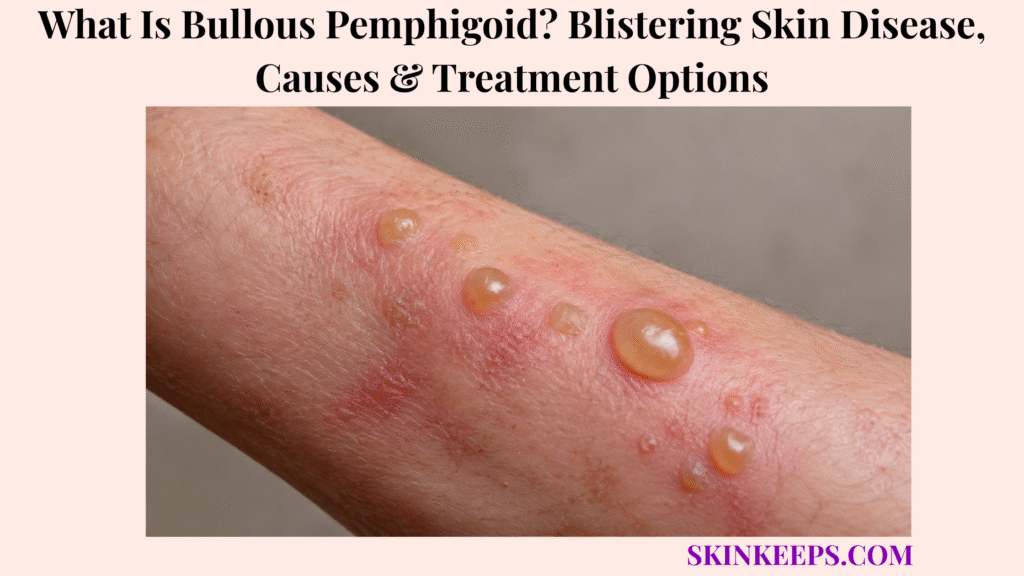
Bullous pemphigoid is an autoimmune blistering disease that can cause intense itching, rash, and large tense fluid-filled blisters.
It is not contagious, and it should not be treated like a simple friction blister, eczema flare, or impetigo. This page covers symptoms, causes, diagnosis, biopsy, treatment options, skin care, infection risk, follow-up, mistakes, and urgent signs.
What Is Bullous Pemphigoid and Why Does It Cause Blisters?
Bullous pemphigoid is an autoimmune blistering disease where the immune system attacks proteins that help attach the outer skin layer to deeper skin, causing inflammation, itching, and tense blisters.
The outer layer of skin is called the epidermis. The deeper layer is called the dermis. Between them is the dermal-epidermal junction, where attachment proteins help hold the skin layers together.
In bullous pemphigoid, autoantibodies target this attachment zone. That weakens the junction, inflammation builds, and fluid collects below the epidermis to form a subepidermal blister.
Why Are Bullous Pemphigoid Blisters Usually Tense?
Bullous pemphigoid blisters are usually tense because the blister forms below the epidermis, leaving a stronger blister roof than many fragile blistering diseases.
This deeper blister level makes the bullae feel firm, tight, and less likely to break easily than flaccid blister diseases.
Tense blisters are an important recognition clue, but they do not confirm the diagnosis by themselves. Testing is still needed because several blistering diseases can overlap visually.
Why Can Itching Appear Before Blisters?
Itching can appear before blisters because early bullous pemphigoid may begin as inflammation, eczema-like rash, or hive-like plaques before tense bullae form.
Some people have severe itch for weeks or months before obvious blisters appear. The early rash may look like atopic dermatitis, dermatitis, or another nonspecific itchy condition.
This pre-blister phase can delay diagnosis. Persistent severe itch plus later tense blisters should change the diagnostic pathway toward dermatology evaluation.
Practical rule: Unexplained intense itch plus tense blisters should be assessed with dermatologist-led testing before treatment is chosen.
What Bullous Pemphigoid Symptoms Appear on Skin?
Bullous pemphigoid can cause intense itch, eczema-like or hive-like patches, tense fluid-filled blisters, raw erosions after blisters break, crusting, pain, and sleep disruption.
Some people have rash and itch before blisters. Others present after large tense bullae appear on inflamed or normal-looking skin.
What Do Early Bullous Pemphigoid Symptoms Look Like?
Early bullous pemphigoid may look like severe itch with eczema-like patches, hives-like raised areas, burning, soreness, or inflamed skin before blisters appear.
The rash may look red or pink on lighter skin. On brown or black skin, inflammation may look brown, purple, grayish, darker, or more textured than nearby skin.
Early bullous pemphigoid can also look like hives, especially when raised itchy plaques come and go before blisters are obvious.
What Do Bullous Pemphigoid Blisters Look Like?
Bullous pemphigoid blisters are often large, tense, fluid-filled bumps that may contain clear, cloudy, yellowish, or bloodstained fluid.
They may appear on inflamed skin or skin that looks nearly normal. The blister roof may feel tight and may not break as easily as fragile infectious or pemphigus-type blisters.
If a blister breaks, it can leave a painful erosion, crust, or raw area that needs protection and infection monitoring.
What Symptoms Suggest Complications?
Complications are more likely when bullous pemphigoid causes increasing pain, pus, cloudy drainage, spreading warmth, fever, dehydration, weakness, confusion, or major treatment side effects.
Broken blisters can become secondarily infected, especially in older adults, people with diabetes, frailty, poor wound healing, or patients receiving systemic steroids or immunosuppressive medicines.
Mouth, eye, or genital sores are not a routine simple-blister issue and should be checked faster.
| Symptom | What User May Notice | Why It Matters |
|---|---|---|
| Severe itch | Intense scratching and sleep disruption | Can start before blisters |
| Eczema-like rash | Red, pink, brown, purple, or darker inflamed patches | Can delay diagnosis |
| Hive-like plaques | Raised itchy areas | Pre-bullous pattern possible |
| Tense blisters | Large firm fluid-filled bubbles | Core BP clue |
| Clear, cloudy, or bloodstained fluid | Blister contents vary | Monitor for infection signs |
| Erosions or crusts | Raw areas after rupture | Infection and wound-care risk |
| Mouth, eye, or genital involvement | Sores or irritation at mucosal sites | Needs faster review |
| Fever, pus, or warmth | Systemic or local infection signs | Prompt medical care |
| Weakness or confusion | Frail older adult may be deteriorating | Urgent care |
Where Does Bullous Pemphigoid Usually Appear?
Bullous pemphigoid often affects the arms, legs, trunk, abdomen, groin, armpits, and skin folds, although distribution can vary.
Blisters may occur on the lower abdomen, thighs, groin, armpits, inner arms, legs, trunk, and flexural or fold areas. They may appear on inflamed skin or on skin that otherwise looks normal.
Mouth or mucous membrane involvement can occur but is less common in classic bullous pemphigoid. Eye, mouth, or genital involvement should prompt faster review.
| Area | Common Clue | Safety Concern |
|---|---|---|
| Lower abdomen | Itchy plaques or tense blisters | Widespread erosions can become infected |
| Thighs / legs | Tense bullae or crusted erosions | Mobility and wound-care burden |
| Groin | Fold-area blisters | Pain, moisture, infection risk |
| Armpits | Fold-area itch or blisters | Friction and dressing difficulty |
| Inner arms | Flexural involvement | Scratching and erosion risk |
| Trunk | Widespread rash or bullae | More extensive disease |
| Skin folds | Blisters along creases | Moisture and friction complications |
| Mouth / mucosa | Less common in classic BP | Needs faster medical review |
| Eyes | Not a typical simple BP presentation | Prompt specialist review |
What Causes Bullous Pemphigoid?
Bullous pemphigoid happens when the immune system mistakenly targets proteins at the junction between the epidermis and dermis, leading to inflammation and blister formation.
The exact reason the immune system starts this response is not always clear. In some people, medicines, medical conditions, light therapy, radiation, or skin injury are reviewed as possible contributors.
What Skin Proteins Are Involved in Bullous Pemphigoid?
The main skin proteins involved in bullous pemphigoid are BP180, also called collagen XVII, and BP230, which help anchor skin layers together.
These proteins are part of the attachment structures that help connect the epidermis to deeper skin. When autoantibodies target them, inflammation weakens that attachment.
The result is a subepidermal split, meaning the blister forms under the epidermis. That deeper split helps explain the tense blister roof.
Why Is Bullous Pemphigoid Not Contagious?
Bullous pemphigoid is not contagious because it is an autoimmune disease, not a bacterial, viral, or fungal infection.
It is not spread by touch, towels, blister fluid, or close contact in the way contagious infections spread.
Open erosions can still become secondarily infected, so wound care and infection monitoring remain important even though the disease itself is not contagious.
Can Medicines or Treatments Trigger Bullous Pemphigoid?
Some medicines, light therapy, radiation, or skin injury have been associated with bullous pemphigoid, so medication and treatment history should be reviewed.
Medication review is especially important in older adults because many patients take several medicines and may have diabetes, kidney disease, neurologic disease, or other conditions.
Do not stop prescribed medicine without clinician guidance. Sudden medication changes can be dangerous and may not solve the blistering disease.
Who Is More Likely to Develop Bullous Pemphigoid?
Bullous pemphigoid most often affects older adults, and treatment planning must consider frailty, infection risk, diabetes, bone health, kidney disease, blood pressure, and other medicines.
Age over 60 is a useful risk context, but younger adults and rare pediatric cases can still occur. A younger person with unexplained tense blisters should not be dismissed only because the condition is more common in older adults.
Neurologic history, multiple medicines, diabetes, chronic kidney disease, prior skin disease, phototherapy, radiation, and poor wound healing may all affect evaluation and treatment planning.
How Is Bullous Pemphigoid Different From Other Blistering Skin Problems?
Bullous pemphigoid can resemble eczema, hives, impetigo, burns, pemphigus vulgaris, drug reactions, herpes infections, and severe blistering reactions, but the pattern of tense blisters, intense itch, older age, and biopsy findings helps separate it.
Several blistering conditions need urgent care, and some have very different treatment pathways. Diagnosis should not end with photo comparison.
How Is Bullous Pemphigoid Different From Bullous Impetigo?
Bullous pemphigoid is different from bullous impetigo because bullous pemphigoid is autoimmune and not contagious, while bullous impetigo is a contagious bacterial infection.
Bullous impetigo often has fragile blisters, yellow drainage, and spread through contact. Bullous pemphigoid often has tense blisters, severe itch, and an autoimmune testing pathway.
Antibiotics may be needed if secondary infection occurs, but antibiotics alone do not treat the autoimmune disease process.
How Is Bullous Pemphigoid Different From Pemphigus Vulgaris?
Bullous pemphigoid is different from pemphigus vulgaris because pemphigus vulgaris usually causes fragile blisters and erosions, often with more prominent mouth involvement.
Bullous pemphigoid blisters are usually tense. Pemphigus vulgaris blisters tend to break more easily and often involve mucous membranes more prominently.
Biopsy and immunofluorescence help distinguish them because treatment intensity and risks can differ.
How Is Bullous Pemphigoid Different From Eczema or Hives?
Bullous pemphigoid can look like eczema or hives at first, but persistent severe itch in an older adult plus later tense blisters should change the diagnostic pathway.
Allergic contact dermatitis may blister after plant, adhesive, cosmetic, rubber, metal, or chemical exposure, while bullous pemphigoid is autoimmune.
Random steroid creams may partly suppress visible inflammation and delay testing if blisters continue spreading without diagnosis.
How Is Bullous Pemphigoid Different From Severe Drug Reactions?
Bullous pemphigoid is different from severe drug reactions such as SJS/TEN because SJS/TEN can cause painful widespread skin detachment, mucosal sores, fever, and rapid systemic illness.
New medicine plus fever, widespread pain, mouth sores, eye symptoms, genital sores, or peeling skin needs urgent care. Not every drug-related blistering eruption is bullous pemphigoid.
Shingles is another blistering condition with a different pattern, often painful and one-sided, and it needs a different evaluation path.
| Condition | Main Clue | Contagious? | Safer Next Step |
|---|---|---|---|
| Bullous pemphigoid | Tense blisters plus intense itch, often older adults | No | Dermatology biopsy/testing |
| Bullous impetigo | Fragile blisters plus bacterial spread or yellow drainage | Yes | Antibiotics plus hygiene control |
| Pemphigus vulgaris | Fragile blisters or erosions, mouth involvement | No | Urgent dermatology testing |
| Eczema / hives | Itch or rash without true tense bullae | No | Reassess if blisters appear |
| Blisters from friction/burn | Rubbing or burn history | No | Treat cause; review if unexplained or widespread |
| Shingles | Painful one-sided blistering pattern | Contagious-risk context | Antiviral evaluation if suspected |
| SJS/TEN | Painful widespread blistering plus mucosa plus medicine link | No | Emergency care |
How Is Bullous Pemphigoid Diagnosed?
Bullous pemphigoid is usually diagnosed through clinical examination plus skin biopsy, direct immunofluorescence, and blood tests for pemphigoid antibodies when available.
Clinical appearance matters, but biopsy and immune testing are central because other blistering diseases can look similar.
What Does a Dermatologist Check During Exam?
A dermatologist checks bullous pemphigoid by reviewing itch severity, rash history, blister tension, distribution, erosions, crusting, mucosal involvement, infection signs, medicines, age, and comorbidities.
The exam may review when itching began, when blisters appeared, where they appeared first, whether mouth, eye, or genital sores are present, and whether fever, pus, odor, spreading warmth, or worsening pain suggests infection.
Medication history matters because some medicines and treatments have been associated with bullous pemphigoid, and steroid treatment risk depends on the patient’s overall health.
Why Do Biopsy and Direct Immunofluorescence Matter?
Biopsy and direct immunofluorescence matter because they confirm the autoimmune blistering pattern and distinguish bullous pemphigoid from impetigo, pemphigus, eczema, drug reactions, and other blistering diseases.
One biopsy may examine routine histology. A second biopsy from nearby non-blistered skin may be used for direct immunofluorescence.
Direct immunofluorescence looks for immune deposits along the basement membrane zone. Blood antibody tests such as BP180 or BP230 testing may support diagnosis and monitoring in selected patients.
When Should Infection Testing Be Considered?
Infection testing should be considered when bullous pemphigoid erosions develop pus, cloudy drainage, fever, increasing pain, spreading warmth, or delayed healing.
Culture may be used when secondary infection is suspected. This is especially relevant when a patient is on systemic steroids, immunosuppressants, or has diabetes, frailty, or poor wound healing.
Not all bullous pemphigoid blisters are infected. Infection testing is targeted to signs of secondary infection.
What Treatment Options Are Used for Bullous Pemphigoid?
Bullous pemphigoid treatment aims to stop new blisters, heal existing erosions, reduce itch, prevent infection, and minimize treatment side effects, especially in older adults.
Treatment is severity-led and clinician-directed. The plan may combine anti-inflammatory treatment, wound care, itch control, infection monitoring, medication-risk review, and slow tapering after control.
When Are High-Potency Topical Steroids Used?
High-potency topical steroids may be used for localized or moderate bullous pemphigoid, and sometimes for more extensive disease when safe application is practical.
They can reduce inflammation, itch, and blister formation. Correct application, amount, body-site caution, and monitoring matter.
Skin thinning, infection risk, and absorption are part of the safety discussion, especially in older adults or when large body areas are treated.
When Are Oral Corticosteroids Used?
Oral corticosteroids may be used when bullous pemphigoid is widespread, severe, rapidly worsening, or difficult to control with topical treatment alone.
Systemic steroids can control inflammation quickly, but older adults need monitoring for infection, diabetes changes, blood pressure, bone loss, mood changes, stomach risk, sleep changes, and medication interactions.
After control, steroid tapering should be clinician-guided. Systemic steroids should not be stopped suddenly without medical guidance.
When Are Steroid-Sparing Treatments Considered?
Steroid-sparing treatments may be considered when bullous pemphigoid is recurrent, severe, long-lasting, steroid-risky, or not controlled enough with first-line therapy.
Selected patients may use doxycycline-based strategies, nicotinamide in some regimens, methotrexate, azathioprine, mycophenolate, or other immunosuppressive medicines.
Specialist options for refractory disease may include rituximab, omalizumab, IVIG, or other targeted approaches depending on the patient, country, severity, and guideline setting.
Where Does Dupilumab Fit in Current Treatment?
Dupilumab may be considered for appropriate adults with bullous pemphigoid in settings where it is approved and clinically suitable.
In the United States, dupilumab labeling includes adult bullous pemphigoid. It does not replace diagnosis confirmation, wound care, infection monitoring, or individualized risk assessment.
Availability and approval status vary by country, so the treatment discussion should stay clinician-guided.
| Treatment Option | Best-Fit Situation | Main Goal | Key Caution |
|---|---|---|---|
| High-potency topical steroid | Localized/moderate disease or topical-first plan | Reduce inflammation and blisters | Application burden, skin thinning, absorption |
| Oral corticosteroid | Widespread/severe/rapidly worsening disease | Fast disease control | High side-effect risk in older adults |
| Doxycycline-based strategy | Selected steroid-risk patients | Lower steroid burden | May be slower or less potent |
| Immunosuppressants | Refractory disease or steroid-sparing need | Reduce steroid exposure | Monitoring and infection risk |
| Biologic/targeted therapy | Eligible patients where approved/appropriate | Target inflammatory pathway | Specialist-led; approval varies |
| Wound/infection care | Broken or infected skin | Heal erosions and prevent complications | Do not ignore infection signs |
| Follow-up/taper plan | Controlled disease | Prevent relapse and side effects | Do not stop steroids suddenly |
How Should Skin and Blisters Be Cared for During Bullous Pemphigoid?
Skin care during bullous pemphigoid should protect blister roofs when possible, cover erosions, reduce infection risk, and avoid trauma while medical treatment controls new blister formation.
Do not pop tense blisters unless a clinician gives specific instructions. Protect blisters from friction, tight clothing, rough fabrics, harsh adhesives, scratching, and strong scrubs.
Use non-stick dressings for open erosions when advised. Keep wounds clean and covered, use gentle bathing and moisturizers as tolerated, keep nails short, and ask for wound-care support if erosions are widespread.
Can Bullous Pemphigoid Come Back or Last for Years?
Bullous pemphigoid may last months to years, and treatment is usually tapered slowly after disease control to reduce relapse risk and limit side effects.
The disease can flare and remit. Some patients need long treatment courses, especially when blisters are widespread, relapsing, or difficult to control safely.
Follow-up monitors itch, new blisters, healing erosions, blood tests, infection signs, medicine side effects, steroid taper safety, and relapse risk. Older adults need careful balance between disease control and medication harm.
| Course Phase | What It Means | What Follow-Up Watches |
|---|---|---|
| Active itch/rash | Severe itch or eczema/hive-like phase | Diagnosis suspicion and symptom tracking |
| Blistering phase | Tense blisters and erosions appear | Biopsy, treatment choice, infection risk |
| Control phase | Fewer new blisters and reduced itch | Medication response and side effects |
| Healing phase | Erosions close and crusts reduce | Wound care, pigment changes, infection |
| Taper phase | Medicines are slowly reduced | Relapse and steroid safety |
| Monitoring phase | Long-term follow-up | Recurrence, comorbidities, medication harm |
What Bullous Pemphigoid Mistakes Should You Avoid?
The biggest bullous pemphigoid mistake is treating widespread tense blisters as simple blisters or eczema without biopsy-based diagnosis.
Do not self-diagnose unexplained widespread blisters. Do not pop or peel blister roofs aggressively, and do not ignore intense itch in an older adult when blisters appear.
Do not use leftover antibiotics for autoimmune blisters, keep using random creams while blisters spread, or stop systemic steroids suddenly. Do not ignore infection signs while using steroids or immunosuppressive treatment.
| Mistake | Why It Is Risky | Better Action |
|---|---|---|
| Calling it eczema only | Delays autoimmune diagnosis | Dermatology biopsy/testing |
| Popping all blisters | Infection and erosion risk | Protect and cover |
| Stopping steroids suddenly | Flare and adrenal-risk concerns | Taper with clinician |
| Ignoring infection | Serious complication risk | Prompt medical review |
| Treating with antibiotics only | Autoimmune disease persists | Cause-specific treatment |
| Ignoring mucosal or eye sores | Higher-risk site involvement | Faster specialist review |
| Delaying care in frailty | Dehydration and infection risk rises | Earlier medical care |
When Should Bullous Pemphigoid Be Checked Urgently?
Bullous pemphigoid needs prompt medical care when blisters are widespread, painful, infected-looking, rapidly worsening, involving the eyes, mouth, or genitals, or occurring with fever, dehydration, weakness, confusion, or major medication side effects.
Frail older adults, people with diabetes, immune suppression, chronic kidney disease, poor wound healing, or systemic steroid use should use a lower threshold for care.
Which Symptoms Need Faster Medical Care?
Faster medical care is needed when bullous pemphigoid causes fever, rapid spread, severe pain, pus, cloudy drainage, bad odor, spreading warmth, large open erosions, eye involvement, mouth or genital sores, dehydration, weakness, dizziness, confusion, or new blistering after a medication.
Spreading redness, warmth, swelling, or darker color change around erosions can signal secondary infection. Older adults may show deterioration through weakness, confusion, poor intake, or dehydration rather than dramatic skin symptoms alone.
Which Patients Should Not Wait?
Frail older adults and people with diabetes, immune suppression, chronic kidney disease, severe heart or lung disease, poor wound healing, or systemic steroid use should seek care earlier for bullous pemphigoid flares.
Nursing-home or bedbound patients, patients with widespread erosions, and people on systemic steroids or immunosuppressants may need urgent wound, infection, hydration, and medication-risk review.
Seek prompt medical care if bullous pemphigoid involves:
What Should You Remember About Bullous Pemphigoid?
The most important thing to remember about bullous pemphigoid is that it is an autoimmune blistering disease, so the safest approach is diagnosis confirmation, inflammation control, skin protection, infection monitoring, and careful treatment follow-up.
Severe itch, tense blisters, widespread erosions, infection signs, mucosal or eye symptoms, and medication side effects should be taken seriously, especially in older adults.
Frequently Asked Questions About Bullous Pemphigoid
Is bullous pemphigoid contagious?
No. Bullous pemphigoid is autoimmune, not a contagious bacterial or viral blister infection, although open skin can become secondarily infected.
What does bullous pemphigoid look like?
It can cause intense itching, rash or hive-like patches, and large tense blisters that do not break easily. Blisters may contain clear, cloudy, yellowish, or bloodstained fluid, and blister color can vary by skin tone.
What causes bullous pemphigoid?
Bullous pemphigoid is caused by an autoimmune attack against proteins that help attach the epidermis to the dermis, especially BP180/collagen XVII and BP230-related structures.
How is bullous pemphigoid diagnosed?
Diagnosis may involve clinical examination, skin biopsy, direct immunofluorescence, and blood tests for pemphigoid antibodies when available. Infection testing may be needed if erosions look infected.
What is the best treatment for bullous pemphigoid?
There is no one best treatment for every person. Treatment depends on severity, age, comorbidities, infection risk, and steroid risk; options may include high-potency topical steroids, oral corticosteroids, steroid-sparing medicines, targeted therapy such as dupilumab where approved and appropriate, and wound care.
Can bullous pemphigoid go away?
It can enter remission, but the course may last months to years, and medicines often need slow tapering with clinician monitoring. Stopping treatment suddenly can trigger problems or relapse.
Can medicine trigger bullous pemphigoid?
Some medicines and treatments have been associated with bullous pemphigoid, but medication changes should be reviewed with a clinician and prescribed medicines should not be stopped without medical guidance.
When should bullous pemphigoid need urgent care?
Prompt care is needed for widespread blistering, fever, infection signs, severe pain, eye, mouth, or genital involvement, dehydration, confusion, rapid worsening, new blistering after medication, or frail older adults with large erosions.
Sources & Evidence About Bullous Pemphigoid
DermNet — Bullous Pemphigoid was used for autoimmune subepidermal blistering disease framing, severe itch, large tense bullae, rash before blisters, flexural and fold distribution, blister fluid variation, treatment categories, and long taper/course framing.
Mayo Clinic — Bullous Pemphigoid Symptoms and Causes was used for itching before blisters, large blisters that do not break easily, skin-fold locations, not-contagious explanation, age-over-60 risk context, medicine/light/radiation associations, complications, course duration language, and when to seek medical care.
StatPearls / NCBI Bookshelf — Bullous Pemphigoid was used for BP180/BP230 mechanism, subepidermal split, direct immunofluorescence, perilesional biopsy, ELISA, differential diagnosis, and treatment categories.
MSD Manual Professional — Bullous Pemphigoid was used for chronic autoimmune blistering disorder framing, older-adult pattern, pruritic bullous lesions, biopsy and immunofluorescence diagnosis, BP180/BP230 antigens, topical/systemic corticosteroids, and long-term maintenance therapy.
British Association of Dermatologists — Pemphigoid Patient Information was used for patient-friendly treatment variability, topical/oral steroid care, steroid-alert card advice, and sick-day-rule context for people on steroid tablets.
DailyMed — DUPIXENT Prescribing Information was used for current U.S. labeling that lists bullous pemphigoid under indications added in 06/2025.
Mayo Clinic — Pemphigus Symptoms and Causes was used only for differential support: pemphigus causes fragile blisters or sores, often involving mucous membranes, and is distinct from bullous pemphigoid.
Educational Disclaimer: This SkinKeeps article is for educational purposes only and does not diagnose or replace medical care. Widespread tense blisters, severe itch with new blisters, fever, infected-looking erosions, pus, cloudy drainage, bad odor, spreading warmth or color change, severe pain, eye involvement, mouth or genital sores, dehydration, weakness, dizziness, confusion, rapid worsening, new blistering after medication, frailty, diabetes, kidney disease, immune suppression, poor wound healing, or major steroid/immunosuppressant side effects should be checked promptly by a qualified healthcare professional or dermatologist. Do not stop systemic steroids or other prescribed medicines suddenly without medical guidance.



