
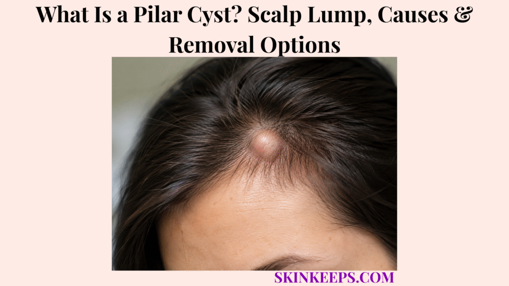
A pilar cyst is a benign keratin-filled cyst arising from the outer root sheath of a hair follicle, usually forming a smooth, firm and mobile lump on the scalp. Also called a trichilemmal cyst and historically a “wen,” it usually lies beneath normal-looking skin, often lacks a visible central punctum and may occur as one lesion or several scalp cysts.
Most pilar cysts are painless and do not require treatment simply because they are present. Removal is reasonable when a cyst becomes painful, repeatedly inflamed or traumatized, enlarges, interferes with hair care, causes cosmetic concern or remains diagnostically uncertain; complete removal of the cyst wall lowers same-lesion recurrence, while rapid or atypical change deserves reassessment.
This article is for educational purposes only and does not constitute medical advice, diagnosis, or treatment. A scalp lump that grows rapidly, becomes persistently painful, repeatedly bleeds or ulcerates, becomes fixed, develops ongoing drainage, repeatedly returns after treatment, or changes substantially from its previous appearance should be evaluated by a licensed dermatologist or qualified healthcare professional because atypical scalp masses may require excision and histopathologic diagnosis.
How Can You Recognize a Pilar Cyst?
A pilar cyst usually feels like a smooth, firm, round or oval mobile lump beneath normal-looking scalp skin and typically has no visible central punctum.
What Does a Pilar Cyst Usually Feel Like?
A typical pilar cyst is a well-defined, firm subcutaneous nodule that can usually be moved slightly beneath the scalp.
The overlying skin usually looks normal and the cyst is commonly painless. Tenderness becomes more likely after rupture, inflammation or repeated pressure from brushing, combing or other trauma.
Where Do Pilar Cysts Usually Develop?
The scalp is the characteristic site for pilar cysts, accounting for the large majority of trichilemmal cysts.
DermNet describes about 90% as occurring on the scalp, although lesions can occasionally occur on the face, neck, trunk or extremities. Scalp location strongly supports the diagnosis but is not an absolute requirement.
Does a Pilar Cyst Have a Central Pore, and What Is Inside It?
Pilar cysts usually lack a visible central punctum and contain dense keratin produced by their trichilemmal cyst lining.
Keratin is structural protein material, not sebum. The cyst wall resembles the outer root sheath of a hair follicle and undergoes trichilemmal keratinization, explaining why calling these lesions “sebaceous cysts” is inaccurate.
Can Someone Have More Than One Pilar Cyst?
Yes; multiple pilar cysts are common, especially in people with a familial predisposition.
One person may develop a single lesion while another has several scalp cysts of different sizes. Multiple lesions and affected relatives make inherited susceptibility more plausible.
Figure 1. The classic pilar cyst is a keratin-filled outer-root-sheath cyst that forms a firm, mobile scalp lump and usually lacks a central punctum.
Why Do Pilar Cysts Develop, and Can They Run in Families?
Pilar cysts develop from outer-root-sheath follicular cells that form a closed keratin-producing cyst, and some people inherit a strong tendency to develop multiple lesions.
How Does a Pilar Cyst Form?
A pilar cyst forms when outer-root-sheath follicular cells create an enclosed cyst whose lining continues producing keratin.
As keratin accumulates, the cyst enlarges gradually beneath the skin. This is a follicular epithelial process rather than a blocked-sebum disorder.
Are Pilar Cysts Caused by Poor Scalp Hygiene?
No; ordinary pilar cysts are not caused simply by dirty hair or inadequate scalp washing.
The lesion originates from follicular epithelial tissue, and inherited susceptibility can play a role. Washing more often does not remove a pre-existing cyst wall or prevent a genetically predisposed person from forming new cysts.
Can Pilar Cysts Be Inherited?
Yes; familial pilar cysts can follow an autosomal-dominant pattern and often appear earlier or as multiple scalp lesions.
Family clusters are well recognized, but sporadic pilar cysts also occur. A patient does not need a known affected parent for a typical scalp cyst to be diagnosed clinically.
What Is the Role of PLCD1 in Hereditary Pilar Cysts?
PLCD1 has been linked to many hereditary trichilemmal-cyst families, although routine genetic testing is not necessary for an ordinary isolated scalp cyst.
A molecular study found inherited PLCD1 susceptibility variants together with a recurrent cyst-specific somatic PLCD1 change in familial lesions, supporting a combined inherited-plus-somatic mechanism. This genetic context explains familial disease without turning PLCD1 testing into a routine clinical requirement.
Figure 2. Pilar cysts can be sporadic, while familial cases often present with multiple scalp cysts and an autosomal-dominant tendency; PLCD1 has been linked to many hereditary families.
How Is a Pilar Cyst Distinguished and Diagnosed?
A typical pilar cyst is usually diagnosed clinically from its scalp location, firm mobile character and absent punctum, while imaging or histopathology is reserved for atypical lesions.
How Is a Pilar Cyst Different From an Epidermoid Cyst or Lipoma?
A pilar cyst strongly favors the scalp and usually lacks a punctum, while an epidermal cyst more often has a visible punctum and a lipoma usually feels softer or more rubbery because it is composed of fat.
Pilar cysts are typically firm and mobile. Epidermoid cysts can occur at many body sites and have different epithelial keratinization, while a lipoma is a benign fatty mass rather than a keratin-filled follicular cyst.
How Is an Inflamed Pilar Cyst Different From an Abscess?
A ruptured pilar cyst can become red, swollen and painful from sterile keratin-induced inflammation, while an abscess represents a true infectious pus collection.
Rupture can trigger intense inflammation without bacteria. Progressive surrounding erythema, purulent drainage, increasing tenderness or systemic symptoms make true infection more concerning, so antibiotics should not be assumed necessary simply because a cyst looks red.
Does a Pilar Cyst Need Ultrasound or Biopsy?
No; classic superficial pilar cysts usually need neither routine imaging nor biopsy, but unusually large, fixed, deep or diagnostically uncertain masses may require further investigation.
Ultrasound or other imaging is selected when depth, complexity or diagnosis is unclear. Histopathology is often available after excision and becomes especially important when growth or morphology is atypical.
When Should a Proliferating Trichilemmal Tumour Be Considered?
A rapidly enlarging, recurrent, ulcerated or otherwise atypical scalp mass deserves histopathologic assessment because a rare proliferating trichilemmal tumour can resemble or arise in association with a pilar cyst.
Proliferating trichilemmal tumours are separate uncommon lesions; most are benign, while malignant variants are rare. Rapid growth does not prove cancer, but it does make continued assumption of an ordinary stable cyst less appropriate.
| Finding | More Suggestive Of | Practical Meaning |
|---|---|---|
| Firm mobile scalp lump, no punctum | Pilar cyst | Typical clinical pattern |
| Visible central punctum | Epidermoid cyst | Different follicular cyst more likely |
| Soft or rubbery subcutaneous mass | Lipoma | Fatty rather than keratin-filled lesion |
| Fluctuant lesion with convincing pus/infection | Abscess | Infectious pathway rather than sterile rupture alone |
| Rapid growth, ulceration, fixation or repeated recurrence | Atypical lesion | Reassess and obtain histology when appropriate |
Does a Pilar Cyst Need to Be Removed?
A stable painless pilar cyst does not require removal, but excision is reasonable when it becomes symptomatic, repeatedly inflamed, enlarging, cosmetically bothersome or diagnostically uncertain.
Can a Small Painless Pilar Cyst Be Left Alone?
Yes; observation is appropriate for a stable, painless and clinically typical pilar cyst that is not causing practical or cosmetic problems.
Ordinary pilar cysts are benign, so surgery is not a requirement for every lesion. Observation remains a legitimate management choice when the diagnosis is secure and the cyst is not troublesome.
When Is Surgical Removal Reasonable?
Removal becomes reasonable when a pilar cyst is painful, repeatedly inflamed or traumatized, enlarging, interfering with hair care, cosmetically unwanted or diagnostically uncertain.
Repeated catching on a comb or brush, pressure discomfort and patient preference are valid reasons to consider treatment. Diagnostic uncertainty is another reason because excision allows histopathologic examination.
How Is a Pilar Cyst Removed, and Why Does the Wall Matter?
Definitive treatment removes both the keratin contents and the cyst wall because residual lining can continue producing keratin and allow the lesion to reform.
Clinicians may use excision, enucleation or selected small-incision approaches according to lesion size and location. Merely emptying the cyst reduces bulk but leaves the keratin-producing lining behind, so it is not equivalent to complete removal.
Should an Inflamed Pilar Cyst Be Removed Immediately or Drained at Home?
Significant acute inflammation can make complete cyst removal more difficult, and a pilar cyst should not be punctured, squeezed or cut open at home.
A clinician may manage inflammation first and perform definitive excision after the tissue settles. Drainage can temporarily decompress a cyst, but if the wall remains the same lesion can reform.
Home manipulation can provoke rupture, bleeding, inflammation, infection and more difficult later excision.
Figure 4. Many typical painless pilar cysts can be observed; when removal is chosen, removing the cyst wall as well as its contents reduces the chance that the same lesion reforms.
What Complications Can Pilar Cysts Cause, and Can They Come Back?
Pilar cysts can rupture, become inflamed or occasionally infected, and the same cyst can recur when its lining remains, while predisposed patients can develop entirely new cysts elsewhere.
What Happens When a Pilar Cyst Ruptures or Becomes Infected?
Rupture releases keratin into surrounding tissue and can cause sudden sterile inflammation, while true bacterial infection is a separate complication that requires evidence of infection rather than redness alone.
Rupture can produce tenderness, pain, redness and swelling. Purulent drainage, progressively spreading erythema or systemic symptoms make bacterial infection more plausible and warrant clinical assessment.
Can a Pilar Cyst Return After Surgery?
Yes; the same pilar cyst can recur if part of the cyst wall remains after treatment.
Complete wall removal lowers recurrence because the keratin-producing epithelial lining has been removed, although no procedure can promise zero recurrence.
Can New Pilar Cysts Develop After Successful Removal?
Yes; people with a familial or multiple-cyst predisposition can develop new pilar cysts elsewhere even after one cyst has been removed completely.
A new cyst on another scalp site is different from the previously treated cyst returning at the same location. Successful excision treats that lesion; it does not erase a person’s underlying tendency to form additional cysts.
Which Changes Need Reassessment Rather Than Routine Observation?
Rapid enlargement, persistent ulceration or bleeding, fixation, repeated recurrence or a new solid component should prompt reassessment rather than continued assumption that the lesion is an ordinary pilar cyst.
Ordinary pilar cysts are overwhelmingly benign, but very large size, fixation, recurrent growth, ulceration or a meaningful change from a person’s other cysts can justify excision and histopathologic assessment.
What Should You Remember About Pilar Cysts?
A pilar cyst is a benign keratin-filled follicular cyst that usually appears as a firm, mobile scalp lump without a central punctum and often needs no treatment unless it becomes bothersome or atypical.
- Pilar cyst and trichilemmal cyst are the same entity.
- The cyst arises from the hair-follicle outer root sheath.
- The scalp is the dominant location, but rare non-scalp lesions occur.
- Typical lesions are smooth, firm, mobile and usually painless.
- Normal-looking overlying skin and absence of a punctum support the diagnosis.
- The cyst contains keratin rather than sebum.
- One or multiple pilar cysts can occur.
- Both sporadic and familial disease occur.
- Familial disease can be autosomal dominant.
- PLCD1 is linked with many hereditary pilar-cyst families but is not a routine test for an ordinary cyst.
- Poor scalp hygiene is not the cause.
- Epidermoid cysts more often have a punctum, while lipomas are softer fatty masses.
- Rupture can cause sterile inflammation without bacterial infection.
- Diagnosis is usually clinical; imaging is selective.
- Atypical lesions deserve histopathologic assessment.
- Stable painless cysts can be observed.
- Removal is driven by symptoms, growth, preference or diagnostic uncertainty.
- Complete cyst-wall removal lowers same-site recurrence.
- Drainage alone is not definitive.
- Home popping or incision should be avoided.
- New cysts can form elsewhere despite successful removal.
- Rapid growth, ulceration, fixation or recurrent atypical change warrants reassessment.
Recognize classic scalp cyst → Distinguish major mimics → Observe if uncomplicated → Remove completely if treatment is needed → Separate inflammation from infection → Reassess atypical growth.
Frequently Asked Questions About Pilar Cysts
The most important pilar-cyst questions concern why these scalp lumps form, how they differ from epidermoid cysts, whether painless lesions need removal, why they can recur and when a changing cyst should be reassessed.
What Causes a Pilar Cyst to Form on the Scalp?
A pilar cyst forms when outer-root-sheath follicular cells create an enclosed lining that produces and accumulates keratin. Pilar cysts can occur sporadically or as part of a familial tendency to develop multiple scalp cysts.
How Can You Tell a Pilar Cyst From an Epidermoid Cyst?
A pilar cyst strongly favors the scalp and usually lacks a central punctum, while epidermoid cysts occur at many sites and more commonly have a visible punctum. Histopathology can make the distinction definitive when tissue is removed.
Does a Painless Pilar Cyst Need to Be Removed?
No; a stable painless pilar cyst can usually be observed if it is clinically typical and not causing practical or cosmetic concern. Removal becomes reasonable for pain, repeated inflammation, growth, hair-care interference, preference or diagnostic uncertainty.
Can a Pilar Cyst Grow Back After Surgical Removal?
Yes; recurrence is more likely when part of the cyst wall remains, while complete excision lowers the chance that the same cyst returns. A predisposed person can still form an entirely new cyst elsewhere.
When Should a Changing Pilar Cyst Be Checked by a Dermatologist?
A pilar cyst should be reassessed if it begins growing rapidly, ulcerates, bleeds, becomes fixed, repeatedly returns or develops another major change. Atypical lesions may need excision and histopathology to exclude a rare proliferating trichilemmal tumour or another diagnosis.
Which Sources Support This Pilar Cyst Guidance?
DermNet — Trichilemmal Cyst — Primary source for outer-root-sheath origin, keratin contents, scalp predominance, firm mobile morphology, absent punctum, familial tendency, rupture inflammation, observation, excision and cyst-wall removal.
DermNet — Cutaneous Cysts and Pseudocysts — Used for broader pilar-versus-epidermoid cyst morphology and cyst classification.
Scientific Reports / PMC — Hereditary Trichilemmal Cysts Are Caused by Two Hits to the Same Copy of PLCD1 — Used narrowly for autosomal-dominant familial disease and the inherited-plus-somatic PLCD1 mechanism.
DermNet — Proliferating Trichilemmal Cyst — Used only for the rare proliferating lesion, association with pre-existing pilar cysts and the need to distinguish atypical enlargement from an ordinary cyst.
DermNet — Malignant Proliferating Trichilemmal Cyst — Used narrowly to preserve the boundary that malignant proliferating lesions are very rare and should not be generalized to ordinary pilar cysts.



