
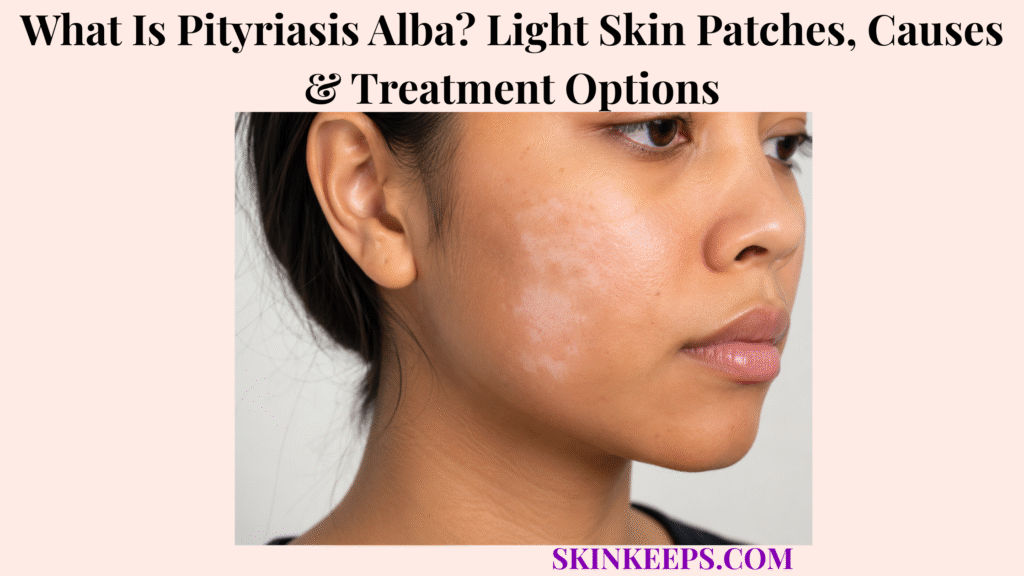
Pityriasis alba is a common benign inflammatory skin condition that causes poorly defined light patches, often with fine scale, especially on the faces of children and adolescents. The patches represent hypopigmentation rather than complete pigment loss, commonly have soft or blurred borders and may appear on the cheeks, chin, neck, shoulders or upper arms.
Pityriasis alba is associated with dry or atopy-prone skin and mild eczematous inflammation rather than fungal infection or melanocyte destruction. Most cases resolve spontaneously, while moisturizer and sun protection reduce dryness and contrast; the inflammation may settle quickly, but normal pigmentation can take months or occasionally longer to return.
This article is for educational purposes only and does not constitute medical advice, diagnosis, or treatment. A light skin patch that becomes sharply chalk-white, rapidly expands, develops significant itching or inflammation, has an unusual distribution, remains progressively changing, or is associated with hair whitening, altered sensation or another unexplained symptom should be evaluated by a licensed dermatologist or qualified healthcare professional because another pigmentary, fungal or inflammatory disorder may be responsible.
How Can You Recognize Pityriasis Alba?
Pityriasis alba usually appears as one or more poorly defined light patches with mild dryness or fine scale, most often on the cheeks and other facial areas.
What Do Pityriasis Alba Patches Look Like and Where Do They Appear?
Pityriasis alba produces round, oval or irregular hypopigmented patches with soft borders, most commonly on the cheeks and chin but sometimes on the neck, shoulders or upper arms.
The patches are lighter than surrounding skin rather than usually pure white. Fine scale can be visible during the active stage, while later lesions often become smooth as inflammation settles.
Does Pityriasis Alba Always Begin as a White Patch?
No; pityriasis alba can evolve from a faint pink or mildly inflamed scaly patch into a lighter scaly patch and finally a smooth residual area of hypopigmentation.
The typical sequence is faint pink/scaly plaque → light scaly patch → smooth pale patch → gradual normal colour return. Families often notice the pale stage only after the earlier inflammatory phase has become subtle or disappeared.
Does Pityriasis Alba Itch or Become More Visible in Certain Seasons?
Pityriasis alba is usually asymptomatic or only mildly itchy, while summer tanning can make pale patches more obvious and winter dryness can make scale more noticeable.
Strong itching is less typical. Greater summer contrast does not automatically mean that the disorder is progressing; surrounding skin may simply tan more than the hypopigmented patch.
Why Is Pityriasis Alba More Noticeable on Darker Skin?
Pityriasis alba often appears more prominent on darker skin because the reduction in pigment creates greater contrast with surrounding normally pigmented skin.
The surrounding skin may deepen with sun exposure while the affected area remains lighter, making the patch easier to see. Increased visibility should not be interpreted automatically as more severe inflammation.
Figure 1. Pityriasis alba can begin as a faint pink scaly patch, become a lighter scaly patch, leave a smooth pale area and then repigment gradually.
Why Does Pityriasis Alba Develop, and Who Is More Likely to Get It?
The exact cause of pityriasis alba is uncertain, but it is strongly associated with dry, atopy-prone skin and mild eczematous inflammation rather than melanocyte destruction.
How Are Dry Skin and Atopic Dermatitis Related to Pityriasis Alba?
Pityriasis alba commonly occurs in people with dry or atopy-prone skin, where impaired barrier function and low-grade inflammation can temporarily reduce visible pigmentation.
Atopic dermatitis is a useful related condition because pityriasis alba is often considered part of the eczema spectrum, but not every child with pityriasis alba has diagnosed atopic dermatitis.
Which Age Group Is Most Commonly Affected?
Pityriasis alba mainly affects children and adolescents, especially during the school-age years, although adults can occasionally develop it.
DermNet and StatPearls describe the condition as particularly common between about ages 3 and 16. This is a typical age pattern rather than an absolute diagnostic boundary.
Does Sun Exposure or Nutritional Deficiency Cause Pityriasis Alba?
Sun exposure mainly increases contrast rather than acting as a proven cause, and no specific vitamin or mineral deficiency has been established as the cause of pityriasis alba.
Tanning of surrounding skin makes the pale patch more visible. Studies examining trace elements or nutritional factors have not established a consistent deficiency mechanism, so supplements should not be presented as treatment unless an independent deficiency has been diagnosed.
Is Pityriasis Alba Contagious or Inherited?
No; pityriasis alba is neither contagious nor considered a hereditary skin disorder.
It does not spread from person to person and is not a fungal disease by default. Family members may share dry or atopic skin tendencies, but pityriasis alba itself is not treated as a simple inherited pigment disorder.
Figure 2. Pityriasis alba is inflammatory hypopigmentation: melanocytes are preserved, while low-grade inflammation and barrier dysfunction temporarily reduce visible pigment.
How Is Pityriasis Alba Distinguished and Diagnosed?
Pityriasis alba is usually diagnosed clinically from its poorly defined, incompletely pigmented, mildly scaly patches, with Wood’s lamp or fungal testing used only when another diagnosis remains plausible.
How Is Pityriasis Alba Different From Vitiligo?
Pityriasis alba usually causes soft-edged incomplete hypopigmentation with fine scale, while vitiligo produces more complete, sharply demarcated depigmentation.
Pityriasis alba is usually light rather than chalk-white and tends to fade spontaneously. Vitiligo can persist or progress and does not typically show the fine eczematous scale seen in active pityriasis alba.
How Is Pityriasis Alba Different From Tinea Versicolor or Other Post-Inflammatory Light Patches?
Tinea versicolor is a fungal disorder more often affecting the trunk and can be confirmed with mycology, while other post-inflammatory hypopigmentation usually follows a recognizable preceding rash or injury.
Pityriasis alba commonly favors the face. Tinea versicolor is associated with Malassezia and more often affects the trunk, shoulders or neck; fungal scraping or KOH can help when the morphology and site make that diagnosis plausible.
Post-inflammatory hypopigmentation generally matches the distribution of an earlier inflammatory eruption rather than the classic facial pityriasis-alba pattern.
How Is Pityriasis Alba Different From Nevus Depigmentosus?
Nevus depigmentosus is usually a congenital or early-life stable hypopigmented patch, while pityriasis alba develops through a mild inflammatory or scaly phase and gradually resolves.
A stable patch present from birth without a pink-to-scaly-to-pale evolution supports a congenital hypopigmented lesion more than pityriasis alba.
When Are Wood’s Lamp, Fungal Testing or Biopsy Needed?
Testing is usually unnecessary in typical pityriasis alba but can help when vitiligo, tinea or another persistent pigment disorder remains possible.
Wood’s lamp examination does not show the striking bright enhancement expected with complete depigmentation and can help define the patch. Fungal scraping or KOH is used only when tinea is plausible, while skin biopsy is rarely needed for atypical or persistent disease.
Biopsy findings are nonspecific and can show mild spongiotic dermatitis with reduced melanin rather than complete melanocyte loss. The broader category of hypopigmentation includes many other causes, so atypical lesions should be reassessed rather than forced into one diagnosis.
| Pattern | More Suggestive Of | Useful Distinction |
|---|---|---|
| Poorly defined pale facial patch ± fine scale | Pityriasis alba | Incomplete hypopigmentation; usually self-resolving |
| Sharply demarcated chalk-white patch | Vitiligo | More complete depigmentation; may persist or progress |
| Scaly trunk/neck patches with fungal evidence | Tinea versicolor | Fungal disease; confirm with mycology when needed |
| Stable congenital or very early patch | Nevus depigmentosus | No classic inflammatory/scaly evolution |
| Pale area matching a previous rash or injury | Post-inflammatory hypopigmentation | Distribution follows preceding inflammation |
How Is Pityriasis Alba Treated?
Most pityriasis alba needs only reassurance, moisturizer and sun protection, while mild anti-inflammatory treatment is reserved for active itchy or scaly lesions.
Does Every Case Need Medical Treatment?
No; asymptomatic pityriasis alba often requires no medical treatment because spontaneous repigmentation is expected.
The condition is benign and does not scar in ordinary cases. Treatment is therefore aimed at comfort, dryness and inflammation rather than forcing immediate pigment normalization.
How Do Moisturizers and Sunscreen Help?
Moisturizers reduce dryness and scale, while sunscreen limits tanning of surrounding skin and therefore reduces the visible contrast of pityriasis alba patches.
Emollients support the skin barrier and can make active scale less noticeable. Sunscreen provides routine ultraviolet protection and reduces contrast but does not directly repigment the lesion.
When Are Mild Topical Corticosteroids or Calcineurin Inhibitors Used?
Low-potency topical corticosteroids or selected steroid-sparing calcineurin inhibitors can be considered when pityriasis alba remains inflamed, itchy or noticeably scaly.
Mild topical corticosteroids can suppress active eczematous inflammation, while tacrolimus or pimecrolimus are selected steroid-sparing options. Facial safety matters, so strong or prolonged topical corticosteroid treatment should not be presented as routine.
Do Antifungals or Aggressive Pigment Treatments Help?
Antifungals do not treat ordinary pityriasis alba unless a separate fungal infection is diagnosed, and aggressive pigment-directed treatment is usually unnecessary because the condition resolves spontaneously.
Fine scale plus pale colour does not prove fungus. The central management principle remains reassurance + barrier care ± mild anti-inflammatory treatment rather than prolonged empiric antifungal or pigment-erasing therapy.
Figure 4. Reassurance, moisturizer and sun protection are enough for many cases; mild anti-inflammatory treatment is added only for active itch, scale or inflammation, while colour often normalizes later.
How Long Does Pityriasis Alba Take to Clear, and Can It Return?
Pityriasis alba usually fades gradually over months, and pigment recovery often continues long after dryness and scaling have already resolved.
How Long Does Pityriasis Alba Usually Last?
Most pityriasis alba patches improve over months to about a year, although some can take two or three years to regain normal colour completely.
These are approximate biological timelines rather than guarantees. StatPearls notes that most cases resolve within about a year, while some persist for several years before full repigmentation.
Why Can Scale Disappear Before the Light Patch?
The eczematous inflammation and dryness can settle relatively quickly, while normal melanogenesis and visible pigment recovery occur more slowly.
The expected sequence is active inflammation resolves → scale disappears → a smooth pale patch remains → pigment gradually returns. Persistent pallor by itself does not automatically mean active disease is still present.
Does Pityriasis Alba Leave Permanent White Skin or Scars?
Ordinary pityriasis alba typically resolves without scarring or permanent depigmentation.
A lesion that becomes completely chalk-white, expands progressively or remains morphologically atypical should prompt reconsideration of the diagnosis rather than being assumed to be unusually stubborn pityriasis alba.
Can Pityriasis Alba Return, and When Should a Patch Be Reassessed?
Pityriasis alba can recur when dry or eczematous skin flares again, but sharply white, expanding, intensely itchy, sensory-altered or persistently changing lesions should be reassessed.
Regular moisturizer, gentle skin care and sunscreen can reduce dryness and contrast. Reassessment is appropriate for progressive expansion, atypical sites, substantial persistent scale, intense itching, altered sensation or continued evolution that no longer fits the expected benign course.
What Should You Remember About Pityriasis Alba?
Pityriasis alba is a benign inflammatory hypopigmentation disorder that produces poorly defined light patches—often on the face of children—and usually resolves gradually without scarring.
- Pityriasis alba is a low-grade inflammatory or eczematous disorder.
- It causes hypopigmentation rather than complete depigmentation.
- Melanocytes remain present.
- Facial patches, especially on the cheeks, are common.
- Borders are usually soft or poorly defined.
- Fine scale can occur during the active stage.
- A faint pink patch can evolve into a light scaly patch and then a smooth pale patch.
- Itch is absent or mild in many cases.
- Darker skin and summer tanning can increase visual contrast.
- Dry or atopy-prone skin is commonly associated, but not every patient has atopic dermatitis.
- Children and adolescents are affected most often.
- Sun exposure is not established as the primary cause.
- No specific nutritional deficiency is established as the cause.
- Pityriasis alba is not contagious and is not considered hereditary.
- Vitiligo is more sharply and completely depigmented.
- Tinea versicolor requires fungal evidence rather than appearance alone.
- Diagnosis is usually clinical.
- Wood’s lamp, fungal testing and biopsy are selective.
- Many cases need only reassurance, moisturizer and sun protection.
- Mild anti-inflammatory therapy is reserved for active itch, scale or inflammation.
- Antifungals are not routine treatment.
- Pigment recovery often takes longer than scale recovery.
- Ordinary pityriasis alba resolves without scarring.
- Recurrence can occur.
- Atypical progressive patches deserve reassessment.
Recognize soft-edged hypopigmentation → Distinguish vitiligo/fungus → Reassure → Repair barrier → Treat mild inflammation if needed → Allow slow repigmentation → Reassess atypical persistence.
Frequently Asked Questions About Pityriasis Alba
The most important pityriasis-alba questions concern why the patches become lighter, how they differ from vitiligo, whether fungus or contagion is involved, which treatments help and how long pigment recovery takes.
What Causes Light Skin Patches in Pityriasis Alba?
Pityriasis alba is thought to result from mild inflammatory and barrier dysfunction that temporarily reduces pigment production without destroying melanocytes. Dry and atopy-prone skin are common associations, but the exact cause remains uncertain.
How Can You Tell Pityriasis Alba From Vitiligo?
Pityriasis alba usually has soft, poorly defined borders and fine scale, while vitiligo more often causes sharply defined, more completely white depigmented patches. Wood’s lamp examination can help when the clinical distinction remains uncertain.
Is Pityriasis Alba a Fungal Infection or Contagious?
No; pityriasis alba is not a fungal infection and does not spread from person to person. Fungal testing is reserved for cases where tinea versicolor or another fungal disorder is clinically plausible.
Which Treatments Help Pityriasis Alba Clear?
Most cases improve with reassurance, moisturizer and sun protection, while mild topical anti-inflammatory treatment can be used when active itching, dryness or scale is present. Low-potency corticosteroids or selected calcineurin inhibitors are options when clinically appropriate.
How Long Does It Take for Normal Skin Colour to Return?
Normal pigmentation usually returns gradually over months, although some patches can remain lighter for one to several years before completely resolving. Scale and inflammation often disappear before the remaining pale colour normalizes.
Which Sources Support This Pityriasis Alba Guidance?
DermNet — Pityriasis Alba — Primary source for the low-grade eczema classification, childhood predominance, lesion evolution, darker-skin contrast, seasonal visibility, atopic association, selective Wood’s lamp/fungal testing/biopsy, topical treatment options, recurrence and prolonged repigmentation.
StatPearls — Pityriasis Alba — Used for ill-defined facial hypopigmented patches, mild scale/pruritus, melanocyte-preservation context, spontaneous resolution, typical 3–16-year age range, moisturizer and low-potency topical corticosteroid management, and months-to-years recovery timing.
American Family Physician — Pigmentation Disorders: Diagnosis and Management — Used selectively for the differential diagnosis of hypopigmented disorders and conservative management framing.



