
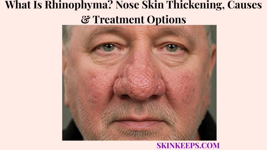
Rhinophyma is a chronic form of phymatous rosacea that progressively thickens and enlarges nasal skin through sebaceous-gland enlargement, connective-tissue overgrowth and fibrosis. Early changes may include enlarged pores, persistent redness and irregular texture, while more established disease can become nodular, lobulated or bulbous and distort the normal nasal contour.
Alcohol is not considered the direct cause of rhinophyma, despite the old and stigmatizing stereotype linking nasal enlargement with drinking. Diagnosis is usually clinical, while treatment separates active inflammatory rosacea from established fibrotic overgrowth: medicines may help selected inflammatory disease, whereas substantial structural tissue usually requires procedural reduction and can recur over time.
This article is for educational purposes only. Progressive nasal thickening, ulceration, persistent bleeding or a changing focal lesion should be professionally evaluated.
What Is Rhinophyma and How Does the Nose Change as It Progresses?
Rhinophyma is progressive phymatous overgrowth of nasal skin that can begin with subtle thickening and enlarged pores and advance to nodular, bulbous tissue that distorts the normal shape of the nose.
How Does Phymatous Rosacea Lead to Rhinophyma?
Rhinophyma appears to develop through chronic inflammatory, vascular, sebaceous and fibrotic changes associated with phymatous rosacea, although its exact biology is not completely established.
A useful cautious model is chronic inflammatory and vascular change → sebaceous-gland enlargement → connective-tissue overgrowth → fibrosis → progressive nasal thickening. This explains why rhinophyma is a structural process rather than simply persistent facial redness.
What Early Changes Can Rhinophyma Cause?
Early rhinophyma may cause increasing skin thickness, enlarged pores, oiliness, persistent redness, visible vessels and irregular nasal texture before obvious enlargement develops.
The early pattern is not identical in every person and does not progress at a predictable rate. Enlarged follicular openings and uneven texture may be more noticeable before the nose becomes visibly bulbous.
What Does Advanced Rhinophyma Look Like?
Advanced rhinophyma can produce bulbous, nodular or lobulated tissue with marked irregularity, asymmetry and distortion of the normal nasal contour.
The nasal tip, alae, sidewalls and lower nasal surface can be involved. Thickened tissue may contain prominent pores, visible superficial vessels and irregular lobules, but appearance alone should not be used to dismiss a distinct new ulcerated or bleeding focal lesion.
Can Rhinophyma Cause Functional Problems?
Severe rhinophyma can become more than a cosmetic problem because bulky tissue may narrow the external nasal openings and interfere with airflow.
Functional burden can coexist with substantial psychosocial impact from progressive facial change. Obstruction is not inevitable, but increasing tissue volume can become clinically important when it changes breathing or normal nasal anatomy.
Figure 1. Rhinophyma is best understood as progressive structural change: enlarged pores and subtle thickening can precede irregular lobulated tissue, bulbous enlargement and, in advanced disease, distortion that may affect airflow.
What Causes Rhinophyma and Which Factors Are Associated With It?
Rhinophyma is strongly associated with phymatous rosacea, but no single lifestyle factor explains every case and alcohol is not considered its direct cause.
Is Rosacea the Main Condition Associated With Rhinophyma?
Yes; rhinophyma is generally regarded as the nasal manifestation of phymatous rosacea, although only a subset of people with rosacea develop substantial tissue overgrowth.
The broader rosacea spectrum includes flushing, persistent facial erythema and inflammatory lesions, while rhinophyma specifically refers to progressive phymatous change of nasal tissue.
Why Does Rhinophyma Occur More Often in Men?
Rhinophyma occurs disproportionately in men, particularly later in adulthood, but the biological reason for this sex difference remains uncertain.
Hormonal influences and differences in sebaceous-gland biology have been proposed, but these should be treated as hypotheses rather than established explanations for why the condition is more common in men.
Does Drinking Alcohol Cause Rhinophyma?
No; alcohol is not established as the direct cause of rhinophyma, although it may trigger flushing in some people with rosacea.
The historic “alcoholic nose” stereotype is inaccurate and stigmatizing. Alcohol may aggravate flushing for some people, but a trigger that changes vascular symptoms is not the same thing as the underlying cause of progressive phymatous tissue overgrowth.
Can Rosacea Triggers Make Rhinophyma Worse?
Rosacea triggers may worsen flushing or inflammatory activity in some people, but avoiding triggers cannot be expected to reverse established fibrotic nasal overgrowth.
Trigger management may help symptom control, yet it is not a structural treatment. Once fibrosis and substantial tissue overgrowth are established, physical tissue reduction becomes more relevant than simply suppressing flushing.
Figure 2. Rhinophyma is linked to phymatous rosacea and progressive tissue overgrowth. Flushing triggers can aggravate rosacea symptoms, but they should not be confused with the direct cause of established fibrotic rhinophyma.
How Is Rhinophyma Diagnosed and Distinguished From Other Nose Growths?
Rhinophyma is usually diagnosed clinically from progressive nasal thickening, enlarged pores, vascular change and contour distortion, while atypical or focal lesions may require biopsy to exclude another diagnosis.
How Does a Dermatologist Diagnose Rhinophyma?
A dermatologist usually diagnoses rhinophyma from the medical history, rosacea background, progression pattern, tissue texture and characteristic nasal appearance.
The examination focuses on how the change developed, which nasal areas are involved, the degree of thickening and asymmetry, and whether the overall pattern is diffuse phymatous overgrowth rather than a separate focal tumor.
What Conditions Can Resemble Rhinophyma?
Benign nasal growths, inflammatory or granulomatous disorders, sebaceous proliferations and some skin cancers can resemble or coexist with rhinophyma.
The differential should remain focused on features that do not fit the expected diffuse phymatous pattern, especially a discrete lesion with a different surface, growth behavior or symptom profile.
Why Is Basal Cell Carcinoma an Important Differential?
Basal cell carcinoma can occur on the nose and may occasionally be hidden within irregular rhinophyma tissue, so a distinct ulcerated, bleeding or changing focal lesion should not automatically be attributed to rhinophyma.
A separate basal cell carcinoma evaluation becomes more relevant when one focal area behaves differently from the surrounding rhinophyma tissue.
When Is Biopsy or Histopathology Needed?
Biopsy or histologic examination is most useful when rhinophyma is atypical, markedly asymmetric, ulcerated, contains a suspicious focal lesion or when tissue has been surgically removed.
Typical rhinophyma is often a clinical diagnosis, so biopsy is not automatically required simply because phymatous thickening is present. Histology becomes more valuable when the pattern is unusual or when another lesion must be excluded.
Figure 3. Typical diffuse rhinophyma is often diagnosed clinically, while a distinct bleeding, ulcerated, changing or unusually asymmetric focal lesion shifts the task toward histologic exclusion of another diagnosis.
How Is Rhinophyma Treated?
Rhinophyma treatment depends on how much active inflammation versus established structural overgrowth is present, with medicines helping selected inflammatory disease and procedures usually needed to reduce significant fibrotic tissue.
Can Medicines Treat Early or Active Rhinophyma?
Medical treatment may help associated rosacea inflammation or selected earlier rhinophyma, but it usually cannot remove substantial established fibrotic tissue.
Rosacea-directed treatment and selected isotretinoin use can be considered by clinicians when active inflammation or sebaceous activity remains important. The key limitation is structural: medicine does not reliably erase major lobulated fibrosis once that tissue has developed.
How Are Surgical Excision and Dermabrasion Used?
Surgical techniques and dermabrasion physically reduce excess phymatous tissue so the nasal contour can be reshaped while preserving appropriate underlying structures.
The principle is excess tissue reduction → contour restoration → bleeding control → healing and re-epithelialization. Technique selection is procedural and clinician-dependent rather than something that can be reduced to a single universally preferred method.
How Does CO₂ Laser Treatment Work?
Ablative CO₂ laser can precisely vaporize or remove hypertrophic rhinophyma tissue while also helping control bleeding during treatment.
Its role is tissue ablation and contouring, with hemostasis as an important procedural advantage. That does not make CO₂ laser automatically superior to excision, electrosurgery or other methods in every case.
Are Electrosurgery or Other Laser Techniques Used?
Yes; electrosurgery, radiofrequency, Er:YAG laser and combined techniques may be selected according to tissue thickness, bleeding control, healing considerations and operator experience.
Rhinophyma treatment is individualized because lesion size, anatomy, fibrosis depth, equipment availability and clinician expertise all influence which approach is most appropriate.
Figure 4. Rhinophyma treatment is selected by inflammation versus structural fibrosis: medicines can support selected active disease, while established overgrowth is generally managed with physical tissue-reduction techniques chosen for anatomy, healing needs and clinician expertise.
How Does Rhinophyma Heal After Treatment, Can It Return, and When Should It Be Reassessed?
After rhinophyma treatment the nasal surface generally heals through re-epithelialization, but redness, pigment change, scarring or recurrence can occur, and new suspicious focal changes still require evaluation.
What Happens During Healing After Rhinophyma Removal?
After tissue reduction, the treated surface gradually re-epithelializes while wound care protects the healing nasal skin.
Healing time and aftercare vary by procedure depth and technique, so wound care is clinician-directed rather than one universal dressing protocol. The goal is controlled resurfacing while preserving function and minimizing avoidable trauma.
Can Treatment Leave Scarring or Pigment Changes?
Yes; prolonged redness, pigment alteration, textural irregularity or scarring can occur after rhinophyma procedures, with risk depending on treatment depth, technique and individual healing.
These outcomes are possibilities rather than certainties, and cosmetic result depends on both the original disease severity and how the tissue heals after reduction.
Can Rhinophyma Grow Back After Treatment?
Yes; recurrence is possible because removing excess tissue does not necessarily eliminate the underlying susceptibility to phymatous rosacea.
Long-term follow-up and ongoing rosacea management remain useful after procedural treatment. No treatment should be described as guaranteeing permanent removal in every person.
When Should Rhinophyma Be Evaluated or Reassessed?
Dermatology assessment is appropriate for progressive nasal enlargement, increasing distortion, airflow obstruction, persistent bleeding, ulceration, marked asymmetry or a new focal lesion that changes within rhinophyma tissue.
Reassessment is also reasonable when recurrent crusting, new nodular growth, functional difficulty or psychosocial burden becomes important. Managing active rosacea may help inflammatory disease, but it should not be presented as guaranteed prevention or reversal of established rhinophyma.
What Should You Remember About Rhinophyma?
Rhinophyma is a benign progressive form of phymatous rosacea that structurally enlarges and distorts nasal skin and often requires procedural tissue reduction once substantial fibrosis is established.
- Rhinophyma is associated with phymatous rosacea.
- It causes progressive nasal tissue overgrowth.
- Fibrosis is an important structural component.
- Sebaceous glands and follicular openings can become enlarged.
- Early disease may begin with subtle thickening and irregular texture.
- Advanced disease can become bulbous, nodular or lobulated.
- The nasal tip, alae and sidewalls can be affected.
- Severe tissue overgrowth can distort the nose and sometimes affect airflow.
- Rhinophyma is benign and non-contagious.
- Alcohol is not established as its direct cause.
- The condition is more common in men, but the reason is not fully established.
- Diagnosis is usually clinical.
- Some benign and malignant lesions can mimic focal changes.
- Basal cell carcinoma must sometimes be excluded from a suspicious focal lesion.
- Biopsy is selective rather than universal.
- Medical treatment can help selected inflammatory disease.
- Substantial established fibrosis usually requires physical tissue reduction.
- Surgical reduction, dermabrasion, electrosurgery and laser techniques are options.
- No single procedure is universally best.
- Healing occurs through re-epithelialization after tissue reduction.
- Redness, pigment change, textural change or scarring can occur.
- Recurrence is possible.
- Rosacea treatment does not guarantee prevention or reversal of established rhinophyma.
- Persistent bleeding, ulceration or a changing focal lesion deserves reassessment.
Phymatous rosacea → progressive tissue overgrowth → confirm rhinophyma → distinguish suspicious lesions → treat inflammation versus structural tissue appropriately → monitor healing and recurrence.
Frequently Asked Questions About Rhinophyma
The main rhinophyma questions concern alcohol, its relationship to rosacea, spontaneous resolution, recurrence and possible nasal obstruction.
Is Rhinophyma Caused by Drinking Alcohol?
No; alcohol is not established as the direct cause of rhinophyma, although it may trigger flushing in some people with rosacea.
Is Rhinophyma a Type of Rosacea?
Yes; rhinophyma is generally regarded as a nasal manifestation of phymatous rosacea.
Can Rhinophyma Go Away Without Treatment?
Established fibrotic rhinophyma generally does not simply disappear on its own, and substantial tissue overgrowth usually requires procedural reduction.
Can Rhinophyma Grow Back After Surgery or Laser Treatment?
Yes; recurrence can occur because treatment removes excess tissue but does not necessarily eliminate the underlying tendency toward phymatous rosacea.
Can Rhinophyma Make It Difficult to Breathe Through the Nose?
Yes; very advanced tissue enlargement can narrow the external nasal openings and interfere with airflow in some people.
Which Sources Support This Rhinophyma Guidance?
DermNet — Rhinophyma — Used for definition, phymatous rosacea association, enlarged sebaceous glands and pores, clinical progression, male predominance, alcohol misconception, treatment categories and basal-cell-carcinoma context.
DermNet — Rosacea — Used for the broader phymatous rosacea relationship and the distinction between rosacea features and rhinophyma-specific structural overgrowth.
Procedural Management of Rhinophyma: A Comprehensive Review — Used for excision, dermabrasion, electrosurgery, CO₂ laser, Er:YAG and the principle that procedural choice depends on technique-specific tradeoffs.
Rhinophyma: A Treatment Review — Used for treatment selection, functional impact and the distinction between medical and structural management.
Rhinophyma: A Systematic Review and Meta-Analysis — Used for contemporary epidemiologic and treatment-outcome context while avoiding claims that one technique is universally superior.



