
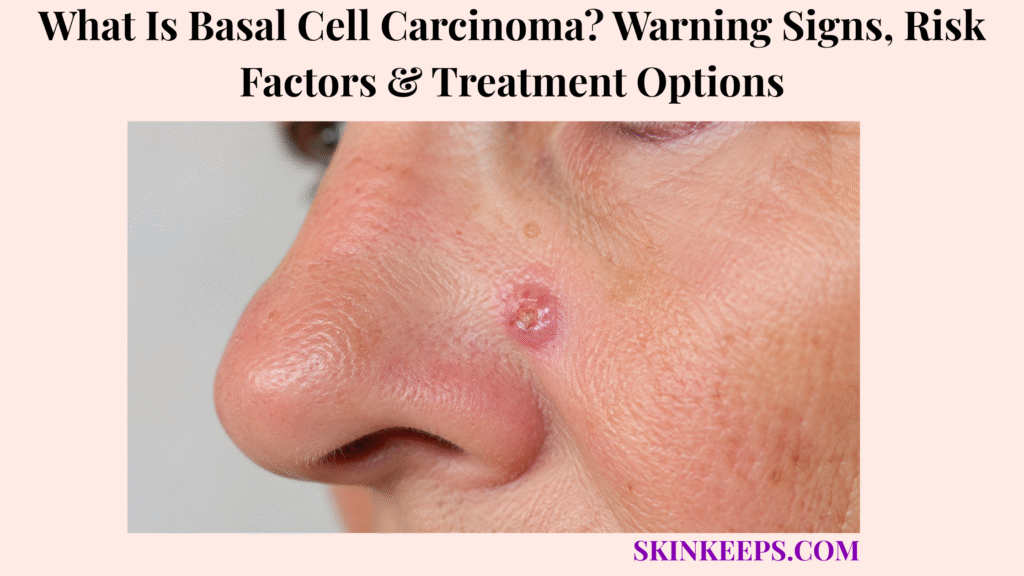
Basal cell carcinoma is a type of skin cancer that often grows slowly but can invade nearby skin and tissue if it is not diagnosed and treated. It is often shortened to BCC.
This page explains warning signs, common locations, risk factors, diagnosis, biopsy, treatment options, follow-up, prevention, mistakes, and when to seek care. BCC can look like a pimple, sore, scaly patch, scar, shiny bump, or pigmented lesion, so persistent or changing spots need professional evaluation.
What Is Basal Cell Carcinoma and How Does It Start?
Basal cell carcinoma is a type of skin cancer that begins in basal cells of the epidermis and often develops after ultraviolet light damages skin-cell DNA.
The epidermis is the outer skin layer. Basal cells sit near the bottom of that layer and help produce new skin cells. When DNA damage and growth-control errors accumulate, abnormal keratinocyte growth can create a persistent cancerous lesion.
BCC is a keratinocyte cancer and a nonmelanoma skin cancer. It is often slow-growing, but it is still cancer and should not be treated as a cosmetic mark.
Why Is Basal Cell Carcinoma Common but Still Serious?
Basal cell carcinoma is common and often slow-growing, but it is still serious because untreated tumors can grow deeper and damage nearby skin, cartilage, nerves, or other tissue.
BCC rarely spreads to distant body parts compared with some other cancers, but “rarely spreads” does not mean safe to ignore. Local invasion can create scarring, disfigurement, tissue loss, and complicated treatment, especially on the nose, eyelids, ears, lips, scalp, hands, and genitals.
Early diagnosis usually makes treatment simpler. Delayed care can make the final repair larger than the visible spot first suggested.
Why Is Basal Cell Carcinoma Not Just Sun Damage?
Basal cell carcinoma is not just sun damage because the lesion already contains cancer cells that require diagnosis and treatment, not sunscreen alone.
UV exposure increases future risk and can damage skin-cell DNA over years. Once a sore bleeds, crusts, heals, and returns, or a shiny bump, scaly patch, scar-like area, or pigmented lesion persists, the priority is evaluation and biopsy when suspicious.
Sun protection helps reduce future UV damage. It does not remove, destroy, or control an existing BCC.
Practical rule: A spot that bleeds, crusts, heals, returns, or keeps growing should not be treated for months as a pimple, scratch, eczema patch, or dry area.
What Basal Cell Carcinoma Warning Signs Can Appear on Skin?
Basal cell carcinoma warning signs can include a shiny or pearly bump, non-healing sore, bleeding or crusting spot, flat scaly patch, scar-like area, pink or red growth, or darker pigmented lesion.
BCC does not have one single look. It can be skin-colored, pink, red, brown, black, blue, gray, glossy, waxy, scaly, or scar-like depending on subtype, body site, skin tone, and how long it has been present.
What Does Nodular Basal Cell Carcinoma Look Like?
Nodular basal cell carcinoma often looks like a shiny, pearly, translucent, skin-colored, pink, red, brown, or darker bump that may bleed, crust, or develop a central ulcer.
Tiny visible blood vessels may appear on the surface. The bump may have a rolled border or a central depression. It commonly appears on the face, head, or neck.
In brown and Black skin, BCC may appear brown, darker, or glossy black rather than pink or pearly, so color alone should never reassure the reader.
What Does Superficial Basal Cell Carcinoma Look Like?
Superficial basal cell carcinoma can look like a flat pink, red, brown, or scaly patch that grows slowly and may be mistaken for eczema, psoriasis, or irritation.
This pattern may appear on the trunk, shoulders, arms, or legs. A single scaly patch that keeps returning, bleeding, crusting, or failing routine treatment deserves evaluation instead of repeated steroid or moisturizer use.
What Does Morpheaform or Scar-Like Basal Cell Carcinoma Look Like?
Morpheaform basal cell carcinoma can look like a waxy, pale, firm, scar-like area with poorly defined borders, sometimes without a history of injury.
Because the edges may be hard to see, treatment planning can be more careful than the surface mark suggests. A scar-like patch that appears without a clear injury or keeps enlarging should not be dismissed.
What Does Pigmented Basal Cell Carcinoma Look Like?
Pigmented basal cell carcinoma can look brown, black, blue, gray, or glossy darker, which means it can be confused with melanoma, mole changes, or seborrheic keratosis.
Dermoscopy and biopsy may be needed because pigment does not tell the full diagnosis. A dark changing lesion, a glossy black bump, or a pigmented lesion with bleeding, crusting, or a rolled border should be checked promptly.
| BCC Sign | What It May Look Like | Why It Matters |
|---|---|---|
| Pearly bump | Shiny, translucent, skin-colored, pink, brown, or glossy black bump | Classic nodular clue |
| Non-healing sore | Sore that persists or returns | Needs evaluation |
| Bleeding/crusting spot | Scabs, bleeds, heals, then returns | Common delay pattern |
| Visible vessels | Tiny surface blood vessels | Supports BCC suspicion |
| Scaly patch | Flat pink, red, brown, or darker patch | Can mimic eczema or psoriasis |
| Scar-like area | Waxy, firm, pale, ill-defined patch | Can hide deeper spread |
| Pigmented lesion | Brown, black, blue, gray, or darker lesion | Can mimic melanoma or benign growths |
Where Does Basal Cell Carcinoma Usually Appear?
Basal cell carcinoma most often appears on sun-exposed areas such as the face, nose, ears, scalp, neck, shoulders, arms, and hands, but it can also occur in less sun-exposed areas.
Common sites include the nose, eyelids, cheeks, ears, lips or skin around the mouth, scalp, neck, shoulders, chest, back, arms, and hands. BCC on a thinning or bald scalp can be harder to see during routine self-checks.
Protected areas such as the genitals are less common sites, but a non-healing sore or changing growth should not be dismissed because it is not sun-exposed. Location matters because treatment must preserve function, tissue, and appearance whenever possible.
| Location | Common Warning Sign | Treatment-Planning Concern |
|---|---|---|
| Nose | Pearly bump, bleeding sore, ulcer | Tissue-sparing and cosmetic/function risk |
| Eyelid | Shiny bump, crusting, non-healing lesion | Eye protection and specialist planning |
| Ear | Crusting, sore, scaly or bleeding spot | Cartilage and recurrence risk |
| Lip / around mouth | Persistent sore or shiny growth | Function and cosmetic planning |
| Scalp | Bleeding or crusting spot, especially on thinning scalp | Harder self-monitoring |
| Neck / shoulders | Scaly patch or pearly bump | Sun-exposed distribution |
| Trunk | Superficial scaly patch | Can mimic eczema or psoriasis |
| Hands / arms | Non-healing sun-damaged lesion | Functional area and sun exposure |
| Protected areas | Unusual sore or growth | Do not dismiss because it is not sun-exposed |
What Risk Factors Increase Basal Cell Carcinoma Risk?
Basal cell carcinoma risk increases with ultraviolet exposure, tanning beds, sunburn history, fair complexion traits, older age, prior skin cancer, immune suppression, radiation exposure, arsenic exposure, and certain inherited syndromes.
Risk factors help decide who needs a lower threshold for checking lesions. They do not mean people without obvious risk factors can ignore a suspicious spot.
How Does UV Exposure Increase Basal Cell Carcinoma Risk?
Ultraviolet exposure increases basal cell carcinoma risk by damaging skin-cell DNA over time, especially with chronic sun exposure, intense intermittent exposure, sunburns, and tanning beds.
Outdoor work, outdoor recreation, high-UV environments, and tanning lamps can increase cumulative damage. UV damage may build for years before a visible lesion appears.
Which Personal Factors Can Increase Basal Cell Carcinoma Risk?
Personal factors that can increase basal cell carcinoma risk include fair skin that burns easily, light-colored eyes, blond or red hair, freckles, older age, prior BCC, and personal or family history of skin cancer.
Darker skin tones have lower incidence, but BCC can still occur and may be diagnosed later if suspicious spots are dismissed. Prior BCC also raises the need to watch for new primary tumors over time.
Which Medical or Environmental Factors Increase Basal Cell Carcinoma Risk?
Medical and environmental BCC risk factors include immune suppression, organ transplant history, prior radiation therapy, arsenic exposure, and inherited syndromes that increase skin-cancer susceptibility.
Basal cell nevus syndrome, also called Gorlin syndrome, and xeroderma pigmentosum can increase risk of multiple or early skin cancers. These are specialist-surveillance situations, not routine sunspot situations.
| Risk Factor | Why It Matters | Prevention / Evaluation Action |
|---|---|---|
| Chronic UV exposure | Accumulated DNA damage | Sun protection and skin checks |
| Tanning beds | Artificial UV exposure | Avoid tanning beds |
| Sunburn history | Marker of UV injury | Watch damaged areas |
| Fair complexion traits | Higher UV sensitivity | Lower threshold for checking spots |
| Darker skin tones | Lower incidence but possible delayed diagnosis | Do not dismiss suspicious lesions |
| Prior BCC | New primary tumors can occur | Follow-up exams |
| Immune suppression | Higher skin-cancer risk | Earlier specialist review |
| Radiation / arsenic | Environmental carcinogen context | Share exposure history |
| Inherited syndromes | Multiple or early tumors possible | Specialist surveillance |
How Is Basal Cell Carcinoma Different From Other Skin Spots?
Basal cell carcinoma can mimic pimples, eczema, psoriasis, scars, moles, seborrheic keratoses, squamous cell carcinoma, or melanoma, so persistent or changing lesions should be checked rather than treated as routine irritation.
Behavior matters. A short-lived bump that resolves is different from a lesion that bleeds, crusts, heals, returns, grows, ulcerates, or resists routine care.
How Is Basal Cell Carcinoma Different From Acne or a Pimple?
Basal cell carcinoma is different from acne because pimples usually improve, while BCC may persist, bleed, crust, ulcerate, or return in the same spot.
A “pimple” on the nose, cheek, eyelid, ear, lip area, or scalp that does not heal after weeks or keeps bleeding should be checked. Not every face bump is BCC, but persistence changes the safety level.
How Is Basal Cell Carcinoma Different From Eczema or Psoriasis?
Basal cell carcinoma can look like eczema or psoriasis when it appears as a persistent scaly patch, but a single patch that does not respond or keeps recurring should be checked.
Superficial BCC can look rash-like. Atopic dermatitis and psoriasis often have broader, recurring inflammatory patterns, while a single stubborn scaly patch may need biopsy consideration.
How Is Basal Cell Carcinoma Different From Melanoma or Squamous Cell Carcinoma?
Basal cell carcinoma is different from melanoma and squamous cell carcinoma, but pigmented BCC and crusted or scaly BCC can overlap visually, so biopsy is the deciding step when a lesion is suspicious.
Melanoma often raises concern through changing pigment, asymmetry, border irregularity, multiple colors, or evolution. Squamous cell carcinoma may look thicker, scaly, crusted, tender, painful, or faster-growing. A clinician should decide when biopsy is needed.
How Is Basal Cell Carcinoma Different From Actinic Keratosis or Seborrheic Keratosis?
Basal cell carcinoma is different from actinic keratosis and seborrheic keratosis because BCC is already skin cancer, while those lesions have different risk and treatment pathways.
Actinic keratosis is a sun-damaged precancer-type lesion mainly tied to the squamous-cell pathway. Seborrheic keratosis is usually a benign waxy or stuck-on growth, but pigmented or irritated lesions may still need dermoscopy or biopsy if uncertain.
| Condition | Common Clue | Why Confusion Happens | Best Next Step |
|---|---|---|---|
| BCC | Pearly bump, non-healing sore, bleeding/crusting, scar-like or scaly patch | Many appearances | Dermatology exam and biopsy if suspicious |
| Acne | Pimple improves or changes over time | Face bumps overlap | Check persistent non-healing bumps |
| Eczema / psoriasis | Itchy or scaly inflammatory plaques | Superficial BCC can look rash-like | Check single persistent patch |
| Actinic keratosis | Rough sun-damaged scaly spot | UV-damaged skin context overlaps | Dermatology evaluation if persistent/changing |
| Seborrheic keratosis | Waxy “stuck-on” growth | Pigmented BCC may mimic benign growth | Dermoscopy/biopsy if uncertain |
| SCC | Scaly, crusted, tender, growing lesion | Both are keratinocyte cancers | Biopsy suspicious lesion |
| Melanoma | Changing pigmented lesion | Pigmented BCC can look dark | Prompt evaluation if changing/pigmented |
How Is Basal Cell Carcinoma Diagnosed or Checked?
Basal cell carcinoma is usually diagnosed by skin examination and biopsy, where a sample of the suspicious lesion is examined to confirm the cancer type and guide treatment.
Photos can help show change over time, but photo appearance alone should not be the diagnostic standard. A dermatologist may use close inspection, dermoscopy, and biopsy when the lesion is suspicious.
What Does a Dermatologist Check With Suspected Basal Cell Carcinoma?
A dermatologist checks a suspected basal cell carcinoma by reviewing lesion size, location, borders, color, bleeding, crusting, ulceration, tiny blood vessels, scar-like texture, and growth history.
Prior skin cancer history, immune suppression, transplant history, radiation exposure, and high-risk locations also matter. High-risk sites include the eyelid, nose, ear, lip, scalp, hand, genital area, and other functionally or cosmetically sensitive sites.
Why Does Biopsy Matter Before Basal Cell Carcinoma Treatment?
Biopsy matters before basal cell carcinoma treatment because it confirms whether the lesion is cancer, identifies key subtype clues, and helps choose the safest treatment.
Biopsy can help separate BCC from melanoma, squamous cell carcinoma, infection, scar tissue, and benign growths. It also helps guide excision, Mohs surgery, topical treatment, radiation, or advanced treatment decisions.
Suspicious lesions should not be frozen, burned, scraped, bleached, or treated with long-term steroid cream at home before diagnosis.
- Suspicious lesion is examined.
- Dermoscopy may be used when helpful.
- Biopsy samples the lesion.
- Histology confirms the cancer type.
- Subtype and risk clues are reviewed.
- Size, depth, location, borders, recurrence, and patient factors guide treatment.
What Treatment Options Are Used for Basal Cell Carcinoma?
Basal cell carcinoma treatment depends on tumor size, depth, subtype, location, borders, recurrence status, patient health, and cosmetic or functional risk.
The goal is to remove, destroy, or control the cancer cells while preserving healthy tissue and function as much as possible. Treatment should be chosen after diagnosis and risk assessment.
When Is Surgical Excision Used for Basal Cell Carcinoma?
Surgical excision is commonly used for many basal cell carcinomas because it removes the tumor with a margin of normal-looking skin that can be checked by pathology.
Excision may be suitable for many low-risk or well-defined lesions. Pathology review helps assess whether margins are clear. Scar planning and repair choice depend on location and size.
When Is Mohs Surgery Used for Basal Cell Carcinoma?
Mohs surgery is often considered for basal cell carcinoma in high-risk locations, recurrent tumors, aggressive subtypes, poorly defined borders, or areas where saving healthy tissue is especially important.
Mohs checks tissue layer by layer while preserving as much normal tissue as possible. It may be especially useful on the nose, eyelids, ears, lips, face, hands, genitals, and other sensitive sites. Not every BCC needs Mohs.
When Are Curettage, Electrodesiccation, Cryotherapy, or Topical Treatments Used?
Curettage, electrodesiccation, cryotherapy, or topical treatments may be used only for selected low-risk BCCs when the tumor type, depth, location, and patient factors make them appropriate.
Topical imiquimod or topical 5-fluorouracil may be considered mainly for selected superficial BCCs under dermatologist guidance. These are not appropriate for deep, aggressive, recurrent, high-risk, or unclear lesions.
Nonsurgical approaches can have lower cure rates than surgery, so selection matters.
When Are Radiation or Advanced Systemic Treatments Considered?
Radiation or advanced systemic treatments may be considered when surgery is not suitable, disease is complex, cancer remains after surgery, or BCC is locally advanced or metastatic.
Hedgehog pathway inhibitors such as vismodegib or sonidegib may be used in selected advanced cases. Immunotherapy such as cemiplimab may be considered in selected advanced settings. These are specialist-led options, not routine treatment for ordinary small BCCs.
| BCC Type / Risk | Likely Treatment Direction | Goal | Key Caution |
|---|---|---|---|
| Low-risk, well-defined BCC | Surgical excision or selected local treatment | Remove or destroy cancer cells | Must be confirmed and risk-assessed |
| Facial/high-risk location | Mohs or tissue-sparing specialist plan | Clear margins while preserving function/cosmesis | Not every face lesion automatically gets same treatment |
| Superficial low-risk BCC | Selected topical, destructive, or surgical options | Treat superficial cancer | Not for deep or aggressive lesions |
| Recurrent BCC | Mohs or specialist-led treatment | Reduce recurrence risk | Prior treatment changes planning |
| Aggressive/ill-defined subtype | Mohs or surgery specialist planning | Define and clear margins | Borders may extend beyond visible lesion |
| Surgery not suitable | Radiation or nonsurgical treatment in selected cases | Control cancer when surgery unsuitable | Cure rates and side effects differ |
| Locally advanced/metastatic BCC | Hedgehog inhibitor, immunotherapy, oncology-led plan | Shrink or control advanced disease | Specialist monitoring required |
How Does Basal Cell Carcinoma Follow-Up Reduce Recurrence Risk?
Follow-up after basal cell carcinoma matters because a person who has had one BCC can develop recurrence at a treated site or another primary skin cancer later.
Follow-up checks the scar, surrounding skin, and other sun-damaged areas. The schedule depends on tumor risk, treatment type, margins, prior skin cancer history, immune status, and dermatologist advice.
AAD guideline highlights recommend annual checks after BCC diagnosis for recurrence or another new primary skin cancer. Some people need more frequent checks based on risk.
How Can Sun Protection Lower Future Basal Cell Carcinoma Risk?
Sun protection cannot treat an existing basal cell carcinoma, but it can reduce additional ultraviolet damage and help lower future sun-related skin-cancer risk.
Use shade, protective clothing, a wide-brim hat, UV-protective sunglasses, and broad-spectrum sunscreen on exposed skin. Reapply sunscreen during outdoor exposure according to the product label and activity level.
Pay attention to ears, nose, lips, scalp, neck, and hands because these areas receive frequent UV exposure. Avoid tanning beds and tanning lamps completely, especially after a prior BCC.
- Seek shade when UV is strong.
- Wear protective clothing and a wide-brim hat.
- Protect ears, nose, lips, scalp, neck, and hands.
- Use broad-spectrum sunscreen on exposed skin.
- Reapply sunscreen during outdoor exposure.
- Avoid tanning beds and tanning lamps.
- Check previously treated areas and new sun-exposed spots.
- Keep dermatologist follow-up after BCC treatment.
What Basal Cell Carcinoma Mistakes Should You Avoid?
The biggest basal cell carcinoma mistake is repeatedly treating a non-healing, bleeding, crusting, pearly, or scar-like skin spot as acne, eczema, a scratch, or dry skin without getting it checked.
Do not pick, scrape, burn, freeze, bleach, or try to remove a suspicious lesion at home. Do not assume a painless lesion is safe. Do not continue steroid cream for months on a single persistent scaly patch without diagnosis.
Delaying evaluation is especially risky for lesions near the eyes, nose, ears, lips, scalp, hands, genitals, or other sites where tissue preservation matters.
| Mistake | Why It Is Risky | Better Action |
|---|---|---|
| Calling it a pimple for months | BCC can mimic acne | Check non-healing bumps |
| Moisturizer-only care | A cancer lesion will not be cured by moisturizer | Seek evaluation for persistent sores |
| Repeated steroid use | May delay diagnosis of a single persistent patch | Biopsy suspicious lesions |
| Ignoring bleeding/crusting | Common warning pattern | Dermatology review |
| DIY freezing/burning | May miss cancer margins and delay care | Get diagnosis first |
| Ignoring high-risk sites | Nose, eyelids, ears, lips, hands, genitals need careful planning | Prompt specialist review |
| Skipping follow-up | New BCCs or recurrence can occur | Schedule skin checks |
When Should Basal Cell Carcinoma Be Checked Promptly?
A skin spot should be checked promptly when it does not heal, bleeds, crusts, grows, forms an ulcer, looks pearly or shiny, develops visible blood vessels, resembles a scar without injury, or changes over time.
A suspicious spot should not be watched for months when it repeatedly scabs, bleeds, returns, grows, or sits on a high-risk area.
Which Basal Cell Carcinoma Warning Signs Need Faster Evaluation?
Faster evaluation is needed for a non-healing sore, recurring scab, recurrent bleeding, pearly or shiny bump, scar-like patch, pigmented changing lesion, or suspicious spot near a high-risk location.
High-risk locations include the eye area, nose, ear, lip, scalp, hand, foot, genital area, and sites where function or appearance could be affected. Immunosuppressed people should have a lower threshold for evaluation.
What Should You Bring to a Basal Cell Carcinoma Appointment?
A helpful basal cell carcinoma appointment starts with photos, timeline, bleeding or crusting history, growth changes, sun exposure history, prior skin cancer history, immune status, medicines, and treatments already tried.
Bring the date the spot first appeared, whether it hurts or itches, whether it heals and returns, and whether it affects eyelid movement, nose shape, lip movement, hand function, or another sensitive site.
Seek prompt care for a spot that is:
What Should You Remember About Basal Cell Carcinoma?
The most important thing to remember about basal cell carcinoma is that it is usually treatable, but it is still skin cancer and needs biopsy-based diagnosis, appropriate treatment, and follow-up.
Slow growth should not create delay. Persistent warning signs, sensitive locations, pigmented changes, bleeding, crusting, ulceration, and treatment-resistant patches should be checked.
Frequently Asked Questions About Basal Cell Carcinoma
Is basal cell carcinoma cancer?
Yes. Basal cell carcinoma is a type of skin cancer. It often grows slowly and rarely spreads, but it can grow deeper and damage nearby tissue if untreated.
What does basal cell carcinoma look like?
Basal cell carcinoma can look like pearly or shiny bumps, non-healing sores, bleeding or crusting spots, flat scaly patches, scar-like areas, visible blood vessels, and pigmented lesions.
Does basal cell carcinoma spread?
BCC rarely spreads to distant body parts compared with many other cancers, but it can grow locally and damage nearby tissue if untreated.
What causes basal cell carcinoma?
Risk is linked with UV exposure from sunlight or tanning beds, DNA damage, fair complexion traits, sunburn history, age, prior skin cancer, immune suppression, radiation exposure, arsenic exposure, and inherited syndromes.
How is basal cell carcinoma diagnosed?
Basal cell carcinoma is diagnosed with skin examination, dermoscopy when useful, and biopsy to confirm the cancer type and guide treatment.
What is the best treatment for basal cell carcinoma?
There is no one best treatment for every BCC. Treatment depends on tumor size, depth, subtype, location, borders, recurrence status, patient health, and cosmetic or functional risk.
When is Mohs surgery used for basal cell carcinoma?
Mohs surgery is often used for high-risk locations, recurrent tumors, aggressive or poorly defined subtypes, and areas where tissue preservation is important, such as the nose, eyelids, ears, lips, face, hands, or genitals.
Can topical creams treat basal cell carcinoma?
Some topical medicines may be used for selected superficial, low-risk BCCs under dermatologist guidance, but they are not appropriate for all BCCs and should not replace biopsy-based diagnosis.
When should a suspicious spot be checked?
A suspicious spot should be checked when it is non-healing, repeatedly bleeding or crusting, pearly, shiny, scar-like, changing, pigmented, in a high-risk location, treatment-resistant, or occurring in an immunosuppressed person.
Sources & Evidence About Basal Cell Carcinoma
American Academy of Dermatology — Basal Cell Carcinoma was used for definition, slow growth and rare spread balanced with deep-growth warning, common symptoms, diagnosis, excision, Mohs surgery, cryosurgery, radiation, advanced medicines, and early-treatment framing.
American Academy of Dermatology — Basal Cell Carcinoma Clinical Guideline was used for risk stratification, optimal biopsy principles, surgery as the cornerstone of treatment, nonsurgical lower-cure-rate caution, early complete treatment, and annual checks after BCC diagnosis.
DermNet — Basal Cell Carcinoma was used for BCC as common locally invasive keratinocyte cancer, most common skin cancer framing, subtypes, multiple primary tumors, pigmented and nonpigmented presentations, risk factors, treatment categories, and follow-up.
Mayo Clinic — Basal Cell Carcinoma Symptoms and Causes was used for sun-exposed locations, head and neck tendency, protected-site possibility, skin-tone variation, lesion types, sore that does not heal, UV/DNA explanation, and rare spread with local invasion risk.
Mayo Clinic — Basal Cell Carcinoma Diagnosis and Treatment was used for skin biopsy, biopsy sample testing, treatment depending on type, location, size, recurrence, follow-up ability, and surgery-first treatment framing.
National Cancer Institute — Skin Cancer Treatment PDQ was used for BCC and SCC as common nonmelanoma skin cancers, BCC burden context, skin-cancer signs, risk factors, and treatment category framing.
StatPearls / NCBI Bookshelf — Basal Cell Carcinoma was used for chronic UV exposure as a principal risk factor, DNA damage, hedgehog-pathway context, rare metastasis, local tissue destruction and disfigurement warning, and the broad management range.
Educational Disclaimer: This SkinKeeps article is for educational purposes only and does not diagnose or replace medical care. A non-healing, bleeding, crusting, ulcerated, pearly, shiny, scar-like, pigmented, changing, painful, high-risk-location, or treatment-resistant skin spot should be checked by a qualified healthcare professional or dermatologist.



