
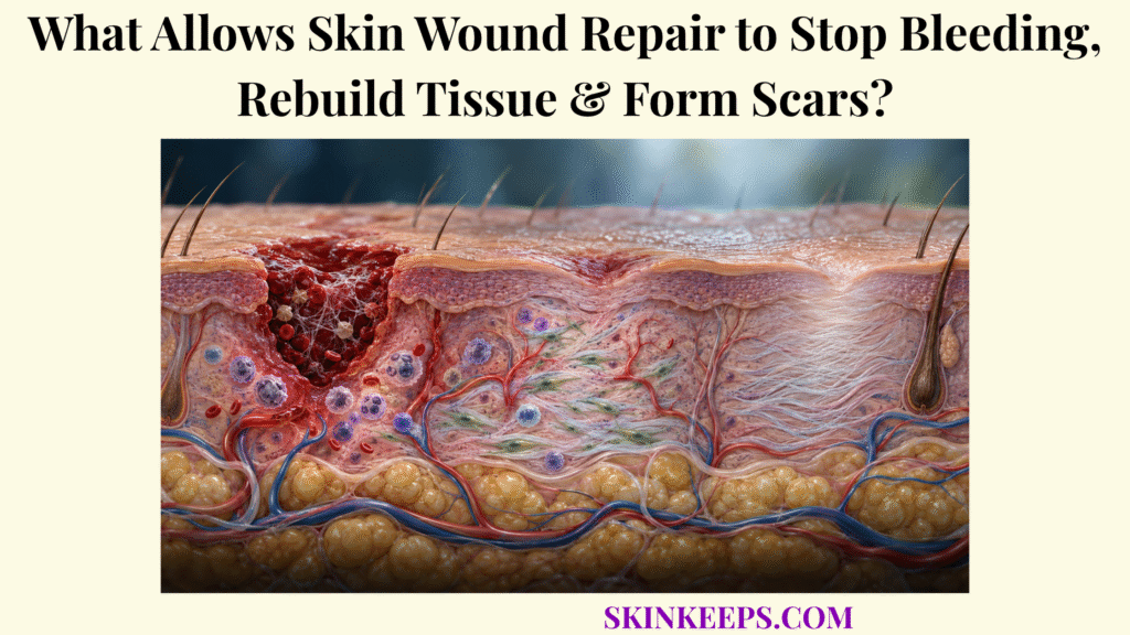
Skin wound repair restores tissue continuity through four overlapping processes: hemostasis stops bleeding, inflammation clears threats and damaged material, proliferation rebuilds vascularized tissue and epidermis, and remodeling reorganizes the repair into a stronger but structurally altered scar.
The wound begins repairing within moments of injury, but each task uses different cells and signals. Clotting, immune cleanup, fibroblast activity, angiogenesis, keratinocyte closure, and collagen remodeling work together, and deeper adult wounds usually heal through repair and scar formation rather than perfect recreation of original skin architecture.
How Does Skin Wound Repair Stop Bleeding After Injury?
Skin wound repair stops bleeding first through rapid vasoconstriction, platelet adhesion and aggregation, and coagulation-driven fibrin formation that converts an unstable platelet plug into a stronger temporary clot. This is the bleeding-control stage explained in skin bleeding control, and it establishes both hemostasis and the first scaffold for later repair.
How Does Vasoconstriction Reduce Blood Flow Immediately After Skin Injury?
Vascular injury triggers a brief local vasoconstrictor response that reduces blood flow while platelets and coagulation mechanisms begin sealing the damaged vessel. Vasoconstriction is an early stabilizing response rather than the whole hemostatic process, so it is quickly followed by platelet activity and fibrin generation.
How Do Platelets Adhere, Activate, and Form a Platelet Plug?
Platelets adhere to exposed subendothelial structures through interactions that include platelet GPIb binding to von Willebrand factor and recognition of exposed collagen, then activate, release granule contents, recruit additional platelets, and aggregate into a primary plug. This primary hemostatic plug reduces bleeding rapidly, but it still needs biochemical reinforcement to resist mechanical disruption.
How Does Coagulation Create a Fibrin Clot That Stabilizes the Wound?
The coagulation system generates thrombin, which converts soluble fibrinogen into fibrin strands that reinforce the platelet plug and produce a more stable hemostatic clot. The resulting temporary barrier is also a provisional matrix, a concept explored in skin clot wound barrier, because it stores signaling molecules and gives incoming repair cells a surface on which to migrate.
How Does the Cutaneous Healing Response Clear Damage and Control Threats?
The cutaneous healing response clears damage through a regulated inflammatory phase in which local signals recruit leukocytes, early neutrophils contribute antimicrobial defense, and macrophages remove debris while coordinating the transition toward tissue rebuilding. This inflammatory choreography, expanded in skin inflammation during repair, is necessary when it is proportionate and self-limited.
How Do Inflammatory Signals Recruit Immune Cells Into the Wound?
Damaged tissue, activated platelets, keratinocytes, mast cells, and other resident cells release mediators that activate local vessels and generate chemotactic signals guiding circulating leukocytes into the wound. Endothelial activation, adhesion, and extravasation allow immune cells to leave the bloodstream and enter the injured tissue where cleanup is needed.
How Do Neutrophils Help Control Microbes During Early Wound Healing?
Neutrophils enter acute wounds early and contribute antimicrobial defense through phagocytosis, granule enzymes, reactive oxygen mechanisms, and other innate responses when microbial threats are present. Their role is important but not identical in every wound, and excessive or persistent neutrophil activity can also amplify protease and oxidant injury.
How Do Macrophages Remove Damaged Tissue and Coordinate the Shift Toward Repair?
Wound macrophages phagocytose microbes, dead cells, and debris and then alter their signaling profile to support angiogenesis, fibroblast activity, keratinocyte migration, extracellular-matrix production, and inflammatory resolution. The broader cell network is covered in skin immune cells in wound repair, and it is safer to describe macrophages as changing through overlapping functional states rather than forcing them into a rigid M1-versus-M2 binary.
How Does Skin Wound Repair Rebuild Damaged Tissue?
Skin wound repair rebuilds damaged dermis during the proliferative phase by combining fibroblast-driven extracellular-matrix deposition, angiogenesis, granulation-tissue formation, and coordinated growth-factor signaling. This is the broader rebuilding task summarized in skin tissue rebuilding, and it reconstructs the wound bed rather than perfectly regenerating original dermal architecture.
How Do Fibroblasts Produce Collagen and Extracellular Matrix?
Fibroblasts migrate into the wound and synthesize fibronectin, proteoglycans, collagen, and other extracellular-matrix components that replace the early fibrin-rich provisional matrix with a stronger repair scaffold. The cell-specific work of skin fibroblasts and collagen production is central here because fibroplasia gives the wound a new matrix in which later remodeling can occur.
Early repair tissue generally contains relatively more type III collagen than mature scar tissue, but remodeling later changes both collagen composition and organization. It is more accurate to think of wound collagen as a continuously rebuilt matrix than as a single static deposit.
How Does Angiogenesis Create New Blood Vessels to Support Healing Tissue?
Angiogenesis restores microvascular supply when endothelial cells sprout from existing vessels, migrate and proliferate into the wound, and form new capillary networks under signals that include VEGF and hypoxia-responsive pathways. These new vessels support oxygen delivery, nutrient supply, and the metabolic demands of a highly active repair environment.
How Does Granulation Tissue Fill the Wound Bed?
Granulation tissue fills the wound with temporary vascularized repair tissue composed mainly of fibroblasts, new capillaries, extracellular matrix, and inflammatory cells. It is not the mature scar; it is the provisional tissue that later becomes less cellular and more fibrotic as remodeling advances.
How Do Growth Factors Coordinate Cell Migration and Tissue Formation?
Growth factors coordinate rebuilding by altering migration, proliferation, matrix production, angiogenesis, and cell-state transitions in fibroblasts, endothelial cells, keratinocytes, macrophages, and other wound cells. PDGF, VEGF, TGF-β, FGF, and EGF/KGF contribute to these networked effects, but no single mediator can explain the whole wound-healing program.
| Phase | Dominant processes | Major cells | Immediate outcome |
|---|---|---|---|
| Hemostasis | Vasoconstriction, platelet plug, coagulation, fibrin stabilization | Platelets, endothelium, coagulation systems | Bleeding controlled and provisional clot formed |
| Inflammation | Threat recognition, leukocyte recruitment, microbial and debris clearance | Neutrophils, macrophages, resident immune cells | Wound cleaned and pro-repair signaling established |
| Proliferation | Fibroplasia, angiogenesis, granulation tissue, re-epithelialization, contraction | Fibroblasts, endothelial cells, keratinocytes, macrophages, myofibroblasts | Wound bed rebuilt and closure progresses |
| Remodeling | Collagen turnover, cross-linking, reorientation, vascular regression, scar maturation | Fibroblasts, myofibroblasts, matrix-remodeling cells | Repair tissue becomes stronger and less cellular |
These phases overlap substantially. Cells and mediators do not obey rigid biological start–stop boundaries.
How Does Epidermal Repair Close the Wound and Restore the Skin Barrier?
Epidermal repair closes the wound when activated keratinocytes migrate across the wound surface, proliferate behind the advancing edge, re-establish epidermal layers, and progressively rebuild barrier architecture. The epithelial side of this process is developed further in keratinocyte epidermis repair, which helps separate surface closure from deeper dermal rebuilding.
How Do Keratinocytes Migrate Across the Wound Surface?
Keratinocytes near the wound edge temporarily loosen normal cell and basement-membrane attachments, reorganize their cytoskeleton, and migrate across the provisional wound matrix until opposing epithelial fronts meet. Migration is essential early in re-epithelialization and should not be replaced conceptually by proliferation alone.
How Do Epidermal Cells Proliferate to Restore Tissue Thickness?
Keratinocytes behind the migrating edge proliferate to replace cells used during wound coverage, while surviving epidermal appendages can provide additional epithelial progenitors when the injury has not destroyed them. Because depth matters, deep full-thickness wounds rely more heavily on wound-edge epithelialization when appendage-associated epithelial sources are lost.
How Does Re-Epithelialization Restore Protection Against Water Loss and External Threats?
Re-epithelialization restores surface continuity first; subsequent keratinocyte stratification, basement-membrane repair, differentiation, and stratum-corneum maturation progressively restore permeability and environmental barrier function. A wound can therefore look closed before the barrier behaves like intact unwounded skin.
How Does Skin Remodeling Strengthen Healed Tissue and Form a Scar?
Skin remodeling strengthens healed tissue by replacing and reorganizing provisional extracellular matrix, increasing collagen cross-linking and alignment, reducing excess cellularity and vessels, and maturing the repair into scar tissue. This longer-term phase of skin wound remodeling explains why a wound can keep changing for months after it has already closed.
How Is Early Collagen Reorganized and Replaced During Remodeling?
During remodeling, early collagen-rich repair matrix is repeatedly degraded and rebuilt, with type III-rich provisional tissue becoming relatively more type I-rich and collagen fibrils becoming cross-linked and reorganized along mechanical stress lines. Matrix metalloproteinases and their inhibitors help regulate this turnover, so remodeling is a dynamic balance rather than a one-time conversion.
How Do Myofibroblasts Contribute to Wound Contraction?
Myofibroblasts generate contractile force through α-smooth-muscle-actin-rich cytoskeletal machinery and transmit that force through extracellular matrix, drawing wound margins inward and reducing the tissue gap. Contraction begins during proliferative repair and overlaps with remodeling rather than starting only after surface closure is complete.
Why Does Scar Tissue Differ Structurally From Uninjured Skin?
Scar tissue differs from uninjured skin because its collagen architecture is more fibrotic and aligned, its extracellular matrix is remodeled differently, and deeper scars often restore fewer normal dermal appendages and microstructural features. The biology favors rapid restoration of continuity and load-bearing strength over perfect recreation of original anatomy.
Why Can Scar Strength Improve Without Fully Matching Normal Skin?
Scar strength improves as collagen concentration, cross-linking, fibril size, and matrix organization increase, but repaired dermis does not completely recreate the original collagen network and therefore generally remains mechanically different from unwounded skin. Standard descriptions often place mature wound tensile strength near roughly four-fifths of normal skin, but the practical message is that healed scars usually become much stronger without fully regaining original strength.
What Can Disrupt the Skin’s Wound-Healing Process?
Skin wound healing can become delayed or abnormal when infection, persistent inflammation, impaired perfusion, chronic hypoxia, mechanical disruption, systemic disease, or excessive fibrotic signaling prevents orderly transition through repair phases. These disruptors do not all act at the same step, but each can block progress toward closure or distort scar maturation.
How Can Infection Prolong Inflammation and Delay Tissue Rebuilding?
Wound infection can delay repair by maintaining microbial and inflammatory signaling, increasing tissue injury and protease activity, consuming metabolic resources, and preventing timely transition from inflammation toward proliferation. Persistent biofilms are especially relevant in chronic wounds because they can maintain inflammatory signaling and resist normal host clearance.
How Can Poor Blood Supply or Inadequate Oxygen Slow Repair?
Poor perfusion can slow wound repair because oxygen and nutrient delivery become inadequate for sustained fibroblast activity, collagen maturation, epithelialization, angiogenesis, and effective antimicrobial defense. Brief acute hypoxia can activate adaptive angiogenic signaling, but persistent severe hypoxia is the harmful pattern.
How Can Repeated Trauma Interfere With Wound Closure?
Repeated friction, tension, pressure, reopening, or mechanical stress can disrupt newly formed epithelium and matrix, prolong inflammatory signaling, and force repairing tissue to restart portions of the healing response. High tissue tension can also promote fibroblast–myofibroblast signaling and contribute to pathological scar formation in susceptible wounds.
Why Can Abnormal Matrix Production Contribute to Hypertrophic Scars or Keloids?
Hypertrophic scars and keloids arise from dysregulated fibroproliferative repair in which inflammatory, mechanical, fibroblast, TGF-β, extracellular-matrix synthesis, and matrix-degradation pathways remain abnormally active or imbalanced. A hypertrophic scar typically remains within the original wound, whereas a keloid grows beyond the original wound boundaries.
Other systemic modifiers can also influence repair, including diabetes, smoking, malnutrition, advanced age, some medications, and immune dysfunction. They matter because wound healing depends on intact perfusion, metabolism, immunity, and tissue integrity rather than on one isolated pathway.
| Healing problem | Main mechanism disrupted | Possible biological outcome |
|---|---|---|
| Persistent infection or biofilm | Inflammation fails to resolve | Chronic inflammatory wound and delayed proliferation |
| Poor perfusion | Oxygen and nutrient delivery | Slower collagen synthesis, epithelialization, and immune defense |
| Chronic hypoxia | Cellular metabolism and extracellular-matrix synthesis | Impaired fibroblast activity, angiogenesis, and repair |
| Repeated trauma or tension | New epithelium and provisional matrix | Reopening, prolonged inflammation, and delayed closure |
| Persistent inflammatory signaling | Transition to proliferation and remodeling | Chronic wound or excess fibrosis |
| Excess fibroblast or myofibroblast activity | ECM synthesis and remodeling balance | Hypertrophic or keloid-type scarring |
| Excessive contraction | Myofibroblast mechanics | Contracture and restricted movement in severe cases |
These are mechanistic patterns, not diagnoses. A non-healing or clearly abnormal wound needs assessment of the actual cause.
What Are the Key Takeaways About Skin Wound Repair?
The key fact about skin wound repair is that successful healing requires each overlapping phase to perform a different task at the right time: bleeding must stop, inflammation must clear danger and resolve, new tissue must form, the epidermis must close, and extracellular matrix must mature without excessive fibrosis.
- Hemostasis starts with brief vasoconstriction that reduces early blood loss.
- Platelets adhere, activate, and aggregate at vascular injury to form a primary plug.
- Coagulation converts fibrinogen into fibrin that reinforces the clot and stabilizes the wound.
- The clot becomes a temporary scaffold and signaling reservoir for incoming repair cells.
- Inflammation recruits neutrophils, macrophages, and resident cells to control threats and remove damaged material.
- Macrophages help move the wound from inflammatory cleanup toward proliferation and tissue rebuilding.
- Fibroblasts produce extracellular matrix and collagen that reconstruct the dermal scaffold.
- Angiogenesis supplies metabolically active repair tissue with vessels, oxygen, and nutrients.
- Granulation tissue is temporary vascularized repair tissue rather than mature scar tissue.
- Keratinocytes migrate and proliferate to restore epidermal continuity through re-epithelialization.
- Myofibroblasts can contract the wound and reduce the open tissue gap.
- Remodeling degrades, replaces, cross-links, and reorganizes collagen over time.
- Deep adult dermal wounds commonly restore continuity through fibrotic repair rather than perfect regeneration.
- Mature scars become stronger with time but generally do not fully equal normal skin.
- Infection, poor perfusion, persistent hypoxia, repeated trauma, or dysregulated fibrosis can disrupt normal healing.
What Common Questions Do People Ask About Skin Wound Repair?
Common questions about skin wound repair focus on how the phases overlap, whether granulation tissue is a scar, why scars remain weaker than normal skin, and how pathological scars differ.
Are the Four Wound-Healing Phases Completely Separate?
No. Hemostasis, inflammation, proliferation, and remodeling overlap, and cells or signals from one phase commonly remain active while the next phase begins.
Is Granulation Tissue the Same as Scar Tissue?
No. Granulation tissue is temporary vascularized repair tissue rich in fibroblasts, new vessels, extracellular matrix, and inflammatory cells; a mature scar develops later through prolonged matrix remodeling.
Can Healed Skin Become as Strong as Uninjured Skin?
Usually not completely. Collagen remodeling substantially increases tensile strength, but mature scar tissue generally retains different matrix architecture and remains mechanically weaker than unwounded skin.
Why Does a Deep Skin Wound Form a Scar Instead of Perfectly Regenerating?
Deep dermal injury removes complex extracellular matrix and skin structures that adult repair usually replaces rapidly with fibroblast-derived collagen-rich tissue, restoring continuity more effectively than recreating the original microanatomy.
What Is the Difference Between a Hypertrophic Scar and a Keloid?
A hypertrophic scar typically remains within the boundaries of the original wound, whereas a keloid characteristically grows beyond those original borders; both reflect abnormal fibroproliferative repair and require clinical assessment for diagnosis.
StatPearls / NCBI Bookshelf — Physiology, Wound Healing: four overlapping phases, immediate hemostasis, fibrin clot formation, granulation tissue, remodeling, and tensile-strength limits.
PMC — Cellular and Molecular Mechanisms of Wound Repair: From Biology to Therapeutic Innovation: current overview of coordinated cell populations, extracellular matrix, and reasons wounds become chronic or fibrotic.
PMC — Skin Acute Wound Healing: A Comprehensive Review: wound-healing sequence, participant cells, angiogenesis, re-epithelialization, and pathological healing boundaries.
PMC — Epithelialization in Wound Healing: A Comprehensive Review: keratinocyte migration, keratinocyte proliferation, and why epithelialization is required for successful closure.
PMC — Immunology of Wound Healing: inflammatory signaling, neutrophils, macrophages, and the transition from inflammatory cleanup toward proliferation.
PMC — Collagen in Wound Healing: collagen turnover, extracellular-matrix remodeling, matrix enzymes, and the chronic-wound implications of persistent inflammation.
Life — Cutaneous Wound Healing: An Update from Physiopathology to Current Therapies: impaired healing, infection, hypoxia, aging, nutrition, systemic modifiers, and the balance of overlapping repair mechanisms.
PMC — Hypertrophic Scarring and Keloids: Epidemiology, Molecular Pathogenesis, and Therapeutic Interventions: distinctions between hypertrophic scars and keloids, abnormal fibroproliferation, and the role of mechanical and profibrotic signaling.
This page is educational and does not diagnose a wound or prescribe treatment. Seek urgent care for uncontrolled significant bleeding, deep or extensive trauma, exposed deep structures, or rapidly spreading infection signs; seek professional assessment for a wound that is worsening, separating, draining persistently, not progressing toward closure, or forming an enlarging or function-limiting scar.



