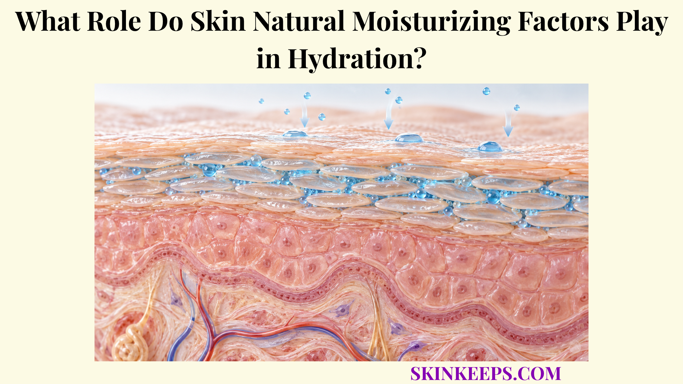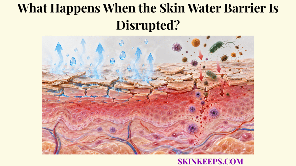
When the Skin Water Barrier is disrupted, the stratum corneum becomes less effective at controlling movement of water and external substances. Transepidermal water loss can rise, hydration can fall, and altered corneocyte and lipid organization can make the outer epidermis less mechanically and chemically resilient.
Barrier disruption also affects more than water balance. Selected irritants may penetrate more readily and activate keratinocyte stress or inflammatory signaling, while inflammation can in turn interfere with normal barrier organization. At the same time, the epidermis activates repair mechanisms that restore extracellular lipids, cellular differentiation, and permeability control.
What Changes First When the Skin Water Barrier Is Disrupted?
The first functional change in Skin Water Barrier disruption is a loss of normal stratum corneum permeability resistance caused by disturbance of its corneocyte–lipid architecture.
The normal barrier depends on mature corneocytes, intact cornified envelopes, controlled corneocyte cohesion, and correctly organized extracellular lipid lamellae. Damage may affect one or several of these elements. The skin water barrier therefore behaves as an integrated permeability system rather than a simple surface coating or one continuous lipid seal.
Descriptions of “gaps” are useful only as a simplified concept. Ordinary barrier dysfunction more often creates less continuous or less effectively organized low-resistance pathways for molecular movement, not large literal holes. Because this architecture normally supports skin water-loss prevention while also limiting entry of many external substances, disruption can alter transport in both directions.
How Does the Skin Water Barrier Increase Transepidermal Water Loss?
A disrupted Skin Water Barrier increases transepidermal water loss because damaged or disorganized stratum corneum structures offer less resistance to passive outward water diffusion.
Water tends to move from deeper, more hydrated tissue toward the drier external environment. An intact stratum corneum strongly slows that movement, especially through its ordered extracellular lipid lamellae. When permeability resistance falls, outward water-vapor flux increases and measured TEWL rises. The detailed mechanism of transepidermal water loss should therefore be understood as a transport process rather than water “pouring through holes.”
TEWL is a flux measurement, not a direct measurement of stratum corneum water content. Higher TEWL can contribute to lower hydration when outward loss exceeds water replacement, but hydration also depends on Natural Moisturizing Factor, environmental humidity, anatomical site, deeper water supply, and other physiological variables.
How Does Skin Water Barrier Damage Dehydrate Corneocytes?
Skin Water Barrier damage can dehydrate corneocytes when increased outward water loss reduces the water available within the stratum corneum.
Corneocyte hydration is determined by several interacting systems: water supplied from deeper tissue, environmental humidity, barrier permeability, extracellular lipid organization, and intracellular hygroscopic solutes. The physiology of how corneocytes retain water therefore cannot be reduced to TEWL alone.
Natural Moisturizing Factor-containing corneocytes require adequate water for normal molecular mobility. The mechanism by which Natural Moisturizing Factor binds water helps the keratin-rich intracellular compartment remain hydrated and flexible. When water content falls sufficiently, stratum corneum tissue can become mechanically stiffer and experience greater drying stress.
Visible roughness or scaling may increase as dehydration becomes more marked. Fissuring or cracking can occur when drying is substantial and mechanical stress or additional barrier abnormalities coexist, but neither outcome is inevitable after every measurable rise in TEWL.
How Does Skin Water Barrier Disruption Affect Barrier Lipids?
Skin Water Barrier disruption can involve both quantitative and structural abnormalities in the extracellular lipids that normally form ordered lamellar membranes between corneocytes.
Ceramides, cholesterol, and free fatty acids are central components of these membranes. Upper differentiating keratinocytes synthesize lipid precursors, package lipids and processing enzymes into lamellar bodies, secrete that material near the stratum granulosum–stratum corneum interface, and then generate mature extracellular lamellar structures. The mechanism by which intercellular lipids limit evaporation depends on this organization as well as on the presence of individual lipid classes.
Barrier dysfunction may therefore involve lower amounts of selected lipids, altered lipid species, impaired processing, abnormal delivery, or disordered lamellar architecture. Lipid disorganization can reduce resistance to water movement and intensify the dehydration challenge, but it should not always be presented as a secondary consequence of water loss because abnormal lipid architecture may be part of the original defect.
| Barrier change | Water-loss effect | Possible visible or functional consequence |
|---|---|---|
| Reduced lipid organization | Lower resistance to outward diffusion | Higher TEWL |
| Reduced stratum corneum hydration | Greater tissue stiffness and drying stress | Roughness or tight-feeling skin |
| Impaired corneocyte shedding | Hydration-dependent surface processing becomes less efficient | Flaking or scaling |
| Severe combined structural disruption | Greater drying and mechanical stress | Fissuring or cracking can occur |
| Increased permeability to selected external substances | Outside-in diffusion resistance is reduced | Stinging, discomfort, or inflammatory signaling in susceptible or sufficiently damaged skin |
Barrier disruption affects water movement, tissue mechanics, and outside-in exposure through related but distinct mechanisms; visible outcomes remain dependent on severity and context.
How Does a Damaged Skin Water Barrier Increase Irritant Entry?
A damaged Skin Water Barrier can increase entry of selected irritants because disruption of the stratum corneum reduces the diffusion resistance that normally limits penetration from the external environment.
The increase is selective rather than universal. Penetration depends on molecular size and chemistry, concentration, formulation or vehicle, contact duration, skin site, temperature and hydration, and the degree of barrier disruption. The broader stratum corneum barrier therefore does not regulate every external substance to the same extent, and TEWL cannot be used as a numerical substitute for the absorption rate of a particular chemical.
With sufficient penetration, irritants may reach viable keratinocytes, disturb cell membranes or signaling pathways, and activate stress responses. This can contribute to stinging, burning, irritation, or discomfort in susceptible or sufficiently damaged skin. Irritant effects represent direct cellular or tissue stress; allergic contact responses require adaptive immune sensitization and are mechanistically distinct.
How Does Skin Water Barrier Failure Trigger Inflammation?
Skin Water Barrier failure can trigger inflammatory signaling because stressed keratinocytes detect tissue disturbance and increased environmental exposure and can release cytokines, chemokines, and other innate immune mediators.
Keratinocytes are not passive structural units. Barrier injury can alter cellular stress signaling, local ionic conditions, and exposure to irritants or microbial products. In response, keratinocytes can signal neighboring epidermal cells, influence immune-cell recruitment, modify antimicrobial responses, and change differentiation programs.
In persistent or inflammatory settings, this can form a bidirectional loop: barrier disruption increases environmental exposure and keratinocyte stress; inflammatory mediators then can alter differentiation, lipid metabolism, or cohesion and further impair barrier function. These linked consequences help explain why skin water loss and irritation can coexist, but a brief transient rise in TEWL does not automatically produce clinical inflammation or chronic disease.
How Does the Skin Water Barrier Repair Itself?
The Skin Water Barrier begins repairing itself rapidly after disruption by increasing lamellar-body secretion and epidermal lipid production, followed by continued keratinocyte differentiation, cornification, and structural renewal.
How Does the Rapid Homeostatic Response Begin?
After acute experimental disruption, pre-existing lamellar bodies in upper differentiating keratinocytes are rapidly secreted so that lipid material and processing enzymes enter the stratum corneum intercellular space. Changes in the epidermal calcium gradient participate in this response; much of the detailed signaling evidence comes from animal and experimental models, so it is best treated as a mechanistic explanation rather than a universal clinical measurement.
How Are New Barrier Lipids Rebuilt?
Keratinocytes increase synthesis of barrier lipids and generate new lamellar bodies. Extracellular processing then helps reconstruct ceramide-rich, cholesterol-containing and free-fatty-acid-containing lamellar membranes. The goal is not to replace one missing lipid in isolation but to restore correctly processed and organized extracellular architecture.
How Does Structural Renewal Continue?
Longer-term recovery includes continued keratinocyte differentiation, cornification, corneocyte maturation, and proliferation where required. These processes rebuild the cellular scaffold and extracellular organization that support normal permeability resistance.
How Is Functional Recovery Recognized?
As permeability resistance improves, TEWL moves toward baseline, stratum corneum water balance becomes easier to maintain, and resistance to outside-in penetration improves. Recovery time varies with the damage mechanism, severity, anatomical site, age, environment, underlying skin condition, and repeated exposure, so there is no single universal repair interval.
What Are the Key Takeaways About Skin Water Barrier Disruption?
Skin Water Barrier disruption changes both inside-to-outside water control and outside-to-inside permeability because the stratum corneum no longer provides its normal level of diffusion resistance.
- Skin Water Barrier disruption increases permeability and usually increases transepidermal water loss.
- Higher water loss can reduce stratum corneum hydration and make corneocytes and the surrounding tissue less flexible.
- Abnormal extracellular lipid quantity, processing, delivery, or lamellar organization can weaken permeability control further.
- Barrier damage can increase penetration of selected irritants, although penetration remains substance- and exposure-dependent.
- Keratinocyte stress and inflammatory signaling can amplify persistent barrier dysfunction.
- Barrier repair begins rapidly with lamellar-body secretion and lipid synthesis and continues through epidermal differentiation and structural renewal.
Frequently Asked Questions
Does a disrupted Skin Water Barrier always mean the skin is visibly damaged?
No. Functional barrier impairment can occur before obvious cracking, scaling, or redness is visible. TEWL and permeability can change without dramatic surface signs.
Is increased TEWL the same as dry skin?
No. TEWL measures outward water flux, whereas dry skin describes a broader clinical or physical state influenced by hydration, Natural Moisturizing Factor, lipids, environmental exposure, and other factors.
Does Skin Water Barrier damage remove all barrier lipids?
No. Dysfunction can involve altered lipid amounts, composition, processing, delivery, or lamellar organization rather than complete lipid loss.
Can a damaged Skin Water Barrier let irritants penetrate more easily?
Yes. Selected substances may penetrate more readily when stratum corneum permeability resistance is reduced, but penetration depends on the substance, formulation, exposure, skin site, and degree of damage.
Can the Skin Water Barrier repair itself?
Yes. The epidermis activates homeostatic repair mechanisms including rapid lamellar-body secretion, increased lipid production and processing, and continued keratinocyte differentiation and renewal. Recovery time varies with the cause and severity of disruption.
Sources & Evidence
Research grounding
Skin Lipid Barrier: Structure, Function and Metabolism — Berdyshev E. Allergy Asthma Immunol Res. 2024;16(5):445–461. Use: Stratum corneum lipid architecture, ceramide-rich lamellae, terminal differentiation, and the barrier to water loss and outside-in penetration.
Regulation of permeability barrier homeostasis — Feingold KR, Denda M. Clin Dermatol. 2012;30(3):263–268. Use: Acute barrier repair, lamellar-body secretion, calcium-gradient signaling, lipid synthesis, and restoration of permeability function.
The correlation between transepidermal water loss and percutaneous absorption: an overview — Levin J, Maibach H. J Control Release. 2005;103(2):291–299. Use: TEWL as a barrier-integrity measure and the important distinction between TEWL and substance-specific percutaneous absorption.
The effects of barrier disruption and moisturization on the dynamic drying mechanics of human stratum corneum — Liu X, German GK. J Mech Behav Biomed Mater. 2015;49:80–89. Use: Lipid-depleted stratum corneum, faster drying, greater stiffness, drying stress, and susceptibility to chapping or cracking.
Skin hydration: a review on its molecular mechanisms — Verdier-Sévrain S, Bonté F. J Cosmet Dermatol. 2007;6(2):75–82. Use: NMF, intercellular lipid organization, TEWL control, stratum corneum hydration, and the distinction between water content and water loss.
Cytokines and the Skin Barrier — Hänel KH, Cornelissen C, Lüscher B, Baron JM. Int J Mol Sci. 2013;14(4):6720–6745. Use: Keratinocyte cytokine signaling and bidirectional interactions between epidermal differentiation, barrier function, and inflammation.
Irritant Contact Dermatitis — a Review — Bains SN, Nash P, Fonacier L. Curr Dermatol Rep. 2022;11:70–78. Use: Barrier disruption, increased irritant permeability, keratinocyte stress signaling, and distinction of irritant mechanisms from allergic responses.
The outer frontier: the importance of lipid metabolism in the skin — Feingold KR. J Lipid Res. 2009;50 Suppl:S417–S422. Use: Lamellar-body biology, epidermal lipid synthesis, extracellular lamellar membranes, and rapid homeostatic responses after permeability-barrier disruption.
Medical/Educational Disclaimer: This article explains normal skin-barrier physiology for educational purposes and is not intended to diagnose or treat a skin condition. Persistent, painful, bleeding, oozing, severely inflamed, infected-looking, or otherwise concerning skin changes should be assessed by a qualified healthcare professional.