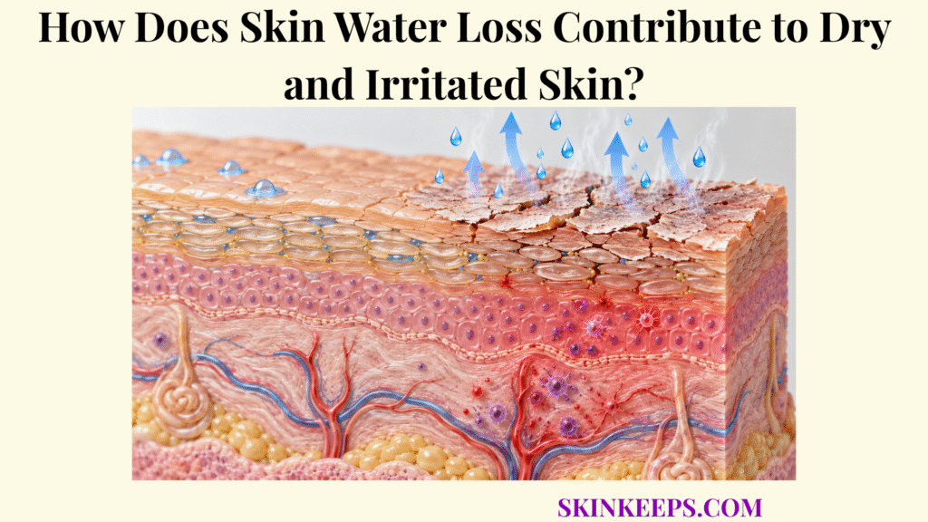
Skin Water Loss contributes to dryness when outward water flux becomes high enough that the stratum corneum cannot maintain its usual hydration. As corneocytes lose water, their keratin-rich structure becomes less flexible and mechanically stiffer, contributing to roughness or scaling and, under more severe or persistent stress, possible fissuring.
Water loss and irritant penetration often share the same underlying cause: an impaired stratum corneum permeability barrier. That barrier defect may simultaneously increase outward water flux and allow selected external substances to enter more readily, while marked dehydration or inflammatory signaling can add further stress.
How Does Skin Water Loss Reduce Stratum Corneum Hydration?
Skin Water Loss reduces stratum corneum hydration when outward water flux exceeds the outer epidermis’s ability to replenish and retain water.
Water moves from deeper hydrated tissue through the epidermis and stratum corneum toward the environment. The skin water barrier slows that movement. Transepidermal water loss (TEWL) is the outward water flux passing through the epidermal barrier and into the surrounding air; the detailed causes of increased transepidermal water loss belong to the separate mechanism page.
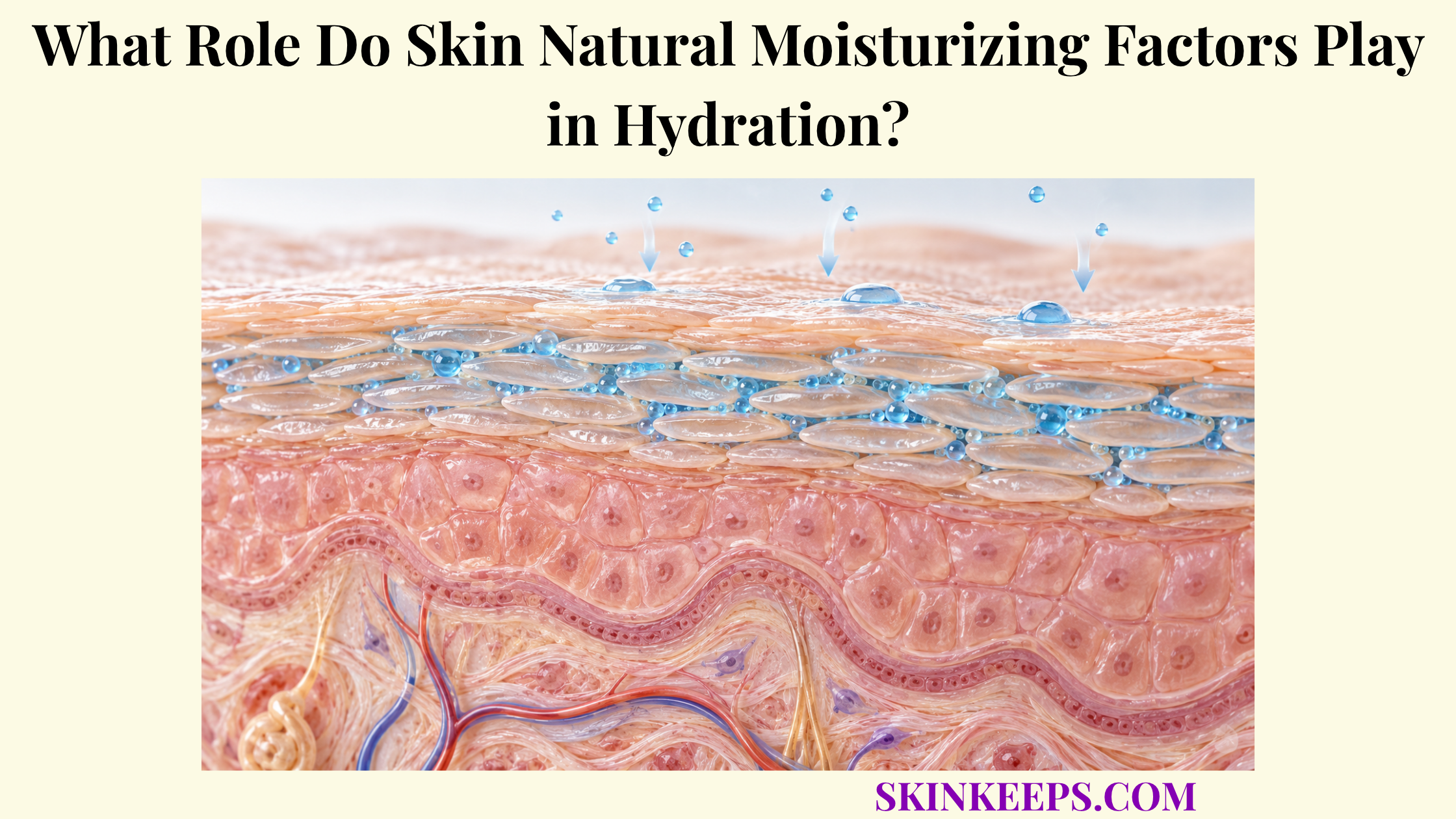
Stratum corneum hydration means the amount and distribution of water retained within the outer epidermis. TEWL measures a rate of water movement, not water content. Hydration also reflects hygroscopic NMF, extracellular lipid organization, environmental humidity, body site, and water supply from deeper tissue. When water content falls, the surface can become less supple or rougher; a sensation of tightness alone cannot establish the cause.
How Does Skin Water Loss Make Corneocytes Dry and Stiff?
Skin Water Loss can make corneocytes and the surrounding stratum corneum mechanically stiffer because water acts as an important plasticizing component of keratin-rich tissue.
A corneocyte is a terminally differentiated epidermal cell that contributes to the cellular framework of the stratum corneum. Water supports keratin molecular mobility, corneocyte swelling and tissue compliance, helping the surface accommodate ordinary movement. The wider process of how corneocytes retain water involves more than outward water movement alone.
Hygroscopic compounds in the intracellular NMF pool associate with water and support flexibility. Understanding how Skin Natural Moisturizing Factor binds water clarifies why a decline in water availability can alter keratin-rich structures without implying that every episode of high TEWL depletes NMF. As drying becomes more marked, reduced molecular mobility and increased tissue stiffness can make roughness, flaking or chapping more likely.
How Does Skin Water Loss Weaken Barrier Integrity?
Skin Water Loss can contribute to further barrier weakness when persistent dehydration alters stratum corneum mechanics, desquamation or surface integrity, but the original increase in water loss usually reflects an existing permeability-barrier defect.
The causal order matters: a primary permeability impairment raises TEWL; hydration may then fall; stiffness and drying stress can increase; and sufficiently severe or repeated mechanical stress may produce additional surface disruption. This is a possible secondary feedback, not a rule that water loss always starts barrier damage or that every dry surface develops fissures. The earlier structural events are addressed in skin barrier disruption.
Water also supports controlled degradation of corneodesmosomes during normal surface shedding. Marked dryness can contribute to retention of surface corneocytes and visible scaling. Intercellular lipid lamellae, rather than NMF, provide the main extracellular resistance to outward diffusion; the separate page on how intercellular lipids limit evaporation explains that permeability role in detail.
How Does Skin Water Loss Increase Irritant Penetration?
Skin Water Loss does not by itself determine irritant penetration; rather, the permeability defect that allows excessive water to escape can also allow selected external substances to cross the stratum corneum more readily.
Increased TEWL and increased irritant penetration are often parallel consequences of barrier disruption rather than a one-way sequence in which water loss directly increases chemical entry. The intact stratum corneum restricts outward movement of water and inward movement of many external substances, but those movements depend on different molecular and exposure properties.
Penetration of an irritant—a substance capable of direct tissue stress or injury under sufficient exposure—depends on its molecular size, polarity, lipid solubility, concentration, vehicle, contact time, anatomical site, hydration and degree of barrier impairment. Selected irritants may reach viable keratinocytes, cell membranes or signaling receptors more readily after disruption, but higher TEWL cannot predict the absorption of every substance. Stinging, burning, redness or discomfort may follow sufficiently intense exposure; none of these sensations specifically proves water loss or a particular diagnosis.
| Water-loss or barrier effect | Barrier or tissue change | Possible skin consequence |
|---|---|---|
| Increased TEWL | Water availability may decline when loss exceeds replenishment. | Dryness can become more apparent. |
| Reduced stratum corneum hydration | Lower tissue compliance and greater stiffness. | Rough texture or reduced suppleness. |
| Altered hydration-dependent desquamation | Surface corneocyte aggregates may be retained. | Scaling or flaking. |
| Severe or persistent drying stress | Greater mechanical strain, sometimes with secondary surface disruption. | Cracking or chapping may occur. |
| Underlying permeability defect | Greater entry of selected irritants, depending on the exposure. | Stinging, discomfort or irritation in susceptible skin. |
| Persistent inflammatory signaling | Barrier homeostasis and normal lipid or protein production can be impaired. | Continuing barrier dysfunction in some inflammatory contexts. |
Water loss contributes directly to dehydration. Irritant penetration primarily reflects the underlying permeability defect that can accompany higher TEWL.
How Does Skin Water Loss Trigger Irritation and Inflammation?
Skin Water Loss is most likely to accompany irritation and inflammation when the barrier disruption causing excessive water loss also stresses keratinocytes or increases their exposure to irritants, microbes or inflammatory signals.
Keratinocytes are both structural barrier cells and immune-active sentinels. Chemical exposure, friction, changes in local ionic conditions or other tissue stress can prompt them to release cytokines, chemokines, antimicrobial mediators and repair signals. Increased entry of selected irritants may amplify these responses if sufficient exposure reaches viable epidermal cells.
In persistent inflammatory contexts, cytokine signaling can alter epidermal differentiation, structural-protein expression, lipid metabolism and barrier organization. This creates a possible feedback loop between barrier impairment and inflammatory signaling; it does not mean that dry skin or a brief TEWL rise automatically causes inflammation. The broader relationship through which skin barrier integrity influences skin immunity includes microbial and immune-cell communication that this water-loss page does not cover in detail.
What Worsens Skin Water Loss and Irritation?
Skin Water Loss and irritation can become more pronounced when a damaged barrier is repeatedly exposed to environmental, chemical, mechanical or inflammatory stress.
How Can Dry Air Change Hydration?
Low ambient humidity can reduce environmental water availability and increase the dehydration challenge, leading to lower stratum corneum water content or greater roughness in some settings. Measured TEWL does not universally rise as humidity falls: human studies are heterogeneous, and epidermal responses depend on duration, conditions and individual factors.
How Can Repeated Washing and Surfactant Exposure Add Stress?
Repeated or aggressive exposure to water and surfactants can disturb proteins, extracellular lipids or water-soluble NMF and affect hydration and permeability. The response depends on surfactant chemistry, formulation, concentration, water temperature, contact time, frequency and baseline skin condition. Ordinary cleansing is not equivalent to repeated harsh exposure.
How Can Irritants and Solvents Affect the Barrier?
Some chemicals can extract lipids, interact with proteins or disturb keratinocyte signaling, increasing permeability or local stress. Effects vary with the specific substance, its concentration, vehicle and exposure duration; different solvents and irritants should not be treated as interchangeable.
When Does Mechanical Exposure Matter?
Repeated friction can disturb superficial corneocytes or add strain to already dehydrated tissue. Force, duration, pressure, local moisture, anatomical site and baseline barrier integrity all modify the response.
How Can Existing Inflammation Prolong the Challenge?
Persistent inflammatory signaling can interfere with normal barrier-protein and lipid production and slow restoration of permeability control. The resulting barrier–inflammation interaction is possible in sustained inflammatory settings, but it is not an inevitable consequence of temporary dryness.
How Does Skin Water Loss Improve as the Barrier Recovers?
Skin Water Loss decreases as the epidermal permeability barrier restores its extracellular lipid architecture and normal stratum corneum organization, increasing resistance to outward water diffusion.
What Initiates the Early Repair Response?
After acute barrier disruption, upper epidermal keratinocytes rapidly secrete preformed lamellar bodies that deliver lipids, lipid precursors and processing enzymes into extracellular spaces. This early lipid-secretory response starts before complete epidermal turnover.
How Do Lipid Membranes Recover?
Epidermal synthesis of cholesterol, fatty acids and sphingolipid precursors increases to support further lamellar-body production. Extracellular processing helps restore the ordered lamellae containing ceramides, cholesterol and free fatty acids, increasing resistance to diffusion rather than forming a simple surface coating.
How Does Structural Renewal Continue?
Keratinocyte differentiation, cornification, mature corneocyte formation and normalization of extracellular organization support longer-term recovery. The timing depends on the type and severity of disruption, repeated exposure, body site, age, environment, inflammation and other individual factors.
What Changes as Water Control Returns?
As permeability resistance improves, elevated TEWL tends toward the individual’s baseline and stratum corneum hydration can recover. Flexibility and dryness-related comfort may improve as water balance returns, though subjective discomfort also has other causes. The larger physiological role of skin water-loss prevention explains how an organized stratum corneum maintains that resistance under normal conditions.
What Are the Key Takeaways About Skin Water Loss?
Skin Water Loss contributes most directly to dryness by reducing the water available within the stratum corneum, while irritation usually reflects the broader barrier dysfunction and environmental exposure that accompany excessive water loss.
- Skin Water Loss can lower stratum corneum hydration when outward loss exceeds water retention and replenishment.
- Lower hydration makes the stratum corneum less flexible and mechanically stiffer, contributing to roughness and scaling.
- Persistent or severe dehydration can increase drying stress and susceptibility to fissuring, but cracking is not inevitable.
- Increased TEWL and increased irritant penetration are often parallel consequences of an impaired permeability barrier rather than a direct cause-and-effect pair.
- Greater irritant exposure and keratinocyte stress can promote inflammatory signaling, while inflammation can further impair barrier homeostasis.
- Environmental dryness, repeated harsh cleansing, irritants, solvents, friction and existing inflammation can intensify barrier stress.
- Barrier recovery reduces water loss by restoring extracellular lipids, normal epidermal organization and permeability resistance.
Frequently Asked Questions
Is Skin Water Loss the same as having dry skin?
No. Skin Water Loss describes outward water flux, whereas dry skin reflects the water content and broader physical state of the stratum corneum, influenced by corneocytes, NMF, lipids, desquamation, mechanics, and environmental exposure.
Does increased Skin Water Loss always cause skin cracking?
No. Reduced hydration can increase stiffness and drying stress, but fissuring typically requires sufficiently severe or persistent dehydration together with mechanical or structural stress.
Does Skin Water Loss directly let irritants enter the skin?
Not exactly. The permeability defect that increases TEWL can also increase outside-in penetration of selected substances. Severe dehydration may add mechanical stress, but outward water loss alone does not determine irritant entry.
Does Skin Water Loss always cause inflammation?
No. Barrier stress and increased environmental exposure can activate inflammatory pathways in susceptible or sufficiently damaged skin, but a temporary rise in water loss does not automatically produce clinical inflammation.
Can Skin Water Loss decrease when the barrier recovers?
Yes. As extracellular lipid organization and normal epidermal structure recover, resistance to outward water diffusion rises and elevated TEWL generally moves toward the individual’s baseline.
Sources & Evidence
Research grounding
Skin hydration: a review on its molecular mechanisms — Verdier-Sévrain S, Bonté F. Journal of Cosmetic Dermatology. 2007. Evidence scope: Differentiate water flux from tissue hydration; identify NMF and lipids as complementary water-handling systems.
Skin hydration: interplay between molecular dynamics, structure and water uptake in the stratum corneum — Mojumdar EH et al. Scientific Reports. 2017. Evidence scope: Hydration-dependent keratin mobility, swelling, and stratum corneum mechanical properties.
The effects of hydration on the topographical and mechanical properties of corneocytes — Journal of the Mechanical Behavior of Biomedical Materials. 2024. Evidence scope: Experimental corneocyte swelling and greater compliance with water uptake; avoid treating single-site experimental results as universal thresholds.
The effects of barrier disruption and moisturization on the dynamic drying mechanics of human stratum corneum — Liu X, German GK. Journal of the Mechanical Behavior of Biomedical Materials. 2015. Evidence scope: Lipid-depleted tissue drying, increased stiffness and drying stress, and possible cracking susceptibility.
Abnormal epidermal barrier in the pathogenesis of contact dermatitis — Proksch E, Brasch J. Clinics in Dermatology. 2012. Evidence scope: The shared permeability defect can increase outward water flux and penetration of selected external irritants; distinguish irritant injury from diagnosis.
The Central Roles of Keratinocytes in Coordinating Skin Immunity — Simmons J, Gallo RL. Journal of Investigative Dermatology. 2024. Evidence scope: Keratinocyte sensing, cytokine and chemokine signaling, and the possibility of inflammatory barrier feedback.
Ambient humidity and the skin: the impact of air humidity in healthy and diseased states — Engebretsen KA et al. Journal of the European Academy of Dermatology and Venereology. 2016. Evidence scope: Environmental dryness can challenge hydration while measured TEWL and adaptation vary across studies.
The regulation and role of epidermal lipid synthesis — Feingold KR. Advances in Lipid Research. 1991. Evidence scope: Rapid preformed lamellar-body secretion, increased epidermal lipid synthesis, and later structural barrier restoration.
Medical/Educational Disclaimer: This article explains skin hydration, water loss and barrier physiology for educational purposes. It is not intended to diagnose or treat dry, irritated, sensitive or inflamed skin. Persistent, painful, bleeding, deeply cracked, oozing, severely inflamed, infected-looking or otherwise concerning skin changes should be evaluated by a qualified healthcare professional.