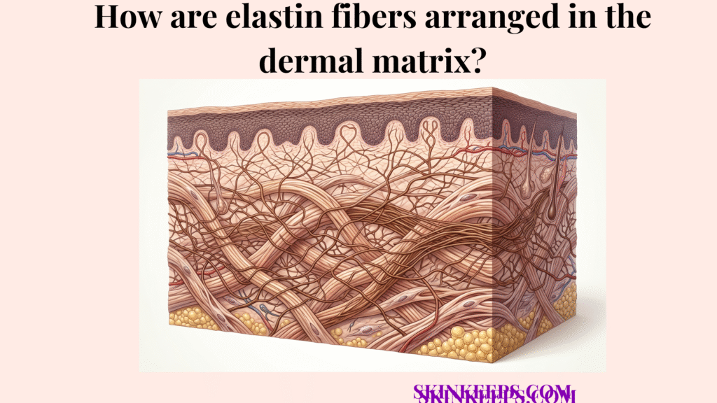
Elastin fibers are arranged in the dermal matrix as a branching elastic network that runs through both the upper and deeper dermis. This network is finer near the superficial dermis and more substantial in the deeper dermis, where it helps the skin stretch and return toward its original shape. Reviews of elastic-fiber biology describe dermal elasticity as depending on integrated networks of thin fibers in the papillary dermis and thick reticular elastic fibers deeper down.
The skin’s elasticity does not come from elastin simply existing in the dermis. It depends on how elastic fibers are distributed, how they connect through the matrix, and how they work alongside collagen to create flexibility without structural collapse.
Understanding dermal elastin fiber arrangement makes it easier to explain why the skin can recoil after movement, why aging changes skin ‘bounce,’ and what actually happens when the elastic network becomes damaged or disorganized.
Why does dermal elastin fiber arrangement matter for skin mechanics?
Dermal elastin fiber arrangement matters because skin recoil depends on how elastic fibers—dermal fibers that help tissue stretch and recoil—are organized through the connective tissue framework.
The skin must stretch without tearing and recoil without staying lax, and elastin helps provide this rebound within the dermal matrix.
This biomechanical property depends on fiber arrangement, not just the total elastin amount alone. Reviews of elastic-fiber biology emphasize that architecture determines elastic performance because fibers must be organized to distribute force through tissue rather than act as isolated strands.
Where is dermal elastin fiber arrangement distributed within the dermis?
Dermal elastin fiber arrangement is distributed across both upper and deeper dermal layers, with each layer contributing differently to elastic behavior.
The papillary dermis—the superficial, finer upper dermal layer—contains a finer elastic network, while the reticular dermis—the deeper, thicker dermal layer with stronger connective architecture—contains a broader, stronger recoil network.
In Lynch et al., 2022, the papillary dermis is described as roughly 60–120 μm thick, while the reticular dermis varies roughly 1000–4000 μm with age; the same study notes that dermal fibers in the reticular dermis are generally thicker and more bundled than in the papillary layer. That makes the dermis a layered recoil system rather than one uniform elastic sheet (Nature, 2022).
How does papillary dermal elastin fiber arrangement differ from reticular dermal elastin fiber arrangement?
Papillary dermal elastin fiber arrangement is finer and lighter, while reticular dermal elastin fiber arrangement is broader and more mechanically dominant.
The papillary dermis contains thinner and more delicate elastic support designed to anchor and buffer the epidermal junction, while the reticular dermis contains a larger recoil network suited to deeper mechanical behavior and tension distribution.
StatPearls describes thinner elastic fibers in the superficial papillary dermis and thicker elastic fibers in the reticular dermis, while elastin reviews describe dermal elasticity as based on thin papillary fibers plus thick reticular elastic fibers working together.
| Dermal layer | Elastin fiber arrangement | Fiber character | Main mechanical role |
|---|---|---|---|
| Papillary dermis | Finer and more delicate | Thin elastic fibers | Surface flexibility and subtle recoil |
| Reticular dermis | Broader and more substantial | Larger elastic network | Deep recoil and mechanical resilience |
How does branching define dermal elastin fiber arrangement?
Branching defines dermal elastin fiber arrangement by turning elastin into a distributed recoil network instead of a set of isolated fibers.
Elastin fibers are arranged as an interconnected network rather than straight, isolated cords, and branching helps force spread more evenly across the dermal matrix, preventing stress from localizing and tearing individual fibers.
This upper-to-deep branching pattern is highly specific: papillary oxytalan fibers—fine microfibrillar elastic-system fibers in the papillary dermis—run more perpendicularly from the dermoepidermal region and branch into a horizontal plexus in the upper reticular dermis, where they transition into elaunin fibers—transitional elastic fibers between papillary and reticular elastic systems. Deeper down, mature elastic fibers form the more substantial reticular elastic network.
How does dermal elastin fiber arrangement work with collagen in the dermal matrix?
Dermal elastin fiber arrangement works with collagen by providing recoil within a matrix that collagen stabilizes structurally.
Elastin allows stretch and return toward original shape, while collagen resists excessive deformation and provides deep structural strength.
Dermal mechanics depend on both systems working together inside the same matrix. The nature of that relationship is visible in imaging and review work showing local alignment similarities between collagen and elastic fibers and describing skin mechanics as a combined collagen–elastin architecture rather than a single-fiber system.
How does dermal elastin fiber arrangement interact with the surrounding dermal matrix?
Dermal elastin fiber arrangement depends on the surrounding matrix because elasticity works best within a hydrated, supportive connective-tissue environment maintained by the ground substance—the hydrated, non-fibrous matrix surrounding dermal fibers.
Elastic fibers are embedded within the ground substance of the dermis, which supports spacing, flexibility, and fluid tissue movement. Dry, depleted tissue reduces the kinetic efficiency of these springs.
Elastin does not function independently; it works within a hydrated connective environment that also contains collagen and extracellular ground material. Standard dermis histology describes elastic fibers, collagen fibers, and amorphous ground substance as the integrated connective components of the dermis.
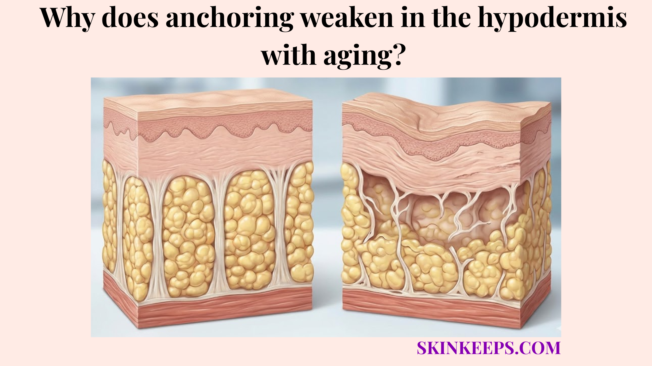
How does healthy dermal elastin fiber arrangement differ from aged or damaged dermal elastin fiber arrangement?
Healthy dermal elastin fiber arrangement supports smoother recoil, while aged or damaged arrangement weakens the skin’s ability to return to shape efficiently.
Healthy dermal organization is more coherent, branching, and mechanically supportive, while aged or damaged arrangement becomes weaker, more fragmented, or disorganized into non-functional clumps.
Chronic sun exposure deeply disrupts elastic-fiber architecture and can produce abnormal elastin-containing material (solar elastosis) in photoaged skin, while intrinsically aged skin shows a progressive loss and degeneration of the normal elastic-fiber network rather than healthy coherent recoil architecture.
| Elastin state | Fiber arrangement | Recoil quality | Visible skin effect |
|---|---|---|---|
| Healthy dermal elastin fiber arrangement | Coherent, branching, supportive | Better rebound | More resilient, springy skin |
| Aged or damaged dermal elastin fiber arrangement | Fragmented, weakened, less organized | Poorer recoil | Laxity, reduced bounce, wrinkling |
Which signs suggest dermal elastin fiber arrangement is weakening?
Dermal elastin fiber arrangement may be weakening when the skin appears less springy, less elastic, and slower to rebound after movement.
Warning signs can include reduced bounce after facial expressions, greater tissue laxity, slower spring-back after applied pressure (such as pillow creases), and fine wrinkling that becomes more persistent.
These visible and functional changes are consistent with weaker recoil architecture rather than simply reduced hydration alone. Photoaging and elastin reviews strongly link abnormal elastic-fiber organization with reduced skin resilience, wrinkling, and laxity.
What factors weaken dermal elastin fiber arrangement?
Dermal elastin fiber arrangement weakens when cumulative damage reduces elastic fiber integrity, network organization, and recoil performance over time.
Major weakening factors include chronic UV exposure, chronological aging, oxidative stress, smoking, ongoing inflammation, and long-term connective-tissue stress. The same factors that degrade collagen density aggressively attack elastin.
UV exposure is especially important because chronic sun damage is a major driver of elastic-fiber disorganization in photoaged skin. Reviews of solar elastosis and elastin biology consistently identify UV exposure as a major, direct disruptor of elastic architecture.
What habits and ingredients help preserve dermal elastin fiber arrangement?
The best preservation strategy for dermal elastin fiber arrangement focuses on protection, lower chronic stress, and long-term tissue stability rather than aggressive short-term correction.
The goal is not to promise rapid elastin rebuilding. The realistic clinical goal is to reduce the environmental and inflammatory stressors that distort or degrade the elastic network over time.
The specific strategies below outline how to protect this delicate architecture.
How does sun protection help preserve dermal elastin fiber arrangement?
Sun protection helps preserve dermal elastin fiber arrangement because UV exposure is one of the strongest disruptors of elastic fiber quality.
Daily SPF helps reduce cumulative elastin damage and preserve the elastic architecture that remains.
Photoprotection is a preservation strategy, not a claim of instant elastic-fiber rebuilding. Photoaging literature consistently identifies chronic UV exposure as a primary cause of abnormal elastic-fiber remodeling.
How do barrier support and lower inflammation help preserve dermal elastin fiber arrangement?
Barrier support and lower inflammation help preserve dermal elastin fiber arrangement because chronic surface stress can contribute to deeper connective-tissue burden over time.
Calmer skin environments are more supportive of long-term structural preservation than chronically irritated conditions.
This section should stay preservation-focused and avoid overpromising direct elastin regrowth from topical calmness alone.
How do long-term supportive routines help preserve dermal elastin fiber arrangement?
Long-term supportive routines help preserve dermal elastin fiber arrangement because consistent, non-aggressive care reduces repeated tissue stress over time.
Harsh routines increase cumulative damage without improving elastic quality.
Preservation works better through stability than through repeated skin aggression. Reviews of damaged elastic networks emphasize protection and long-term reduction of injury rather than repeated aggressive correction as the more credible preservation strategy.
Problem: dermal elastin fiber arrangement is weakening
Implication: recoil and elastic resilience are becoming less stable
Solution: reduce cumulative damage, protect the skin, and maintain a lower-stress skin environment
What are the key takeaways about dermal elastin fiber arrangement?
Dermal elastin fiber arrangement forms a branching recoil network throughout the dermis.
By securing both superficial flexibility and deep resilience, this architectural map preserves structural memory.
Summary Points
- Dermal elastin fiber arrangement forms a branching recoil network throughout the dermis
- The upper dermis contains a finer elastic pattern, while the deeper dermis contains a broader recoil network
- Elastin works with collagen and the surrounding matrix to create flexible but stable skin mechanics
- Aging and UV damage weaken the arrangement and reduce visible rebound over time
- The best support strategy focuses on preservation, not quick-fix claims of rebuilding
FAQs About Dermal Elastin Fiber Arrangement
Are elastin fibers only found in the deep dermis?
Elastin-related fibers are present in both the papillary and reticular dermis, but the upper dermis contains finer oxytalan and elaunin patterns while the deeper dermis contains broader mature elastic fibers.
What makes papillary dermal elastin different from reticular dermal elastin?
Papillary fibers are finer and more delicate to support surface flexibility, while reticular fibers are thicker and more mechanically dominant for deep recoil.
Does elastin work alone to create skin elasticity?
Elastin supports recoil, but it requires the structural framework of collagen and the hydrated environment of ground substance to produce stable dermal mechanics.
Why does UV damage affect elastin arrangement so much?
Chronic UV exposure physically disrupts elastic-fiber architecture and promotes the accumulation of abnormal elastotic material, crippling the skin’s ability to rebound.
What daily steps can you take to help preserve dermal elastin fiber arrangement?
Daily steps can help preserve dermal elastin fiber arrangement by lowering cumulative damage and keeping the connective environment more stable over time.
The most useful support comes from consistency and protection rather than aggressive short treatment cycles designed to trigger forced repair.
Daily Structural Preservation Checklist
Skin elasticity depends on organized elastic architecture, not just the word ‘elastin’ on a label.
Build your routine around long-term structural preservation if your goal is skin that stays more elastic, more responsive, and more resilient over time.