Keratinocytes lose their nuclei during maturation so they can transform from living epidermal cells into flattened, densely packed corneocytes that form the skin’s tough outer protective barrier. Removing the nucleus helps the cell make room for structural proteins, flatten more efficiently, and contribute to a more waterproof surface.
This is one of the most unusual transitions in human biology: a living cell gives up its internal control center without collapsing into debris. When this process fails, scaling, roughness, and visible barrier weakness can appear as the skin’s surface architecture is compromised by retained internal bulk.
Understanding why keratinocytes lose their nuclei during maturation makes it easier to explain how the skin becomes structurally strong, why retained nuclei signal abnormal maturation, and what actually helps support this conversion.
Why do keratinocytes lose their nuclei during maturation in the first place?
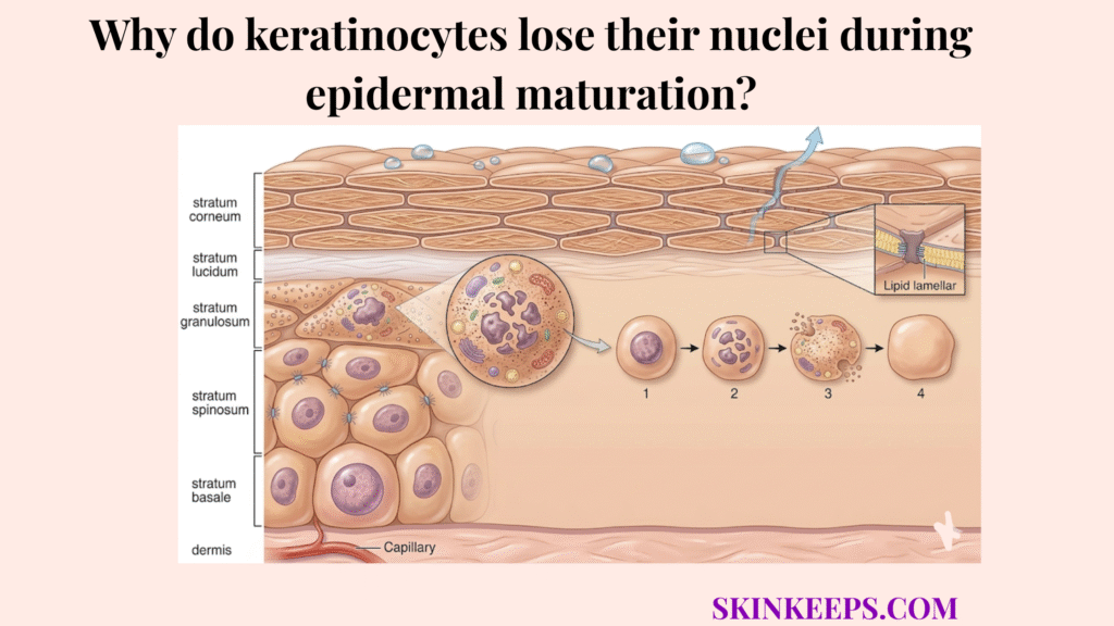
Keratinocytes lose their nuclei during maturation because the skin surface requires thin, highly compacted cells to maintain a functional barrier, and that degree of flattening is not possible while cells remain bulky with active nuclei and organelles.
A nucleus makes the cell too volumetric and metabolically active for the flat-stacking architecture required in the stratum corneum. To achieve the necessary density, the cell must undergo a programmed “clearing” phase that removes space-consuming internal structures.
Essentially, the skin is trading live-cell machinery for barrier efficiency. This controlled deconstruction ensures that the final corneocyte is a pure structural unit, optimized for physical resistance rather than biological activity.
Key takeaway: Keratinocytes lose their nuclei during maturation because the skin surface needs thin, flattened, highly compacted cells rather than metabolically active living cells.
How does losing the nucleus help a keratinocyte become a protective surface cell?
Losing the nucleus helps a keratinocyte become a protective surface cell by freeing internal space for dense keratin-based packing, allowing the cell to flatten more completely and stack tightly into a water-resistant barrier layer during the process of keratinization.
Removing the nucleus reduces internal bulk and makes cell flattening mechanically possible. Without this removal, the outer skin layer would remain too thick and porous, significantly increasing water loss and reducing environmental defense.
This structural simplification improves how surface cells stack, seal, and resist water loss. The resulting “bricks” of the skin barrier are thus engineered for maximum strength and minimum volume.
Key takeaway: Keratinocytes lose their nuclei during maturation because this structural simplification helps them become flatter, denser, and better suited for barrier duty.
How do enzymes and intracellular cleanup pathways ensure that keratinocytes lose their nuclei during maturation?
Enzymes and intracellular cleanup pathways ensure that keratinocytes lose their nuclei during maturation through a tightly regulated dismantling process that breaks down nuclear material and removes unneeded organelles while preserving the cell’s structural framework.
The cell is not collapsing randomly; it is selectively dismantling specific internal parts while preserving the shell needed for barrier formation. This controlled self-digestion is a hallmark of terminal differentiation.
The cleanup machinery involves both genetic degradation and organelle recycling, ensuring that the cell transitions into a dead unit that is still functionally useful to the body.
How do DNase-related enzymes help keratinocytes lose their nuclei during maturation?
DNase-related enzymes help keratinocytes lose their nuclei during maturation by degrading nuclear DNA so the cell can eliminate genetic material without destroying the structural proteins required for the final barrier cell.
DNase1L2 is especially relevant to epidermal terminal differentiation because it is strongly linked to nuclear DNA removal in the later stages of maturation. Epidermal studies identify DNase1L2 as essential to DNA degradation during corneocyte formation and show reduced expression in parakeratotic epidermis (jidonline.org).
How does autophagy help keratinocytes lose their nuclei during maturation?
Autophagy helps keratinocytes lose their nuclei during maturation by removing organelles and internal bulk so the cell can complete its transition into a simplified, flattened barrier unit.
Autophagy functions as a cleanup and remodeling system, not just a starvation response. This process helps the cell shed unnecessary internal complexity while preserving the protein-rich framework needed for the final corneocyte. Recent review literature describes terminal differentiation as involving the destruction and removal of many if not all organelles (pmc.ncbi.nlm.nih.gov).
| Deconstruction Pathway | Main Target | Preserved Outcome |
|---|---|---|
| DNase-related Degradation | Nuclear material (DNA) | Loss of genetic bulk |
| Autophagy | Organelles & clutter | Simplified, flatter cell |
| Preserved Element | Keratin framework | Mechanical surface strength |
How does the granular layer complete the transition in which keratinocytes lose their nuclei during maturation?
The granular layer completes the transition in which keratinocytes lose their nuclei during maturation because it acts as the decisive zone where terminal differentiation intensifies, organelles disintegrate, and the final steps of cornification are triggered.
This layer concentrates the late steps of maturation, including keratohyalin biology, lipid release, and organelle breakdown. It functions as the skin’s biological “exit lane,” where cells prepare for their final role at the surface.
The granular layer is where the cell stops functioning like a living keratinocyte and starts becoming a barrier-specific corneocyte. This transformation is irreversible and defines the quality of the barrier seal.
How does nucleus loss affect barrier strength and surface appearance?
Nucleus loss affects barrier strength and surface appearance by allowing corneocytes to stack in a more uniform, compact pattern that creates a stronger physical seal and a smoother-looking skin surface. This compaction is the primary mechanism that prevents barrier disruption from increasing TEWL.
Once nuclei are removed, the outer cells can flatten and overlap more evenly. This dense stacking is what makes the skin appear radiant and reflective rather than dull and uneven.
This affects both barrier function and visible texture: smoother compaction usually looks less scaly, less rough, and more refined. Successful de-nucleation is therefore a prerequisite for a “glowy” complexion.
How does normal maturation compare with parakeratosis when keratinocytes lose their nuclei during maturation?
Normal maturation results in the complete absence of nuclei in surface cells, known as orthokeratosis, whereas parakeratosis occurs when keratinocytes fail to lose their nuclei during maturation and reach the surface with retained nuclear material.
Orthokeratosis reflects completed terminal differentiation, while parakeratosis signals an incomplete and disordered cycle. This failure often stems from an abnormally rapid turnover speed.
In psoriasis, keratinocyte maturation and turnover are commonly described as accelerating from roughly 28 days to about 3–5 days, which helps explain why nuclear removal can fail and parakeratosis becomes visible (msdmanuals.com).
| Process State | Nucleus Status | Barrier Quality | Visible Result |
|---|---|---|---|
| Normal (Orthokeratosis) | Absent | Stronger & Organized | Smooth, Compact |
| Abnormal (Parakeratosis) | Retained | Weaker & Porous | Scaling, Roughness |
How can you tell when the cycle in which keratinocytes lose their nuclei during maturation is not completing properly?
You can tell when the cycle in which keratinocytes lose their nuclei during maturation is not completing properly by the presence of persistent scaling, rough-textured buildup, and recurring barrier fragility.
Failed de-nucleation often appears as abnormal surface retention rather than simple dryness alone. The skin feels “built up” yet remains sensitive and easily irritated.
Repeated scaling and rough buildup can point to disrupted maturation, especially when moisturizers alone do not normalize the surface. This indicates a biological failure in the maturation program itself.
Warning Signs of Disordered Maturation
What nutrients and actives support the cycle in which keratinocytes lose their nuclei during maturation?
Specific nutrients and active ingredients support the cycle in which keratinocytes lose their nuclei during maturation by regulating differentiation speed and maintaining the biochemical environment required for successful terminal maturation.
Some ingredients regulate the pace of differentiation, while others support the enzymatic environment the cell needs to complete maturation. The goal is not faster shedding at all costs, but more complete and orderly differentiation.
How do retinoids support the cycle in which keratinocytes lose their nuclei during maturation?
Retinoids support the cycle in which keratinocytes lose their nuclei during maturation by binding to nuclear receptors that normalize differentiation and help prevent the disordered maturation patterns associated with hyperproliferative skin.
Retinoids are useful here because they regulate keratinocyte differentiation rather than merely scraping away surface buildup. Retinoid review literature consistently describes these compounds as regulators of keratinocyte proliferation and differentiation (pmc.ncbi.nlm.nih.gov).
How do zinc and calcium support the cycle in which keratinocytes lose their nuclei during maturation?
Zinc and calcium support the cycle in which keratinocytes lose their nuclei during maturation by helping maintain the enzymatic and differentiation conditions required for orderly terminal maturation.
Calcium acts as a major differentiation signal in the upper epidermis, while zinc supports enzyme-dependent biology more broadly. Zinc is recognized as a cofactor for more than 300 enzymatic reactions in biology, which is why zinc sufficiency matters when the skin is trying to complete complex differentiation programs (sciencedirect.com).
| Maturation Problem | Surface Consequence | Support Strategy |
|---|---|---|
| Disordered differentiation | Scaling & rough buildup | Regulate with Retinoid support |
| Poor enzymatic environment | Incomplete maturation | Support with cofactors like Zinc |
| Barrier instability | Fragility & irritation | Reduce stripping and preserve Barrier balance |
What daily habits help keratinocytes lose their nuclei during maturation successfully?
You can help keratinocytes lose their nuclei during maturation successfully by avoiding aggressive routines that destabilize the upper epidermis and by maintaining the hydration and barrier balance required for orderly differentiation.
Routine aggression can disrupt terminal maturation even when the goal is smoother skin. The best habits protect the environment in which differentiation finishes correctly, allowing the skin to complete its biological deconstruction program.
Daily De-Nucleation Support Protocol
Closing insight: Healthy surface skin depends on controlled completion, not just rapid turnover. The goal is to help keratinocytes finish their maturation program properly, not simply force the outer layer to shed faster.
Build your routine around balanced differentiation support if your goal is a smoother surface, stronger barrier, and more normal completion of the maturation cycle.
What are the key takeaways regarding why keratinocytes lose their nuclei during maturation?
The key takeaways regarding why keratinocytes lose their nuclei during maturation center on the need for cellular compaction in barrier defense and the visible consequences of failed nuclear removal.
- ● Keratinocytes lose their nuclei so they can flatten and become effective barrier cells.
- ● DNA degradation and autophagy remove internal bulk while preserving structural frameworks.
- ● Normal completion produces orthokeratotic cells, while failure causes parakeratotic scaling.
- ● The best support strategy combines differentiation regulation with barrier preservation.
Ultimately, when you support the flow of your skin’s maturation escalator, radiance follows naturally.