
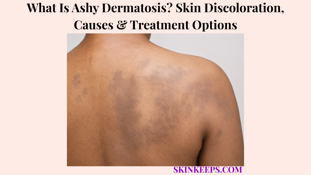
Ashy dermatosis is an acquired pigmentary disorder that causes gray, blue-gray, slate-gray, or ashy-brown flat patches on the skin. It is also called erythema dyschromicum perstans, or EDP.
This page explains symptoms, locations, possible causes, diagnosis, treatment limits, safe daily support, mistakes, and when dermatology care is needed. Ashy dermatosis should not be treated as ordinary uneven tone, melasma, post-inflammatory hyperpigmentation, drug pigmentation, or a bleaching-cream problem.
What Is Ashy Dermatosis and Why Is It Called Erythema Dyschromicum Perstans?
Ashy dermatosis, also called erythema dyschromicum perstans, is an acquired pigmentary condition that causes well-defined gray, blue-gray, slate-gray, or ashy patches.
The patches are usually macules or patches, meaning flat areas of discoloration rather than raised plaques. The gray tone can reflect pigment deeper in the skin, which is why it may look different from ordinary brown marks.
EDP is not dirt, dryness, poor hygiene, or a simple cosmetic uneven-tone issue. It needs diagnosis-first evaluation because several pigmentation disorders can look similar.
Why Does “Ashy” Describe the Skin Color Pattern?
“Ashy” describes the gray, blue-gray, slate-gray, or violaceous-brown color quality that can make the patches look different from ordinary brown hyperpigmentation.
The patches may be round, oval, or irregular and often have well-circumscribed borders. Some early lesions may have a reddish or slightly raised border, but that early phase is not required and may never be noticed.
Why Is Ashy Dermatosis Not the Same as Ordinary Hyperpigmentation?
Ashy dermatosis is not ordinary hyperpigmentation because the color often has a gray dermal quality and the pattern may appear on both sun-exposed and sun-protected skin.
Ordinary hyperpigmentation usually has a clearer trigger such as acne, rash, burn, injury, hormones, sun exposure, or irritation. Ashy dermatosis often needs rule-out thinking for drug pigmentation, melasma, lichen planus pigmentosus, post-inflammatory hyperpigmentation, and systemic pigmentation.
| Feature | Ashy Dermatosis | Ordinary Hyperpigmentation |
|---|---|---|
| Color quality | Gray, blue-gray, slate-gray, violaceous-brown | Often brown or dark brown |
| Depth clue | Dermal pigmentation quality | Often epidermal or mixed depending on cause |
| Location | Trunk, proximal limbs, neck, sometimes face | Depends on trigger |
| Trigger history | Often unclear | Often follows sun, inflammation, hormones, or injury |
| Treatment response | Often slow and resistant | Depends on cause and depth |
| Main risk | Misdiagnosis and over-treatment | Wrong cause can still delay care |
Practical rule: Gray, blue-gray, or slate-gray flat patches should be evaluated by pattern and history before bleaching, peels, or laser treatment.
What Ashy Dermatosis Symptoms Appear on Skin?
Ashy dermatosis usually appears as gray, slate-gray, blue-gray, or violaceous-brown flat patches that may be round, oval, irregular, symmetrical, or sometimes one-sided.
The patches are usually not painful and may have little or no scale, itch, or open skin. Mild itch can occur in some cases, but symptoms such as pain, ulceration, heavy scale, or rapid change should prompt rule-out evaluation.
What Do Early Ashy Dermatosis Patches Look Like?
Early ashy dermatosis patches may begin with a faint reddish or erythematous edge, but that early phase is not always seen.
The border may be slightly raised in some early lesions. Over time, the color may settle into gray, blue-gray, slate-gray, or violaceous-brown discoloration.
What Do Established Ashy Dermatosis Patches Look Like?
Established ashy dermatosis patches are usually flat gray or blue-gray areas with round, oval, or irregular shapes and little or no scale, pain, or open skin.
The patches can persist for years in some people. Changing, symptomatic, or unusual lesions should not be assumed to be EDP without a dermatologist checking for mimics.
| Patch Feature | What the Reader May Notice | Diagnostic Value |
|---|---|---|
| Gray / blue-gray color | Ashy or slate-like tone | Supports EDP pattern |
| Flat macule or patch | Discoloration without major elevation | Helps separate from raised rash |
| Well-defined edge | Border can be clear | Supports pattern recognition |
| Early reddish border | May appear in early lesions | Not required for diagnosis |
| Trunk/proximal limb pattern | Patches away from face-only distribution | Helps separate from melasma |
| Little/no symptoms | Usually not painful or intensely itchy | Symptoms suggest rule-outs |
| Persistence | May remain for years | Sets expectations |
Where Does Ashy Dermatosis Usually Appear?
Ashy dermatosis commonly affects the trunk, proximal arms or legs, neck, and sometimes the face, with patches that may appear on both sun-exposed and sun-protected skin.
The trunk can include the back, chest, or abdomen. Proximal extremities means areas closer to the body, such as upper arms and upper legs.
Face-only pigmentation needs careful differential diagnosis because melasma, Riehl melanosis, drug pigmentation, lentigines, and other pigment disorders may look similar.
| Area | Commonity Clue | What Else to Rule Out |
|---|---|---|
| Trunk | Common EDP area | PIH, drug pigmentation, lichen planus pigmentosus |
| Proximal arms | Common extension area | PIH, drug pigmentation, lichenoid pigmentation |
| Proximal legs | Possible distribution | PIH, drug pigmentation, systemic pigmentation |
| Neck | Can be involved | Lichen planus pigmentosus, Riehl melanosis, contact triggers |
| Face | Less frequent but possible | Melasma, Riehl melanosis, drug pigmentation, lentigo |
| Sun-protected skin | Supports non-melasma thinking | Drug/systemic causes still need review |
What Causes Ashy Dermatosis?
The exact cause of ashy dermatosis is unknown, but proposed associations include genetic susceptibility, contact allergy, chemical exposure, infections, and medication reactions.
Cause uncertainty is one reason diagnosis matters. A gray-brown patch may look like EDP but still come from a medicine, prior inflammation, contact exposure, lichen planus pigmentosus, or systemic pigmentation pattern.
Why May Ashy Dermatosis Be Linked With Immune or Lichenoid Inflammation?
Ashy dermatosis may overlap with lichenoid or interface inflammation, which is one reason dermatologists may compare it with lichen planus pigmentosus.
Biopsy findings can overlap, so diagnosis should use the clinical pattern, distribution, symptoms, oral or genital findings, medication history, and product exposure history. Oral, genital, nail, or hair clues may shift attention toward classic lichen planus or related conditions.
Why Should Medications and Exposures Be Reviewed?
Medications and exposures should be reviewed because drug pigmentation, fixed drug eruption, cosmetics, hair dyes, fragrances, and contact allergens can mimic or contribute to gray-brown discoloration.
A medication-triggered skin reaction may resemble a drug rash pattern or fixed drug eruption when the same site recurs after exposure. Medication review may reveal a reversible cause, but prescribed medicines should not be stopped without clinician guidance.
| Possible Contributor | Clue | What Dermatologist May Ask |
|---|---|---|
| Genetic susceptibility | Family/ethnic pattern, earlier onset | Family background and onset age |
| Contact allergy | Cosmetics, hair dye, fragrance, topical exposure | Product timeline and patch-test need |
| Chemical exposure | Work, environment, dye, chemical contact | Exposure history |
| Viral/infectious theory | Reported association, not proven cause | Illness history only if relevant |
| Medication reaction | New or long-term medication timing | Drug timeline and fixed-site recurrence |
| Lichenoid inflammation | Gray-brown patches, mucosal signs, biopsy overlap | Oral/genital exam and biopsy context |
Who Is More Likely to Develop Ashy Dermatosis?
Ashy dermatosis has been reported across skin colors and ethnic groups, but it is described more often in darker-skinned patients and appears more frequent in young adults, with women affected more often than men.
Latin American and Indian populations are reported more often in some dermatology references. Lighter skin and various ethnic backgrounds can still be affected, so demographics should never be used to dismiss a possible diagnosis.
- Darker skin tone context.
- Young-adult onset context.
- Female predominance in some descriptions.
- Latin American or Indian population reports.
- Lighter skin still possible.
- Any age possible.
- Distribution and history matter more than demographics alone.
How Is Ashy Dermatosis Different From Other Skin Discoloration Conditions?
Ashy dermatosis must be separated from melasma, post-inflammatory hyperpigmentation, lichen planus pigmentosus, drug pigmentation, fixed drug eruption, and systemic causes of diffuse pigmentation.
Color, location, symptoms, medication timing, prior rash history, oral or genital findings, and systemic symptoms all help prevent wrong treatment.
How Is Ashy Dermatosis Different From Melasma?
Ashy dermatosis is different from melasma because melasma usually favors sun-exposed facial areas and brown pigmentation, while ashy dermatosis often has a gray or blue-gray tone and may involve the trunk or proximal limbs.
A face-only pattern should be evaluated carefully because melasma, Riehl melanosis, drug pigmentation, lentigines, and other pigment disorders can overlap visually.
How Is Ashy Dermatosis Different From Lichen Planus Pigmentosus?
Ashy dermatosis can overlap with lichen planus pigmentosus, so dermatologists may use distribution, symptoms, mucosal signs, history, and biopsy context to separate them.
Lichen planus pigmentosus may have brown-gray or ashy macules and may favor face, neck, folds, or sun-exposed and flexural sites. Oral or genital lichen planus clues can change the evaluation.
How Is Ashy Dermatosis Different From Drug Pigmentation or Fixed Drug Eruption?
Ashy dermatosis is different from drug pigmentation or fixed drug eruption because medication timing, repeated same-site recurrence, and long-term drug exposure can point to a medication-related cause.
Drug pigmentation may appear after ongoing medicine exposure. Fixed drug eruption often recurs in the same site after a medicine exposure and may leave darker pigmentation afterward.
| Condition | Color Clue | Location Clue | History Clue |
|---|---|---|---|
| Ashy dermatosis | Gray, blue-gray, slate-gray, violaceous-brown | Trunk, neck, proximal limbs, sometimes face | Often asymptomatic and slow |
| Melasma | Brown or gray-brown | Face and sun-exposed areas | Sun/hormone pattern |
| Post-inflammatory hyperpigmentation | Brown or dark marks | Prior rash, acne, burn, injury sites | Inflammation before pigment |
| Lichen planus pigmentosus | Brown-gray or ashy | Face, neck, folds, sun-exposed/flexural sites | Lichenoid or mucosal clues possible |
| Drug pigmentation | Gray, brown, blue, or mixed | Depends on medication | Medication timing or long-term use |
| Fixed drug eruption | Dark recurrent patches | Same site after drug exposure | Repeats after same medicine |
| Systemic pigmentation | Diffuse or mucosal darkening possible | Wider body/mucosal pattern | Fatigue, dizziness, weight loss, systemic clues |
How Is Ashy Dermatosis Diagnosed or Checked?
Ashy dermatosis is diagnosed from the skin pattern, history, medication review, and sometimes biopsy, especially when discoloration is unusual, spreading, symptomatic, or could represent another pigment disorder.
A dermatologist may diagnose a classic pattern clinically, but many gray-brown pigmentation problems need rule-out evaluation before treatment.
What History Matters Most With Ashy Dermatosis?
Ashy dermatosis history matters because timing, spread, symptoms, medicines, products, prior rash, sun pattern, mucosal signs, and systemic symptoms can point toward EDP or a mimic.
Useful details include when the patches started, how quickly they spread, whether itch, pain, scale, redness, or prior rash appeared, and whether new medicines, supplements, cosmetics, fragrances, dyes, hair dyes, or chemicals were used.
Sun exposure pattern, family or ethnic background, oral or genital symptoms, nail changes, hair changes, fatigue, weight loss, dizziness, and previous treatments can also guide diagnosis.
When Might Biopsy or Additional Evaluation Be Needed?
Biopsy or additional evaluation may be needed when the diagnosis is unclear, discoloration changes rapidly, distribution is unusual, mucosal areas are involved, medication reaction is possible, or melanoma-like or systemic pigmentation must be ruled out.
A biopsy may show basal-layer vacuolar degeneration in early lesions and pigmentary incontinence with dermal melanophages in established lesions. These findings can support the pattern, but biopsy is not always mandatory and may not separate every mimic by itself.
- Photos over time.
- Timeline of onset and spread.
- Areas affected.
- Itch, pain, scale, redness, ulceration, or prior rash.
- Current and past medicines.
- Supplements.
- Cosmetics, fragrances, hair dyes, and topical products.
- Sun exposure pattern.
- Oral, genital, nail, or hair changes.
- Fatigue, weight loss, dizziness, or systemic symptoms.
- Prior treatments and any irritation after use.
What Treatment Options Are Used for Ashy Dermatosis?
Ashy dermatosis is often resistant to treatment, so the main goals are diagnosis confirmation, mimic exclusion, trigger review, gradual cosmetic improvement, and safe expectation-setting.
Treatment should not be framed as quick lightening. The pigment may be deeper, the cause is unclear, and aggressive irritation can worsen discoloration.
Which Topical or Procedure-Based Treatments May Be Considered?
Topical or procedure-based treatments for ashy dermatosis may be considered by a dermatologist, but response is unpredictable and irritation can worsen pigmentation.
Selected cases may involve topical corticosteroids for early or inflammatory activity, topical calcineurin inhibitors such as tacrolimus, sunscreen, carefully selected chemical peels, pigment lasers, or phototherapy approaches.
Laser or peel treatment should come after diagnosis and pigment-risk counseling, especially in darker skin tones where irritation can worsen pigmentation.
Which Systemic Treatments Have Been Reported for Ashy Dermatosis?
Systemic treatments have been reported for ashy dermatosis, but evidence is limited and these medicines are not casual cosmetic therapy.
Clofazimine has been reported in small studies or case series. Other systemic medicines such as dapsone, griseofulvin, hydroxychloroquine, isoniazid, or corticosteroids have been reported in limited cases, not as routine treatment for everyone.
Systemic therapy needs specialist judgment because side effects, monitoring, interactions, pregnancy context, and the uncertain benefit-risk balance matter.
Why Are Treatment Results Unpredictable?
Treatment results are unpredictable because the cause is unclear, pigment may sit deeper in the skin, and some cases remain stable for years while others slowly improve.
Some cases may eventually clear, while others persist. Treatment expectations should focus on safety, rule-out accuracy, irritation avoidance, and gradual monitoring rather than guaranteed clearing.
| Option | Goal | Best-Fit Situation | Limitation / Caution |
|---|---|---|---|
| Observation | Confirm stability and avoid harm | Mild, stable, asymptomatic cases | Does not lighten quickly |
| Sunscreen | Reduce added contrast and sun-triggered darkening | Exposed areas | Does not treat dermal pigment directly |
| Topical corticosteroids | Reduce inflammation in selected early/inflammatory cases | Dermatologist-guided | Long-term misuse can harm skin |
| Topical calcineurin inhibitors | Anti-inflammatory support in selected cases | Specialist-guided, sensitive areas | Evidence limited |
| Chemical peels | Cosmetic improvement in selected patients | Carefully selected cases | Irritation/pigment worsening risk |
| Pigment lasers | Target pigment in selected cases | Specialist-guided after diagnosis | Risk of worsening in darker skin; not first-step care |
| Clofazimine | Reported systemic option | Selected resistant cases | Side effects, monitoring, limited evidence |
| Cosmetic camouflage | Appearance support | Distress or visible areas | Does not treat the condition |
How Can Daily Skin Care Support Ashy Dermatosis Safely?
Daily skin care cannot reliably cure ashy dermatosis, but it can reduce irritation, prevent added post-inflammatory pigmentation, and protect skin from extra discoloration contrast.
Use gentle cleanser, avoid harsh scrubs, and avoid strong bleaching mixtures unless a dermatologist specifically prescribes a plan. Sunscreen can help reduce contrast on exposed areas, but it does not directly remove dermal pigment.
Pause suspected fragrance, hair dye, cosmetic, or topical triggers if they seem connected to flares. Cosmetic camouflage is reasonable if it is non-irritating and helps confidence.
- Use a gentle cleanser.
- Avoid scrubs, acids, and harsh bleaching mixtures unless prescribed.
- Use sunscreen on exposed areas.
- Avoid picking, rubbing, or irritating patches.
- Pause suspected fragrance, hair dye, cosmetic, or topical triggers.
- Patch test new products when skin is reactive.
- Photograph patches under consistent lighting.
- Use non-irritating cosmetic camouflage if desired.
- Review worsening or spreading changes with a dermatologist.
What Ashy Dermatosis Mistakes Should You Avoid?
The biggest ashy dermatosis mistake is treating gray-brown patches with aggressive lightening products before confirming that the discoloration is truly ashy dermatosis.
Strong bleaching creams, steroid mixtures, harsh peels, random acids, and unplanned lasers can irritate skin and worsen pigmentation. Skipping medication history, systemic symptoms, oral or genital clues, or changing lesions can also delay the correct diagnosis.
| Mistake | Why It Fails | Better Action |
|---|---|---|
| Using strong bleaching first | Irritation can worsen pigmentation | Confirm diagnosis first |
| Calling it melasma automatically | Pattern and color may differ | Check distribution and history |
| Ignoring medicines | Drug pigmentation may be reversible | Review medication timeline |
| Expecting quick results | EDP is often treatment-resistant | Set long-term expectations |
| Random laser treatment | Pigment worsening risk | Use specialist-guided treatment |
| Skipping systemic review | Internal or drug causes may be missed | Share symptoms and health history |
When Should Ashy Dermatosis Be Checked by a Dermatologist?
Ashy dermatosis should be checked by a dermatologist when gray, blue-gray, or brown patches are new, spreading, unclear in cause, symptomatic, cosmetically distressing, linked with medicine changes, or different from ordinary hyperpigmentation.
Dermatology review is especially useful before lightening creams, peels, lasers, or systemic medicines are considered.
Which Discoloration Signs Need Faster Evaluation?
Gray or brown discoloration needs faster evaluation when it spreads quickly, becomes symptomatic, involves mucosal areas, changes irregularly, or appears with systemic symptoms.
Warning signs include pain, itch, scale, ulceration, inflammation, oral or genital changes, nail or hair changes, fatigue, weight loss, dizziness, medication-associated onset, or very dark irregular lesions.
An irregular changing pigmented lesion should not be treated as EDP without evaluation because conditions such as lentigo maligna require a different diagnostic pathway.
What Should You Bring to an Ashy Dermatosis Appointment?
A useful ashy dermatosis appointment starts with photos, timeline, medication list, product exposures, prior rash history, sun pattern, mucosal symptoms, and systemic symptoms.
Do not stop prescribed medicines, bleach aggressively, or start strong acids before the visit unless a clinician has already advised that plan.
What Should You Remember About Ashy Dermatosis?
The most important thing to remember about ashy dermatosis is that it is a gray-toned acquired pigmentation disorder where correct diagnosis and realistic treatment expectations matter more than aggressive lightening.
The exact cause is unknown, similar conditions must be ruled out, and treatment can be slow or unpredictable. Safe care focuses on diagnosis, trigger review, irritation avoidance, sunscreen where useful, and specialist-guided treatment decisions.
Frequently Asked Questions About Ashy Dermatosis
Is ashy dermatosis the same as erythema dyschromicum perstans?
Yes. Ashy dermatosis is a common name for erythema dyschromicum perstans, also called EDP or ashy dermatosis of Ramirez.
What does ashy dermatosis look like?
Ashy dermatosis can look like well-defined gray, blue-gray, slate-gray, ashy-brown, or violaceous-brown flat patches, often on the trunk, proximal extremities, neck, and sometimes face.
What causes ashy dermatosis?
The exact cause is unknown, but proposed associations include genetic susceptibility, contact allergy, chemicals, infections, and medication effects.
Is ashy dermatosis the same as melasma?
No. Melasma usually favors brown facial patches in sun-exposed areas, while ashy dermatosis often has a gray or blue-gray tone and may affect trunk or proximal limb areas.
Can ashy dermatosis go away?
Some cases may eventually clear, but ashy dermatosis can persist unchanged for years, so timelines are unpredictable.
What is the best treatment for ashy dermatosis?
There is no one best treatment for every case. Treatment is often difficult and may include dermatologist-guided topical anti-inflammatory therapy, sunscreen, cosmetic support, selected procedures, or rarely systemic therapy, but results are unpredictable.
Should I use bleaching creams for ashy dermatosis?
Unsupervised bleaching is not recommended. Harsh lightening products, steroid mixtures, or strong peels can irritate the skin and worsen pigmentation if the diagnosis is wrong or the skin reacts.
When should ashy dermatosis need biopsy?
Biopsy may be considered when the diagnosis is unclear, distribution is unusual, pigmentation spreads rapidly, lesions change concerningly, medication reaction is suspected, mucosal findings appear, or lichen planus pigmentosus, drug pigmentation, melanoma-like pigmentation, or systemic causes need to be ruled out.
Sources & Evidence About Ashy Dermatosis
DermNet — Erythema Dyschromicum Perstans was used for synonyms, acquired dermal macular hyperpigmentation definition, gray patches, face/neck/trunk distribution, early reddish border, demographic context, unknown cause, differential diagnoses, biopsy findings, treatment resistance, and reported treatment options.
American Family Physician — Ashy Dermatosis was used for diagnostic reasoning around slate-gray or violaceous macules, trunk/proximal extremity distribution, medication history, general health history, oral/genital examination, biopsy logic, treatment limitations, and small clofazimine evidence context.
PubMed Central / Peer-Reviewed Case Literature was used as background context for the overlap between erythema dyschromicum perstans and lichen planus pigmentosus, histology limitations, and limited evidence around topical or phototherapy approaches where case-level evidence exists.
PubMed Treatment Reports was used as background context for small treatment reports involving clofazimine, dapsone, griseofulvin, hydroxychloroquine, or other systemic options, with emphasis that these are specialist-guided and not casual cosmetic therapy.
DermNet — Postinflammatory Hyperpigmentation was used for differential context when pigmentation follows acne, rash, burn, injury, or irritation.
DermNet — Melasma was used for differential context when brown facial pigmentation favors sun-exposed areas rather than gray-toned trunk or proximal-limb patches.
DermNet — Drug-Induced Pigmentation was used for medication-history and drug-pigmentation differential context.
Educational Disclaimer: This SkinKeeps article is for educational purposes only and does not diagnose or replace medical care. New, spreading, rapidly changing, painful, itchy, scaly, ulcerated, mucosal, medicine-linked, irregular, or systemic-symptom-associated pigmentation should be checked by a qualified healthcare professional or dermatologist.



