
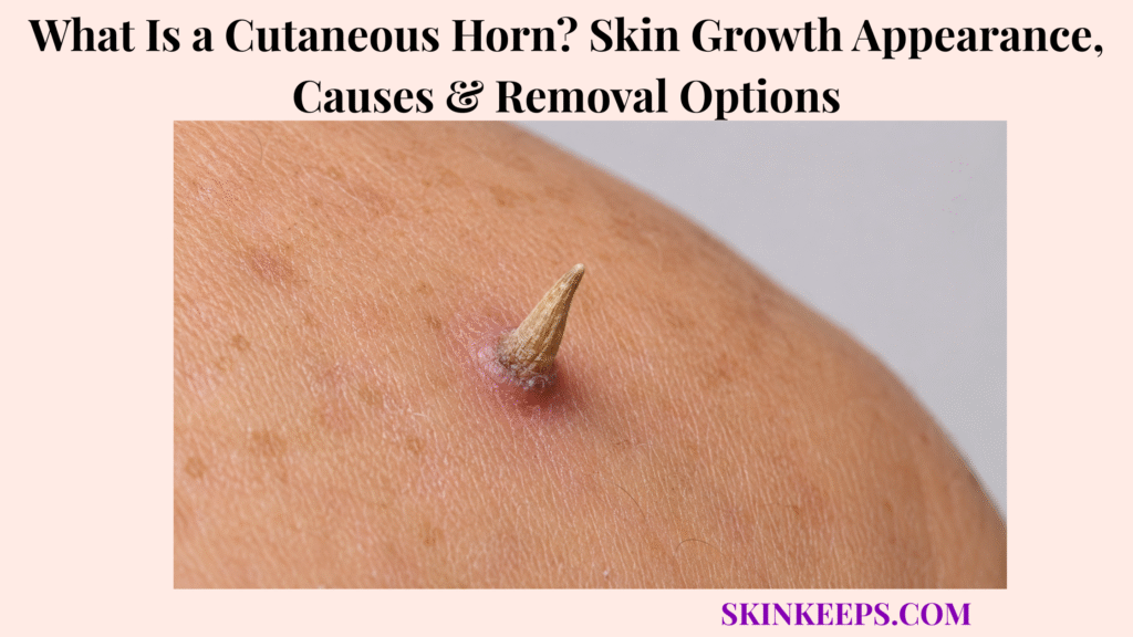
A cutaneous horn is a hard cone-shaped or spike-like projection from the skin made of compact keratin.
It is not a final diagnosis by itself because the base may be benign, precancerous, or cancerous. This page covers appearance, common sites, causes, base-lesion risk, diagnosis, biopsy, removal, aftercare, recurrence, unsafe home removal, and warning signs.
What Is a Cutaneous Horn and Why Does It Grow From Skin?
A cutaneous horn is a hard cone-shaped or spike-like projection from the skin made of compact keratin, the same structural protein found in the outer skin layer, hair, and nails.
The medical term is cornu cutaneum, which means skin horn.
The visible horn describes the shape. The important medical question is what lesion sits at the base and why it produced excess keratin.
Why Is a Cutaneous Horn Also Called Cornu Cutaneum?
A cutaneous horn is also called cornu cutaneum, a medical term that describes the horn-like shape rather than the underlying diagnosis.
It may be short, long, straight, curved, white, yellow, tan, brown, gray, dark, rough, nail-like, or crusted.
The visible part is compact keratin, but the base determines whether the cause is benign, precancerous, or cancerous.
Why Does the Base Matter More Than the Horn Tip?
The base matters more than the horn tip because the tip is mostly compact keratin, while the base contains the living lesion that produced the horn.
A horn-like projection may arise over benign lesions, precancerous lesions, or skin cancers.
Cutting only the top does not diagnose the cause. Biopsy or histology must include the base when the cause is uncertain or risk features are present.
Practical rule: Do not treat a cutaneous horn as a cosmetic spike; safe care depends on checking the base.
What Does a Cutaneous Horn Look Like on Skin?
A cutaneous horn usually looks like a hard, rough, cone-shaped, spike-like, nail-like, or horn-like growth projecting from the skin surface.
The horn may be straight or curved, small or large, dry or crusted, and attached to a base that may look quiet or visibly inflamed.
What Color and Texture Can a Cutaneous Horn Have?
A cutaneous horn can be white, yellow, tan, brown, gray, or darker and usually feels hard, dry, rough, crusted, or nail-like.
It may look like a small animal horn, a nail-like spike, a cone, a thick crust, or a compact keratin column.
Color does not prove whether the base is benign or cancerous.
What Can the Base Look Like?
The base of a cutaneous horn may look normal, pink, red, brown, darker, inflamed, thickened, tender, ulcerated, bleeding, crusted, scaly, wide, or firm.
A quiet base can still need assessment if the diagnosis is uncertain.
A painful, wide, firm, inflamed, bleeding, ulcerated, or rapidly growing base should be checked faster.
What Symptoms Can Happen With a Cutaneous Horn?
A cutaneous horn may cause no symptoms, but it can also become tender, painful, inflamed, itchy, irritated, bleeding after trauma, or recurrent after partial removal.
It may catch on clothing, shaving tools, jewelry, hats, masks, or combs.
Pain, inflammation, bleeding, ulceration, rapid growth, or regrowth after removal increases the need for dermatology review.
| Feature | Typical Clue | Why It Matters |
|---|---|---|
| Shape | Cone, spike, nail-like, horn-like projection. | Suggests compact keratin buildup. |
| Texture | Hard, dry, rough, crusted. | Keratin material. |
| Base | Flat, inflamed, thick, ulcerated, bleeding, wide, or firm. | Base decides diagnosis. |
| Symptoms | May be painless, tender, painful, itchy, or irritated. | Pain or inflammation can increase concern. |
| Course | Persistent, growing, or recurrent. | Needs assessment and removal planning. |
Where Do Cutaneous Horns Usually Appear?
Cutaneous horns often appear on sun-exposed skin, especially the face, ears, scalp, nose, forearms, backs of hands, and other chronically sun-damaged areas.
They can also appear on the neck, lips, trunk, limbs, or almost any skin site.
Location does not diagnose the base, but horns on sun-exposed areas deserve careful review because sun-damaged keratinocyte lesions can sit underneath.
| Site | Common Clue | Why Review May Matter |
|---|---|---|
| Face | Sun-exposed keratin growth. | High-risk visible site. |
| Nose | Chronic sun exposure. | Higher premalignant/malignant base concern. |
| Ear / pinna | Sun-exposed cartilage area. | SCC, AK, or BCC mimic concern. |
| Scalp | Sun-exposed in thinning hair. | Often chronically sun-damaged. |
| Forearm | Outdoor sun exposure. | Actinic damage context. |
| Back of hand | Chronic UV and trauma exposure. | Common keratinocyte lesion site. |
| Neck | Sun exposure and irritation. | Base still needs diagnosis. |
| Trunk / limbs | Less classic but possible. | Do not ignore growth or symptoms. |
What Causes a Cutaneous Horn?
A cutaneous horn forms when an underlying skin lesion produces excessive compact keratin, creating a raised horn-like projection above the skin.
The horn shape is only the surface clue. The underlying base may be benign, precancerous, or cancerous.
Which Benign Lesions Can Form a Cutaneous Horn?
Benign lesions such as seborrheic keratosis, viral warts, epidermal nevi, trichilemmoma, or other benign keratinizing growths can produce a cutaneous horn.
Benign is possible, but it should not be assumed from appearance alone.
A wart-like or stuck-on look does not remove the need to check a growing, painful, bleeding, wide-based, or recurrent horn.
Which Precancerous Lesions Can Form a Cutaneous Horn?
Precancerous lesions such as actinic keratosis, Bowen’s disease, and other chronically sun-damaged keratinocyte lesions can form a cutaneous horn.
A precancerous base needs complete treatment and monitoring, not tip-only clipping.
Sun-exposed sites such as the face, nose, ears, scalp, forearms, and backs of hands deserve careful assessment.
Which Skin Cancers Can Appear Under a Cutaneous Horn?
Skin cancers such as squamous cell carcinoma, basal cell carcinoma, keratoacanthoma-type lesions, and rare malignancies can appear at the base of a cutaneous horn.
The horn shape alone cannot prove or exclude cancer.
Base histology is what tells whether the lesion is benign, precancerous, or malignant and what treatment is needed next.
| Underlying Base Type | Examples | Why It Matters |
|---|---|---|
| Benign | Wart, seborrheic keratosis, epidermal nevus. | Removal may be simpler if confirmed. |
| Precancerous | Actinic keratosis, Bowen’s disease. | Needs complete treatment and monitoring. |
| Malignant | SCC, BCC, keratoacanthoma-type lesion, rare tumors. | Requires cancer-specific management. |
| Unclear | Atypical keratin growth. | Biopsy or histology required. |
Who Is More Likely to Develop a Cutaneous Horn?
Cutaneous horns are more concerning when they occur in older adults, men, sun-damaged skin, immunosuppressed people, or high-risk sites such as the face, ears, scalp, nose, forearms, and backs of the hands.
Risk features do not diagnose cancer, but they lower the threshold for faster dermatology review.
- Older age.
- Male sex.
- Long-term sun exposure.
- Sun-damaged skin.
- Fair skin or history of sunburn.
- Prior actinic keratoses or skin cancer.
- Immunosuppression.
- Outdoor work.
- Horn on face, ear, scalp, nose, forearm, or back of hand.
- Wide or firm base.
- Low height-to-base ratio.
- Pain, inflammation, ulceration, or bleeding.
- Recurrent horn after removal.
How Is a Cutaneous Horn Different From Other Skin Growths?
A cutaneous horn can resemble a wart, skin tag, seborrheic keratosis, actinic keratosis, nail-like growth, keratoacanthoma, squamous cell carcinoma, or basal cell carcinoma, so the base and pathology matter.
The goal is not to scare every reader. The goal is to avoid unsafe home treatment and false reassurance from appearance alone.
How Is a Cutaneous Horn Different From a Wart?
A wart can become thick and keratotic, but a horn-like projection does not prove the growth is a wart.
Some warts produce compact keratin and may look horn-like.
A persistent, painful, bleeding, fast-growing, sun-exposed, or atypical wart-like horn in an adult should be checked before wart remover or home freezing is used.
How Is a Cutaneous Horn Different From a Skin Tag?
A skin tag is usually soft and flexible, while a cutaneous horn is hard, keratinous, and spike-like.
Skin tags often hang from a narrow stalk and are usually soft or flesh-colored.
A firm base, crusting, bleeding, pain, rapid growth, or thick keratin spike should not be dismissed as a skin tag.
How Is a Cutaneous Horn Different From Actinic Keratosis or SCC?
Actinic keratosis may be rough and scaly, while squamous cell carcinoma may be tender, thick, crusted, bleeding, ulcerated, or fast-growing; either can sit at the base of a cutaneous horn.
A horn on sun-damaged skin may represent a thick keratin response over a precancerous or cancerous keratinocyte lesion.
Biopsy or histology is needed when risk features are present because appearance alone cannot reliably separate these possibilities.
How Is a Cutaneous Horn Different From Seborrheic Keratosis?
Seborrheic keratosis is usually waxy, stuck-on, or verrucous, but it can become thick enough to look horn-like.
Some benign keratoses can grow compact keratin.
Do not assume a horn is benign because it resembles seborrheic keratosis, especially if it is painful, bleeding, ulcerated, fast-growing, inflamed, or recurrent.
| Condition | Main Clue | Why Confusion Happens | Safer Next Step |
|---|---|---|---|
| Cutaneous horn | Hard compact-keratin projection. | Many base lesions are possible. | Remove or sample base. |
| Wart | Rough viral growth. | Can be keratotic or horn-like. | Confirm if atypical. |
| Skin tag | Soft flexible growth. | May protrude. | Check if hard, crusted, painful, or bleeding. |
| Actinic keratosis | Rough sun-damaged patch. | Can produce a horn. | Treat or biopsy if thick or suspicious. |
| SCC | Tender, crusted, growing, bleeding, ulcerated lesion. | Can sit under a horn. | Prompt biopsy. |
| BCC | Pearly, ulcerated, non-healing lesion. | Can crust or ulcerate. | Dermatology evaluation. |
| Seborrheic keratosis | Stuck-on waxy lesion. | May become thick and keratotic. | Check changing lesions. |
How Is a Cutaneous Horn Diagnosed?
A cutaneous horn can be recognized by its appearance, but the important diagnosis comes from examining the base of the horn under a microscope.
Clinical appearance identifies the horn-like shape. Histology identifies the underlying lesion.
What Does a Dermatologist Check During Examination?
A dermatologist checks the horn’s size, height, base width, firmness, tenderness, inflammation, bleeding, ulceration, growth speed, location, sun damage, immune status, and skin-cancer history.
The exam may also consider whether the lesion looks like SCC, BCC, wart, actinic keratosis, seborrheic keratosis, keratoacanthoma, or another skin growth.
Photos over time can help show growth speed, trauma, bleeding, crusting, or recurrence.
Why Should Biopsy or Excision Include the Base?
Biopsy or excision should include the base because the horn tip is mostly compact keratin, while the base contains the underlying lesion that determines risk and treatment.
Pathology tells whether the base is benign, precancerous, or malignant.
If only the tip is removed, the cause may remain unknown, recurrence may occur, and a serious base lesion may be missed.
When Is Faster Biopsy More Important?
Faster biopsy is more important when a cutaneous horn has a painful, wide, inflamed, bleeding, ulcerated, fast-growing, recurrent, or sun-exposed base.
Older age, immune suppression, previous skin cancer, and location on the face, ear, nose, scalp, forearm, or back of hand also increase concern.
Faster review does not mean the horn is automatically cancer; it means the base needs timely diagnosis.
- Photos showing growth over time.
- When the horn first appeared.
- Whether it is growing quickly.
- Whether the base hurts, bleeds, crusts, or ulcerates.
- Whether it catches on clothing, shaving, or jewelry.
- Any home clipping, wart acid, freezing, or removal attempts.
- Sun exposure history.
- Prior actinic keratoses or skin cancers.
- Immune suppression, transplant medicines, or cancer treatment history.
- Whether the horn regrew after previous removal.
- Any previous pathology reports.
What Removal Options Are Used for a Cutaneous Horn?
Cutaneous horn removal usually involves removing the horn and sampling or excising its base so the tissue can be examined and the underlying lesion can be treated correctly.
Removal is diagnosis-linked, not cosmetic-only. The right approach depends on the base, risk features, location, and need for pathology.
When Is Shave Removal or Shave Biopsy Used?
Shave removal or shave biopsy may be used for selected small or superficial-appearing horns when enough of the base can be sampled for pathology.
The sample must include adequate base tissue for diagnosis.
Shave removal may be inadequate if invasive cancer is suspected, margins are needed, or the base is wide, firm, painful, bleeding, ulcerated, or rapidly growing.
When Is Surgical Excision Used?
Surgical excision is used when the base is suspicious, wide, firm, painful, bleeding, ulcerated, fast-growing, recurrent, or located on high-risk sun-exposed skin.
Excision may allow fuller base assessment and margin review when needed.
Suspected SCC, BCC, precancerous disease, or recurrent horn should be managed with diagnosis quality first, not cosmetic convenience first.
When Are Curettage, Cautery, Cryotherapy, or Laser Considered?
Curettage, cautery, cryotherapy, or laser may be considered for selected confirmed low-risk lesions, but destructive treatment should not replace biopsy when cancer is possible.
These methods can destroy tissue that might otherwise help diagnosis.
Diagnosis should be secure before destructive treatment is used for a horn-like lesion.
What Happens If the Base Shows Skin Cancer or Precancer?
If the base shows skin cancer or precancer, treatment changes from simple horn removal to lesion-specific care based on the pathology result.
Actinic keratosis may need lesion-directed or field treatment. Bowen’s disease or SCC in situ needs complete treatment and follow-up.
Invasive SCC or BCC requires cancer-specific management, margin review, and follow-up based on pathology.
| Situation | Removal Direction | Key Caution |
|---|---|---|
| Small low-risk horn | Shave biopsy or excision with base pathology. | Do not remove tip only. |
| Suspicious base | Excision or biopsy with adequate depth. | Rule out SCC or BCC. |
| Confirmed benign lesion | Destructive or surgical options may fit. | Diagnosis first. |
| Precancerous base | Complete treatment + sun-damage plan. | Follow-up needed. |
| Cancerous base | Cancer-specific excision or management. | Margins and risk assessment matter. |
| Recurrent horn | Re-biopsy or excise base. | Check missed or persistent pathology. |
Should a Cutaneous Horn Be Removed at Home?
A cutaneous horn should not be clipped, cut, burned, frozen, pulled off, or treated with acid at home because that can cause bleeding, infection, scarring, recurrence, and missed cancer at the base.
Do not use nail clippers, scissors, blades, wart remover, home freezing kits, heat, acids, tying methods, or pulling.
If it catches, snags, or bleeds, cover it lightly and arrange medical assessment. Destroying the base before diagnosis can remove the chance for useful pathology.
| DIY Action | Why It Is Unsafe | Better Action |
|---|---|---|
| Clipping the tip | Misses base diagnosis. | Biopsy/removal with pathology. |
| Wart acid | Burns healthy skin and may delay diagnosis. | Confirm diagnosis first. |
| Home freezing | Destroys tissue before pathology. | Medical assessment. |
| Pulling or picking | Bleeding, infection, and scarring risk. | Protect and review. |
| Ignoring regrowth | Missed underlying lesion. | Reassess base. |
How Should Skin Be Cared for After Cutaneous Horn Removal?
After cutaneous horn removal, care usually focuses on keeping the wound clean, protected, and monitored while waiting for the pathology result.
Follow the clinician’s wound-care instructions, keep the area covered as advised, and do not pick the scab.
Ask when pathology results will return, whether margins or further treatment are needed, and whether follow-up is needed for precancerous or cancerous findings.
- Follow clinician wound-care instructions.
- Keep the area clean and covered as advised.
- Do not pick the scab.
- Watch for bleeding, pus, spreading redness, warmth, swelling, or fever.
- Ask when pathology results will return.
- Confirm whether margins or further treatment are needed.
- Use sun protection on exposed skin.
- Attend follow-up if pathology shows precancer or cancer.
Can a Cutaneous Horn Come Back?
A cutaneous horn can come back if the underlying base lesion is not fully treated or if a new keratin-producing lesion develops in the same sun-damaged area.
Tip-only removal may leave the base behind, which can allow regrowth.
Recurrence should be checked and may need repeat biopsy or excision, especially if it returns quickly, bleeds, hurts, widens, or appears on sun-exposed skin.
Recurrence map: Tip removed only → base remains → horn regrows or new lesion appears → reassess base → repeat biopsy or excision if needed → treat the pathology result.
What Cutaneous Horn Mistakes Should You Avoid?
The biggest cutaneous horn mistake is treating it as a harmless cosmetic spike without checking the base.
Do not remove only the tip, assume it is a wart, use home acid, freeze or burn it before diagnosis, or ignore pain, bleeding, ulceration, rapid growth, or a wide base.
Do not delay care for horns on the face, ear, scalp, nose, forearms, or backs of hands, and do not skip the pathology result after removal.
| Mistake | Why It Fails | Better Action |
|---|---|---|
| Clipping the horn tip | Misses base pathology. | Remove or sample base. |
| Calling it a wart | Cancer or actinic keratosis can mimic. | Confirm diagnosis. |
| Home acid/freezing | Destroys diagnostic tissue. | Clinician biopsy first. |
| Ignoring high-risk sites | More cancer-associated bases occur on sun-exposed sites. | Faster dermatology review. |
| Ignoring pain or bleeding | Suspicious base signs may be missed. | Prompt evaluation. |
| Skipping pathology result | Treatment may be incomplete. | Follow the report. |
| Ignoring recurrence | Base may remain or new lesion may form. | Reassess and biopsy if needed. |
When Should a Cutaneous Horn Be Checked by a Dermatologist?
A cutaneous horn should be checked by a dermatologist or clinician, especially if it is growing, painful, bleeding, ulcerated, inflamed, recurrent, wide-based, located on sun-exposed skin, or occurring in an older or immunosuppressed person.
A horn-like growth should also be checked if the diagnosis is uncertain or if earlier home or office removal did not include useful pathology.
Which Cutaneous Horn Signs Need Faster Review?
Faster review is needed when a cutaneous horn grows quickly, hurts, bleeds, ulcerates, has an inflamed or wide base, recurs after removal, or appears on a high-risk sun-exposed site.
High-risk sites include the face, ear, nose, scalp, forearm, and back of hand.
History of actinic keratoses, prior skin cancer, immune suppression, transplant medicines, or cancer treatment also lowers the threshold for faster dermatology assessment.
What Should You Bring to the Appointment?
A useful appointment includes photos over time, the date the horn appeared, symptoms at the base, prior removal attempts, sun exposure history, immune-risk history, and any previous pathology reports.
Tell the clinician whether it bleeds, hurts, crusts, ulcerates, catches on clothing, grows quickly, or returned after removal.
Bring details about prior wart remover, home freezing, clipping, shaving, acids, cautery, or pathology reports if any were done.
Seek dermatology review if the horn has:
- Rapid growth.
- Pain or tenderness.
- Bleeding.
- Ulceration.
- Inflamed or swollen base.
- Wide or firm base.
- Low height-to-base ratio.
- Thick surrounding scale.
- Recurrence after removal.
- Face, ear, nose, scalp, forearm, or back-of-hand location.
- Prior actinic keratoses or skin cancer.
- Immune suppression.
- Uncertain diagnosis or failed home/removal attempts.
What Should You Remember About a Cutaneous Horn?
The most important thing to remember about a cutaneous horn is that it is a visible keratin growth, but the safest approach is to diagnose and treat the underlying base lesion.
The horn itself may look dramatic, but the base is what guides risk, removal choice, pathology follow-up, and long-term monitoring.
- A cutaneous horn is made of compact keratin.
- It is also called cornu cutaneum.
- It can arise from benign, precancerous, or cancerous lesions.
- The horn’s appearance alone cannot reliably prove the cause.
- The base must be sampled or examined histologically when risk or uncertainty is present.
- Removal should not be DIY.
- Treatment depends on pathology.
- High-risk sites include sun-exposed areas such as the face, ears, scalp, nose, forearms, and backs of hands.
- Pain, bleeding, ulceration, wide base, rapid growth, recurrence, older age, or immune suppression should prompt faster review.
Frequently Asked Questions About Cutaneous Horns
Is a cutaneous horn cancer?
Not always. A cutaneous horn is a keratin projection that can arise from benign, precancerous, or cancerous lesions, so the base must be checked.
What does a cutaneous horn look like?
It usually looks like a hard cone-shaped, spike-like, nail-like, or horn-like projection from the skin, often white, yellow, tan, brown, gray, darker, or crusted.
What causes a cutaneous horn?
An underlying skin lesion produces excess compact keratin. Possible bases include benign growths, warts, seborrheic keratosis, actinic keratosis, Bowen’s disease, squamous cell carcinoma, basal cell carcinoma, and other lesions.
Why does the base of a cutaneous horn need biopsy?
The base contains the underlying lesion, and the visible horn alone cannot reliably distinguish a benign horn from skin cancer. Histology of the base guides treatment.
Can I remove a cutaneous horn at home?
No. Cutting, pulling, burning, freezing, or using acid at home can miss cancer at the base, cause bleeding or infection, destroy tissue needed for diagnosis, and lead to recurrence.
What is the best removal option for a cutaneous horn?
There is no one best removal option for every case. Removal depends on the base lesion and risk features; options may include shave biopsy, surgical excision, curettage, cautery, cryotherapy, or cancer-specific excision, but tissue diagnosis is essential when malignancy is possible.
Can a cutaneous horn come back after removal?
Yes, especially if only the tip was removed or the underlying base lesion was not fully treated. Recurrence should be checked and may need repeat biopsy.
When should a cutaneous horn be checked quickly?
Faster review is needed for rapid growth, pain, tenderness, bleeding, ulceration, inflammation, wide base, recurrence, sun-exposed location, older age, immune suppression, or history of skin cancer.
Sources & Evidence About Cutaneous Horn
DermNet — Cutaneous Horn was used for definition, compact keratin, benign/premalignant/malignant bases, horn-base histology, and the lack of reliable clinical features to rule out malignancy.
StatPearls / NCBI Bookshelf — Cutaneous Horn was used for classification by underlying histopathology, need for excision with histopathological analysis, and sun-exposed location context.
PubMed — A Histopathological Study of 643 Cutaneous Horns was used for the 38.9% malignant/premalignant and 61.1% benign base pathology split, plus risk associations with older age, male sex, high-risk sun-exposed sites, wider base, and low height-to-base ratio.
PubMed — Cutaneous Horn: A Retrospective Histopathological Study of 222 Cases was used for supporting evidence that cutaneous horns can hide benign, premalignant, or malignant base lesions.
PMC Review — Cutaneous Horn in Premalignant and Malignant Conditions was used for the broad list of benign, premalignant, and malignant conditions associated with cutaneous horns and the importance of base histopathology.
DermNet CME — Squamous Cell Carcinoma was used for the teaching point that cutaneous horn may correspond histologically to hyperkeratotic actinic keratosis or well-differentiated squamous cell carcinoma.
Educational Disclaimer: This SkinKeeps article is for educational purposes only and does not diagnose or replace medical care. A cutaneous horn or horn-like skin growth that is growing, painful, tender, bleeding, ulcerated, inflamed, wide-based, recurrent, sun-exposed, non-healing, uncertain, or occurring in an older adult, immunosuppressed person, or person with prior actinic keratoses or skin cancer should be checked by a qualified healthcare professional or dermatologist. Do not cut, clip, burn, freeze, pull, acid-treat, or remove a cutaneous horn at home. Biopsy or histology of the base may be needed to rule out precancerous or cancerous disease.



