
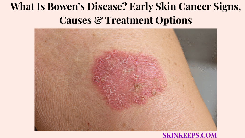
Bowen’s disease is squamous cell carcinoma in situ, an early form of skin cancer in which abnormal squamous cells are still confined to the outer skin layer.
It can look like eczema, psoriasis, fungus, or a dry scaly patch, but a persistent, enlarging, bleeding, painful, ulcerated, or thickening patch needs evaluation. The sections below cover signs, causes, risk factors, diagnosis, biopsy, treatment options, follow-up, prevention, mistakes, and prompt-care signs.
What Is Bowen’s Disease and Why Is It Called Squamous Cell Carcinoma in Situ?
Bowen’s disease is squamous cell carcinoma in situ, meaning cancerous squamous cells are present in the epidermis but have not invaded deeper tissue.
Squamous cells are flat cells in the outer skin layer. The epidermis is the top layer of skin, while the dermis is the deeper layer below it.
“In situ” means the abnormal cells remain in their original layer. For Bowen’s disease, that means the cancerous cells are confined to the epidermis.
Why Is Bowen’s Disease Early Skin Cancer?
Bowen’s disease is early skin cancer because the abnormal squamous cells are limited to the outer layer of skin rather than growing into deeper tissue.
It is usually slow-growing and treatable, but it still needs treatment or monitoring because progression can occasionally occur.
The safest framing is calm but firm: early diagnosis gives more treatment choices, while long delay can allow the patch to thicken, ulcerate, bleed, or become harder to treat.
Why Is Bowen’s Disease Not the Same as Invasive SCC?
Bowen’s disease is not the same as invasive SCC because Bowen’s disease is confined to the epidermis, while invasive squamous cell carcinoma has grown deeper into the skin.
Ulceration, lump formation, rapid growth, increasing pain, thickening, or bleeding can raise concern that a patch may not be only surface-level disease.
Biopsy helps confirm squamous cell carcinoma in situ and helps rule out deeper invasion when the patch is suspicious or unclear.
Practical rule: Bowen’s disease is early and treatable, but a persistent scaly patch should be diagnosed before it is treated as a routine rash.
What Bowen’s Disease Signs Can Appear on Skin?
Bowen’s disease often appears as a persistent scaly, crusted, red, pink, brown, or sore-looking patch that slowly enlarges and does not clear like a typical rash.
It may be a single patch, though more than one patch can occur. It can look dry, rough, inflamed, crusted, or slightly raised.
What Does Early Bowen’s Disease Look Like?
Early Bowen’s disease may look like a flat or slightly raised patch with scale, crust, roughness, dryness, or red, pink, brown, or darker discoloration.
On lighter skin, it may look red, pink, or orange-red. On brown or black skin, it may look darker brown, reddish-brown, purple-brown, grayish, or more textured than nearby skin.
Slow enlargement matters because a patch that keeps expanding is different from a temporary irritation that clears.
What Symptoms Can Bowen’s Disease Cause?
Bowen’s disease often causes no symptoms beyond a persistent patch, but it may itch, feel sore, bleed, crust, form a non-healing surface, or ulcerate.
Lack of pain does not make a persistent patch safe. A painless patch can still need biopsy if it is non-healing, enlarging, crusting, or suspicious.
Bleeding, thickening, lump formation, ulceration, increasing pain, or a non-healing crust needs prompt review.
Why Can Bowen’s Disease Be Mistaken for Eczema or Psoriasis?
Bowen’s disease can be mistaken for eczema or psoriasis because it can look like a scaly rash, especially when it is flat, red, dry, or crusted.
Atopic dermatitis often has a broader itchy pattern, while Bowen’s disease may appear as one persistent patch that does not clear as expected.
Psoriasis often has a repeating plaque pattern, but a single non-clearing scaly patch should be reassessed before long-term steroid-only treatment continues.
| Sign | What the Reader May Notice | Why It Matters |
|---|---|---|
| Persistent scaly patch | Rough patch that does not clear | Classic concern pattern |
| Red, pink, brown, or darker patch | Skin-tone-dependent color | May not look identical on all skin tones |
| Crusting | Dry crust or repeated scab | Non-healing surface needs review |
| Slow enlargement | Patch expands over time | Suggests persistent lesion rather than temporary rash |
| Bleeding | Bleeds with minor trauma or without clear cause | Needs evaluation |
| Open sore or ulcer | Surface breaks down | Possible invasive SCC warning |
| Lump or thickening | Raised or firm area within patch | Possible deeper growth warning |
| Pain or tenderness | New or increasing discomfort | Prompt review sign |
Where Does Bowen’s Disease Usually Appear?
Bowen’s disease can appear anywhere on the skin, but it is often found on sun-damaged areas such as the face, scalp, ears, neck, hands, arms, and lower legs.
It can also appear on the trunk, genital or perianal skin, the nail unit, fingers, previously radiated skin, scarred skin, or chronically damaged skin.
Location cannot diagnose the patch by itself. A persistent patch on the lower leg, face, ear, lip, eyelid, genital skin, nail unit, or radiated/scarred skin needs careful assessment because treatment planning can be more complex.
| Location | Common Clue | Treatment-Planning Concern |
|---|---|---|
| Lower legs | Persistent scaly patch on sun-damaged or fragile skin | Healing can be slower; treatment selection matters |
| Face | Pink, red, brown, or scaly patch | Cosmetic and tissue-sparing planning |
| Scalp | Sun-exposed patch, especially with hair thinning | Harder self-monitoring |
| Ears | Sun-damaged high-caution site | Check crusting, bleeding, or thickening |
| Neck / hands / arms | Chronic UV exposure pattern | New lesions may appear elsewhere |
| Trunk | Can occur away from classic sun sites | Do not dismiss based on location |
| Genital / perianal skin | HPV-related or specialist-pathway context possible | Needs specific evaluation |
| Nail unit / finger | HPV or nail-unit SCC in situ context | Specialist diagnosis is important |
| Radiated or scarred skin | Prior injury or radiation history | Higher caution and individualized planning |
What Causes Bowen’s Disease?
Bowen’s disease is linked with long-term ultraviolet exposure, immune suppression, previous radiotherapy, arsenic exposure, chronic skin damage, and in some sites, human papillomavirus infection.
Not every case has one clear cause. The main safety point is that persistent or suspicious patches need diagnosis, not guessing.
How Does Long-Term Sun Exposure Contribute to Bowen’s Disease?
Long-term sun exposure can contribute to Bowen’s disease by damaging skin-cell DNA over time, especially on chronically sun-exposed skin.
Older age, outdoor work, outdoor recreation, tanning-bed exposure, repeated sunburn, and long-term cumulative ultraviolet exposure can increase the chance of keratinocyte skin cancers.
Sun-exposed areas such as the face, scalp, ears, neck, hands, arms, and lower legs are common sites, but Bowen’s disease can also occur elsewhere.
How Do Immune Suppression and Medical History Affect Risk?
Immune suppression and certain medical histories can increase Bowen’s disease risk because damaged or abnormal skin cells may be less effectively controlled.
Risk can be higher with organ transplant medicines, long-term immune suppression, some immune diseases, previous radiotherapy, chronic irritation, chronic scarring, and chronic skin injury.
Immunosuppressed patients may need closer follow-up because recurrence and new keratinocyte cancers can be more likely.
When Can HPV or Arsenic Be Relevant?
HPV or arsenic can be relevant in selected Bowen’s disease contexts, but they should not be assumed as the cause of every lesion.
HPV may matter more for genital, perianal, nail-unit, or finger-area SCC in situ. Arsenic exposure is now uncommon in many settings but has been historically linked with Bowen’s disease.
Ionising radiation and chronically damaged skin can also matter in selected cases. Genital, perianal, or nail-unit lesions usually need a more specific specialist pathway.
| Risk Factor | Clue | What Clinician May Ask | Prevention / Follow-Up Implication |
|---|---|---|---|
| Long-term UV exposure | Sun-damaged skin, outdoor history | Sunburn, tanning, outdoor work | Sun protection and skin checks |
| Older age | Cumulative skin damage | History of new scaly patches | Lower threshold for review |
| Fair or easily burned skin | Burns easily in sun | Skin type and sunburn history | Strong UV protection |
| Immune suppression | Transplant, immune disease, medicines | Medication and immune history | Closer follow-up |
| Prior radiotherapy | Lesion in treated field | Past cancer or radiation treatment | Individualized care |
| HPV-related site | Genital, perianal, nail, or finger context | Site-specific history | Specialist pathway |
| Arsenic exposure | Rare environmental or occupational history | Historic exposure | Risk review if relevant |
| Chronic scarring or injury | Scarred or chronically damaged skin | Wounds, scars, inflammation | Prompt assessment of changes |
Who Is More Likely to Develop Bowen’s Disease?
Bowen’s disease is more likely in people with significant lifetime sun exposure, older age, fairer skin that burns easily, prior skin cancer, immune suppression, radiotherapy history, or certain HPV-related contexts.
Risk does not mean blame. It simply helps decide when a persistent patch should be checked sooner.
How Is Bowen’s Disease Different From Other Skin Conditions?
Bowen’s disease can resemble eczema, psoriasis, actinic keratosis, superficial basal cell carcinoma, invasive squamous cell carcinoma, fungal rash, or melanoma, so persistent or changing patches should be checked.
Diagnosis matters because treatment for a rash is not the same as treatment for squamous cell carcinoma in situ.
How Is Bowen’s Disease Different From Eczema or Psoriasis?
Bowen’s disease is different from eczema or psoriasis because it often appears as one persistent scaly patch that does not clear, while eczema and psoriasis usually have broader, recurring, or pattern-based features.
A patch that does not clear with appropriate rash treatment needs review. Long-term steroid-only use on an undiagnosed cancer-like patch can delay diagnosis.
How Is Bowen’s Disease Different From Actinic Keratosis?
Bowen’s disease is different from actinic keratosis because actinic keratosis is usually considered a precancerous sun-damage lesion, while Bowen’s disease is squamous cell carcinoma in situ.
Both can be rough, scaly, and sun-damage-related. A thicker, larger, persistent, bleeding, or suspicious rough patch may need biopsy to clarify the diagnosis.
How Is Bowen’s Disease Different From Invasive SCC?
Bowen’s disease is confined to the epidermis, while invasive squamous cell carcinoma grows deeper and may appear as a lump, ulcer, painful area, bleeding lesion, or thick crusted growth.
Dermatology review and biopsy determine the difference when the patch is thick, painful, bleeding, ulcerated, rapidly enlarging, or hard to classify.
| Condition | Common Clue | Why Confusion Happens | Safer Next Step |
|---|---|---|---|
| Bowen’s disease | Persistent scaly or crusted patch | Looks like rash or sun damage | Biopsy if suspicious or unclear |
| Eczema | Itchy inflammatory rash | Red scaly patches overlap | Review single non-clearing patch |
| Psoriasis | Thicker plaques, repeated sites | Scale overlaps | Diagnose before long-term treatment |
| Actinic keratosis | Rough sun-damaged spot | Same sun-damage spectrum | Check persistent, thick, or bleeding lesions |
| Invasive SCC | Lump, ulcer, pain, bleeding, thick crust | May develop from SCC in situ | Prompt biopsy |
| Superficial BCC | Pink scaly patch | Can mimic Bowen’s disease | Dermoscopy or biopsy |
| Fungal rash | Ring-like or scaly rash | Scale and redness overlap | Review if persistent or atypical |
| Melanoma | Changing pigmented lesion | Pigmented Bowen’s can confuse diagnosis | Urgent review if evolving |
A pink scaly patch can sometimes resemble basal cell carcinoma, while a darker or irregular changing patch may raise a separate melanoma safety concern. Biopsy is the decision point when appearance is unclear.
How Is Bowen’s Disease Diagnosed or Checked?
Bowen’s disease is suspected from the appearance and history of a persistent scaly patch, but diagnosis is commonly confirmed with a skin biopsy when the patch is suspicious or unclear.
A clinician may examine the patch with the naked eye and with dermoscopy, a tool that magnifies skin patterns. Dermoscopy can support assessment, but biopsy is needed when the diagnosis is uncertain or invasion must be ruled out.
What Does a Dermatologist Check With Suspected Bowen’s Disease?
A dermatologist checks suspected Bowen’s disease by assessing the lesion’s size, location, scale, crust, bleeding, soreness, ulceration, thickness, border, number of lesions, risk factors, and prior treatment response.
The visit may review sun damage, immune suppression, previous skin cancers, actinic keratoses, radiotherapy history, genital or nail involvement, medication list, and whether eczema, psoriasis, or fungal treatments failed.
Why Does Biopsy Matter Before Treatment?
Biopsy matters because it confirms squamous cell carcinoma in situ, rules out invasive SCC, separates Bowen’s disease from similar rashes or cancers, and guides treatment choice.
A biopsy can help decide whether excision, curettage, cryotherapy, topical treatment, photodynamic therapy, radiotherapy, Mohs surgery, laser, or observation is appropriate.
Biopsy also prevents cosmetic or destructive treatment from being chosen before the depth and diagnosis are understood.
What Treatment Options Are Used for Bowen’s Disease?
Bowen’s disease treatment may include excision, curettage, cryotherapy, topical 5-fluorouracil, imiquimod, photodynamic therapy, radiotherapy, Mohs surgery in difficult sites, laser in selected settings, or observation in selected low-risk cases.
Treatment is not one-size-fits-all. The best option depends on lesion size, thickness, site, healing risk, recurrence, immune status, patient health, cosmetic impact, and whether invasive SCC is suspected.
When Is Surgical Excision Used for Bowen’s Disease?
Surgical excision may be used when a Bowen’s disease lesion is smaller, well-defined, recurrent, suspicious for invasion, or needs complete removal with pathology.
Excision removes the lesion and allows tissue examination. It can be helpful when the diagnosis or depth needs confirmation, but it can leave a scar and requires wound healing.
When Are Curettage, Cautery, or Cryotherapy Used?
Curettage, cautery, or cryotherapy may be used for selected superficial Bowen’s disease lesions when the site, size, and healing risk make them appropriate.
Curettage and cautery remove and destroy abnormal surface tissue. Cryotherapy freezes the abnormal area and can cause blistering, crusting, pigment change, and slower healing.
Lower-leg lesions need caution because healing may be slower, especially in older adults or people with fragile skin, swelling, poor circulation, or wound-healing problems.
When Are Topical Treatments Used?
Topical treatments may be used for selected superficial, broad, multiple, or surgery-avoidance cases when invasion has been ruled out and follow-up is reliable.
Topical 5-fluorouracil and imiquimod are options in selected cases. They can cause inflammation, soreness, redness, crusting, and irritation during treatment.
They should not be used as self-treatment for an undiagnosed patch, especially when the lesion is thick, painful, bleeding, ulcerated, or suspicious for invasion.
When Are Photodynamic Therapy, Radiotherapy, Mohs, or Laser Considered?
Photodynamic therapy, radiotherapy, Mohs surgery, or laser may be considered when lesion site, patient health, cosmetic needs, surgical suitability, or difficult anatomy affects treatment planning.
Photodynamic therapy may be used for selected superficial or cosmetic-sensitive lesions. Radiotherapy may be considered when surgery is unsuitable in selected patients.
Mohs surgery may be used in difficult or tissue-sparing sites such as near the eye. Laser is less common, and availability varies.
| Treatment Option | Best-Fit Situation | Main Goal | Key Caution |
|---|---|---|---|
| Excision | Defined lesion, recurrence, suspicion of invasion | Remove lesion and assess pathology | Scar and healing tradeoff |
| Curettage / cautery | Selected superficial lesions | Remove or destroy abnormal cells | Operator and site dependent |
| Cryotherapy | Selected thin lesions | Freeze abnormal cells | Blistering, pigment change, slow healing |
| 5-FU cream | Selected superficial patch or field | Topical cancer-cell treatment | Inflammation and adherence |
| Imiquimod | Selected superficial lesions | Immune-driven clearance | Irritation and monitoring |
| PDT | Selected superficial or cosmetic sites | Treat surface disease | Pain, availability, recurrence |
| Radiotherapy | Surgery unsuitable in selected patients | Non-surgical control | Site and healing effects |
| Mohs surgery | Difficult or tissue-sparing sites | Clear margins while preserving tissue | Specialist procedure |
| Observation | Frail, elderly, or low-risk selected cases | Monitor low-progression-risk lesions | Requires follow-up |
How Do Location and Healing Risk Affect Bowen’s Disease Treatment?
Bowen’s disease treatment is not one-size-fits-all because healing risk, cosmetic impact, recurrence risk, lesion size, immune status, and site all affect the best option.
Lower legs may heal slowly. Face, eyelid, lip, ear, genital, perianal, and nail-unit lesions need careful planning because tissue preservation, function, comfort, and cosmetic outcome matter.
Large lesions may need staged, topical, photodynamic, or surgical planning. Immunosuppressed patients may need closer follow-up. Suspected invasion makes biopsy or excision more important before surface-only treatment.
| Location / Situation | Treatment Concern | Planning Note |
|---|---|---|
| Lower leg | Slower healing, fragile skin, circulation issues | Avoid casual destructive treatment |
| Face | Cosmetic impact and tissue preservation | Precise diagnosis and cosmetic planning |
| Eyelid / near eye | Difficult anatomy | Specialist or Mohs consideration |
| Ear / lip | Higher-caution site | Prompt biopsy if thick, bleeding, or ulcerated |
| Genital / perianal skin | HPV or specialist pathway possible | Specialist assessment |
| Nail unit | Complex differential and function concerns | Specialist biopsy planning |
| Large lesion | More treatment burden | Topical, PDT, staged, or surgical planning |
| Immunosuppressed patient | Recurrence and new cancer risk | Closer follow-up |
| Suspected invasion | Depth concern | Biopsy or excision priority |
Can Bowen’s Disease Come Back or Progress?
Bowen’s disease can recur after treatment, and untreated or persistent lesions can occasionally progress into invasive squamous cell carcinoma, so follow-up and monitoring matter.
New lesions can also develop on sun-damaged skin. Immunosuppressed patients may need closer monitoring because keratinocyte cancers can behave more persistently.
A treated area that becomes scaly, crusted, bleeding, raised, painful, thickened, or ulcerated should be checked.
How Can Sun Protection Reduce Future Bowen’s Disease Risk?
Sun protection cannot treat an existing Bowen’s disease patch, but it can reduce additional ultraviolet damage and help lower the risk of future keratinocyte skin cancers.
Protect high-exposure sites such as the scalp, ears, face, hands, arms, and lower legs. Avoid tanning beds because they add ultraviolet damage.
Regular skin checks matter after Bowen’s disease or other keratinocyte skin cancers because new lesions can appear elsewhere.
What Bowen’s Disease Mistakes Should You Avoid?
The biggest Bowen’s disease mistake is repeatedly treating a persistent scaly patch as eczema, psoriasis, fungus, or dry skin without confirming the diagnosis.
Do not ignore a patch that persists or enlarges. Do not keep using steroid cream on one non-clearing patch without review.
Do not scrape, burn, freeze, peel, or acid-treat suspicious patches at home. Do not choose cosmetic treatment before ruling out invasive SCC, and do not skip follow-up after treatment.
| Mistake | Why It Is Risky | Better Action |
|---|---|---|
| Calling it eczema for months | Delays skin-cancer diagnosis | Biopsy persistent patch if suspicious |
| Steroid-only treatment | Can mask or delay recognition | Reassess non-clearing patch |
| DIY freezing or burning | Misses diagnosis and depth | Dermatology treatment |
| Ignoring ulceration | Possible invasive SCC warning | Prompt review |
| Choosing cosmetic treatment first | May miss invasive disease | Diagnose before treatment |
| Skipping follow-up | Recurrence and new lesions possible | Keep monitoring plan |
| Ignoring immune risk | Higher recurrence or new-cancer risk | Earlier dermatologist review |
When Should Bowen’s Disease Be Checked Promptly?
A Bowen-like skin patch should be checked promptly when it is persistent, enlarging, bleeding, crusting, ulcerated, painful, thickening, forming a lump, not responding to rash treatment, or appearing in a high-risk person.
Prompt review does not mean panic. It means a dermatologist or qualified clinician should assess whether biopsy, treatment, or monitoring is needed.
Which Signs May Suggest Invasive SCC or Higher Risk?
Signs that need faster review include an open sore, new lump, raised thick area, rapid growth, increasing pain, bleeding, persistent crust, non-healing wound, firmness, tenderness, or a lesion on a high-risk site.
High-risk sites include the lip, ear, face, genital skin, and nail unit. Immunosuppressed patients should also use a lower threshold for review.
What Should You Bring to a Dermatology Appointment?
A useful Bowen’s disease appointment starts with photos, timing, change history, symptoms, treatments tried, sun history, immune status, prior skin cancer history, and site-specific concerns.
Bring a medication list, radiotherapy history, prior biopsy reports, and details about genital, perianal, or nail involvement if relevant.
Seek prompt review if a patch is:
What Should You Remember About Bowen’s Disease?
The most important thing to remember about Bowen’s disease is that it is squamous cell carcinoma in situ, so the safest approach is early recognition, biopsy confirmation, appropriate treatment, and monitoring.
A persistent scaly patch should not be ignored simply because it looks dry, eczema-like, psoriasis-like, or painless.
Frequently Asked Questions About Bowen’s Disease
Is Bowen’s disease cancer?
Yes. Bowen’s disease is squamous cell carcinoma in situ, an early form of skin cancer in which abnormal squamous cells are confined to the outer skin layer.
What does Bowen’s disease look like?
It may look like a persistent red, pink, brown, darker, scaly, crusted, sore-looking, or slowly enlarging patch that can resemble eczema, psoriasis, fungus, or dry skin.
Can Bowen’s disease turn into invasive squamous cell carcinoma?
It can occasionally progress into invasive squamous cell carcinoma, which is why dermatologists usually treat or monitor it rather than ignoring it.
What causes Bowen’s disease?
Risk factors include long-term UV exposure, older age, immune suppression, previous radiotherapy, rare arsenic exposure, HPV in selected sites, chronic scarring, and chronic skin damage.
How is Bowen’s disease diagnosed?
Diagnosis is based on clinical examination and often confirmed with biopsy when the appearance is suspicious, unclear, or invasion must be ruled out.
What is the best treatment for Bowen’s disease?
There is no one best treatment for every lesion. Treatment depends on lesion size, site, healing risk, recurrence, patient health, and whether invasive SCC is suspected. Options include excision, curettage, cautery, cryotherapy, 5-FU, imiquimod, photodynamic therapy, radiotherapy, Mohs surgery in difficult sites, laser in selected settings, or observation in selected low-risk cases.
Does Bowen’s disease come back after treatment?
It can recur after treatment, and new lesions can appear on sun-damaged skin. Follow-up depends on treatment method, lesion risk, immune status, and dermatologist guidance.
When should a Bowen-like patch be checked promptly?
Prompt review is needed for ulceration, bleeding, rapid growth, pain, thickening, lump formation, non-healing crust, genital or nail involvement, immunosuppression, recurrence, or failure to clear with appropriate rash treatment.
Sources & Evidence About Bowen’s Disease
British Association of Dermatologists — Squamous Cell Carcinoma in Situ was used for SCC in situ definition, occasional progression to invasive SCC, long-term sun exposure and immunosuppression risk, biopsy/excision logic, lower-leg healing caution, treatment options, and prompt review signs.
Cancer Research UK — Bowen’s Disease was used for very early non-melanoma skin cancer framing, epidermis-only disease, red scaly patch symptoms, ulceration concern, diagnosis pathway, treatment factors, 5-FU/imiquimod, surgery, cryotherapy, Mohs, laser, follow-up, and skin protection.
DermNet — Intraepidermal Squamous Cell Carcinoma was used for terminology, risk factors, clinical features, common sites, nail-unit context, diagnosis, biopsy confirmation, treatment approaches, recurrence caution, prevention, and the cautious progression context.
British Skin Foundation — Bowen’s Disease was used for SCC in situ explanation, symptoms, diagnosis, treatment options, lower-leg healing caution, self-care, sun-safety points, and prompt review signs such as bleeding, ulceration, or lump formation.
MSD Manual Professional — Squamous Cell Carcinoma in Situ was used for biopsy-based diagnosis, resemblance to psoriasis, dermatitis, and dermatophyte infection, risk factors similar to SCC, and treatment categories including curettage/electrodesiccation, excision, topical chemotherapeutics, electrocautery, or cryosurgery.
DermNet — Cutaneous Squamous Cell Carcinoma was used only for broader invasive SCC differential context, including invasive SCC warning patterns and risk framing.
Educational Disclaimer: This SkinKeeps article is for educational purposes only and does not diagnose or replace medical care. A persistent, enlarging, bleeding, crusted, ulcerated, painful, thickened, lump-forming, recurrent, non-healing, genital, nail-unit, lip, ear, face, immune-risk, or rash-treatment-resistant patch should be checked by a qualified healthcare professional or dermatologist.



