
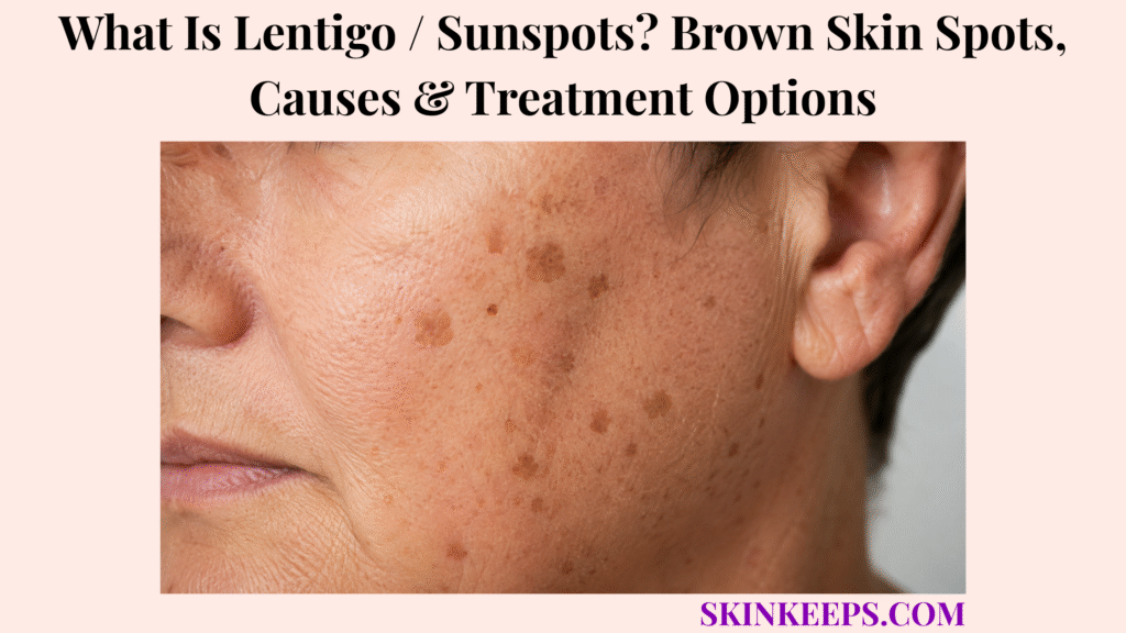
Solar lentigines are benign, persistent brown spots that develop mainly on chronically ultraviolet-exposed skin. They most often appear on the face, backs of the hands, forearms, shoulders, upper chest and upper back, becoming more common as lifetime UV exposure accumulates, although younger people with substantial exposure can also develop them.
Solar lentigines themselves are harmless, but melanoma, lentigo maligna, pigmented actinic keratosis and other lesions can resemble an ordinary sunspot. A new, unusual or changing brown spot should therefore be diagnosed before freezing, laser treatment or pigment-lightening treatment is used.
A new or changing brown lesion should be assessed if it becomes asymmetrical, develops irregular borders or several colours, noticeably enlarges, itches, hurts, bleeds or looks markedly different from surrounding spots. Cosmetic destruction of an undiagnosed atypical pigmented lesion can delay the correct diagnosis.
How Can You Recognize a Solar Lentigo or Sunspot?
A solar lentigo is usually a persistent, sharply defined tan-to-brown flat spot on chronically sun-exposed skin.
What Does a Solar Lentigo Look Like?
A typical solar lentigo is a flat or minimally raised, sharply bordered tan-to-dark-brown lesion with relatively uniform pigmentation.
Solar lentigines range from a few millimetres to several centimetres, and their borders can be gently scalloped. The surface is usually smooth, although mild dryness or scale can appear. A thickened, waxy or increasingly rough lesion raises a different diagnostic question.
Where Do Sunspots Usually Develop?
Solar lentigines most often develop on chronically UV-exposed areas such as the face, hands, forearms, shoulders, upper chest and upper back.
Temples, nose and backs of the hands are frequent sites because their distribution mirrors cumulative photodamage rather than random body placement.
How Do Solar Lentigines Look Across Different Skin Tones?
Solar lentigines occur across skin tones, although their colour contrast and visual prominence vary with background pigmentation.
They may appear tan to dark brown on lighter skin and medium to deep brown on darker skin. Border pattern, lesion structure and change over time are more useful than expecting one universal colour.
Do Sunspots Fade During Winter?
Solar lentigines generally persist through winter rather than disappearing when ultraviolet exposure decreases.
Some lesions lighten slightly, but year-round persistence helps distinguish a solar lentigo from a freckle, which more commonly darkens during UV exposure and fades when exposure falls.
Figure 1. A typical solar lentigo is a persistent flat brown macule on chronically UV-exposed skin. Diagnostic safety depends on stability and pattern; changing or irregular lesions should be assessed before cosmetic treatment.
Why Do Solar Lentigines Develop, and Are All Lentigines Sunspots?
Solar lentigines develop mainly from cumulative ultraviolet damage, but the broader term lentigo includes pigmented lesions that are not caused by sun exposure.
How Does Sun Exposure Create a Solar Lentigo?
Cumulative ultraviolet radiation produces epidermal and pigmentary changes that create persistent localized brown pigmentation known as solar lentigo.
Repeated UV exposure produces photodamage, increases localized melanin production and retention, and alters the sun-damaged epidermis. These changes create a persistent pigmented macule rather than a temporary tanning response.
Who Is More Likely to Develop Sunspots?
Solar lentigines are more common with increasing age and cumulative ultraviolet exposure, particularly after substantial lifetime sun exposure.
- Increasing age and lifetime UV exposure.
- Repeated outdoor sun exposure.
- Previous sunburn.
- Tanning-bed exposure.
- Selected UV phototherapy exposure.
- Lighter phototypes are frequently affected, although solar lentigines occur across skin tones.
Is a Lentigo the Same as a Sunspot?
No; a solar lentigo is one UV-associated type within the broader lentigo category.
Lentigo simplex can arise without the cumulative sun-damage pattern that defines a solar lentigo, while other lentigines occur in genetic, environmental or medical contexts. The shared word lentigo therefore does not mean every lentigo is a sunspot.
Are Sunspots Dangerous?
Typical solar lentigines are benign and are not dangerous themselves.
They do mark cumulative UV exposure, and the main safety issue is diagnostic overlap: melanoma in situ, lentigo maligna, pigmented actinic keratosis and other lesions can resemble a sunspot. Atypical lesions should be diagnosed before cosmetic destruction.
Figure 2. Solar lentigines develop through cumulative ultraviolet exposure, photodamage and persistent epidermal pigment change. Age increases opportunity for this damage to accumulate, but younger people with substantial UV exposure can also develop sunspots.
How Are Solar Lentigines Different From Freckles, Moles and Melanoma?
Solar lentigines differ from freckles, moles, keratoses and melanoma by their persistence, morphology, texture and pattern of change over time.
How Is a Sunspot Different From a Freckle?
A solar lentigo usually persists year-round, whereas a freckle commonly darkens with ultraviolet exposure and fades when exposure decreases.
Freckles are often smaller and more seasonally variable. Freckles / ephelides reflect a different pigmentation pattern from persistent solar lentigines associated with longer-term photodamage.
How Is a Solar Lentigo Different From a Mole?
A solar lentigo is primarily a flat UV-associated pigmented lesion, whereas a mole is a localized proliferation of melanocytes that may be flat or raised.
Appearance can overlap, so an atypical melanocytic nevus / mole may need dermoscopic assessment rather than visual self-classification. Most moles are benign.
How Is a Sunspot Different From Seborrhoeic Keratosis or Actinic Keratosis?
Seborrhoeic keratoses are often thicker and waxier, while actinic keratoses are commonly rough or scaly; a typical solar lentigo remains primarily a flat pigmented macule.
A seborrhoeic keratosis often develops a stuck-on, waxy or thicker surface that is less typical of a flat solar lentigo.
An actinic keratosis usually emphasizes rough or scaly photodamaged texture, although pigmented variants can visually overlap with lentigines.
When Could a Brown Sunspot Actually Be Melanoma?
A brown spot requires melanoma assessment when it becomes asymmetrical, develops irregular borders or multiple colours, changes over time or looks unlike the person’s other lesions.
- A — Asymmetry: one half differs from the other.
- B — Border: irregular, scalloped or poorly defined.
- C — Colour: several colours within one lesion.
- D — Diameter: many melanomas exceed 6 mm, although melanoma can be smaller.
- E — Evolving: change in size, shape, colour or overall appearance.
Itching, pain, bleeding or a lesion that looks markedly different from nearby spots also deserves assessment for melanoma. The ABCDE framework identifies warning signs; it does not prove a diagnosis.
What Is Lentigo Maligna?
Lentigo maligna is melanoma in situ arising mainly on chronically sun-damaged skin and is not a benign solar lentigo.
It often presents as slowly enlarging irregular pigmentation on chronically sun-damaged skin. The shared word lentigo can be misleading, so lentigo maligna must remain clearly separated from ordinary benign solar lentigines.
| Lesion | Persistence | Typical Texture / Shape | Major Distinguishing Clue |
|---|---|---|---|
| Solar lentigo | Persistent year-round | Flat or minimally raised, sharply pigmented | Stable UV-associated macule. |
| Freckle | Often fades when UV exposure falls | Small flat macule | Seasonal darkening and fading. |
| Mole | Usually persistent | Flat or raised melanocytic lesion | Melanocytic pattern; morphology varies. |
| Seborrhoeic keratosis | Persistent | Often thicker, waxy or stuck-on | Surface texture differs from a flat lentigo. |
| Actinic keratosis | Persistent | Usually rough or scaly | Precancerous keratinocytic lesion on photodamaged skin. |
| Melanoma | Usually persists and evolves | May be flat or raised and irregular | Asymmetry, border/colour variation, evolution or outlier appearance. |
How Are Solar Lentigines Diagnosed?
Typical solar lentigines are usually diagnosed clinically, while atypical or uncertain lesions may require dermoscopy or biopsy before treatment.
What Does a Dermatologist Look For?
A dermatologist assesses a suspected solar lentigo by examining its history, border, pigmentation, texture, location, stability and relationship to surrounding sun damage.
- Duration and growth history.
- Colour pattern.
- Border definition.
- Surface texture.
- Anatomical location.
- Surrounding photodamage.
- Comparison with the person’s other pigmented lesions.
A typical stable lesion can often be diagnosed clinically.
How Does Dermoscopy Help Diagnose a Solar Lentigo?
Dermoscopy helps distinguish a solar lentigo from melanocytic and keratotic lesions by revealing pigment and surface structures not seen as reliably with the naked eye.
A dermatoscope magnifies and illuminates skin structures, helping clinicians assess melanocytic lesions, pigmented keratoses and possible melanoma. Dermoscopy improves pattern recognition but does not guarantee that a lesion is benign.
When Is a Biopsy Needed for a Suspected Sunspot?
Biopsy is considered when a presumed sunspot has an uncertain diagnosis or develops features suspicious for melanoma or another skin cancer.
Asymmetry, multiple colours, enlargement, evolution, a markedly different appearance or persistent diagnostic uncertainty can justify tissue diagnosis. A suspicious pigmented lesion should be diagnosed before laser, cryotherapy or bleaching treatment because destroying visible pigment first can delay recognition.
Figure 3. Typical stable solar lentigines may be diagnosed clinically. Dermoscopy supports evaluation when the pattern is uncertain, while suspicious or changing lesions may require biopsy before any cosmetic pigment treatment is considered.
How Are Solar Lentigines Treated, and How Can New Sunspots Be Prevented?
Confirmed benign solar lentigines do not require medical treatment, but topical or procedural treatments can fade them while consistent UV protection reduces new lesions and recurrence.
Do Solar Lentigines Need Treatment?
No; confirmed benign solar lentigines can safely be left untreated because treatment is cosmetic rather than medically necessary.
Treatment depends on cosmetic concern, location, skin tone, cost, expected downtime and the risk of post-treatment pigmentation change. Leaving a confirmed benign lesion alone is a valid choice.
Which Topical Treatments Can Fade Sunspots?
Selected topical pigment treatments can gradually fade confirmed solar lentigines, although improvement usually requires repeated use over weeks or months.
Prescription retinoids, hydroquinone, cysteamine, mequinol, azelaic acid and selected alpha-hydroxy-acid preparations can be considered in appropriate settings. Irritation and pigment change must be monitored, and no topical method guarantees complete or permanent clearance.
How Do Cryotherapy, Laser and IPL Treat Solar Lentigines?
Cryotherapy, pigment-targeting lasers and IPL reduce solar lentigines by damaging or selectively targeting the pigmented tissue responsible for the visible spot.
- Cryotherapy freezes targeted pigmented tissue.
- Pigment-specific lasers target melanin.
- IPL can treat multiple areas of photodamage.
- Chemical peels or resurfacing can reduce visible pigmentation.
Procedural risks include crusting, irritation, scarring, temporary darkening and hypopigmentation. Pigmentary complications deserve particular consideration in darker skin tones, so no single procedure is universally best.
Can Sunspots Return After Treatment?
Yes; treated solar lentigines can recur, and new lesions can form because cosmetic removal does not eliminate the underlying effects of cumulative UV exposure.
Pigment can recur within a treated area while additional lesions develop in surrounding photodamaged skin. Sustained photoprotection therefore remains part of long-term treatment even after a good cosmetic response.
How Can New Solar Lentigines Be Reduced?
Consistent photoprotection reduces additional solar lentigines by limiting further ultraviolet damage to exposed skin.
- Limit intense UV exposure.
- Seek shade.
- Wear protective clothing and a broad-brimmed hat.
- Avoid tanning beds.
- Use broad-spectrum, water-resistant sunscreen with SPF 30 or higher.
- Reapply sunscreen as appropriate for exposure.
When Should an Existing Sunspot Be Rechecked?
A previously stable sunspot should be reassessed if it changes in size, shape, colour, border, texture or symptoms.
- Enlargement.
- Shape change.
- New colour variation.
- Unusual border.
- Becoming significantly raised.
- Repeated crusting.
- Itching or pain.
- Bleeding.
- Looking different from surrounding spots.
| Step | Decision |
|---|---|
| 1. Confirm benign lesion | Do not destroy an atypical pigmented lesion before reasonable diagnostic certainty. |
| 2. Decide whether treatment is desired | Solar lentigines are benign, so treatment is optional. |
| 3. Choose a fading pathway | Use a suitable topical or procedural option based on lesion, skin tone and risk. |
| 4. Protect from UV | Use shade, clothing and broad-spectrum SPF 30+ sunscreen to reduce new photodamage. |
| 5. Monitor | Reassess recurrence or any new change in size, shape, colour, border, texture or symptoms. |
What Should You Remember About Lentigo / Sunspots?
Solar lentigines are benign persistent UV-associated brown spots, but a lesion should be confirmed as benign before cosmetic treatment if its appearance is atypical or changing.
- Cumulative ultraviolet exposure drives solar lentigines.
- Face, hands, forearms, shoulders, upper chest and upper back are common locations.
- Solar lentigines persist more than ordinary freckles.
- Solar lentigo is one subtype within the broader lentigo category.
- Diagnosis comes before cosmetic destruction.
- Treatment is optional after benign confirmation.
- Topical and procedural treatments can fade lesions but do not guarantee permanent clearance.
- Photoprotection reduces new lesions and recurrence.
- Previously stable lesions still need reassessment if they change.
The decision pathway is recognize → differentiate → confirm → treat if desired → protect → monitor.
Frequently Asked Questions About Lentigo / Sunspots?
The most common questions about solar lentigines concern their UV cause, differentiation from other brown spots, treatment and recurrence.
What Causes Solar Lentigines or Sunspots?
Solar lentigines are caused mainly by cumulative ultraviolet exposure that produces persistent localized changes in epidermal pigmentation. Repeated sunlight, tanning-bed exposure and other substantial ultraviolet exposure can contribute to photodamage, increased melanin retention and the formation of stable brown macules.
How Can You Tell a Sunspot From a Freckle?
A solar lentigo usually persists throughout the year, whereas a freckle commonly darkens with sun exposure and fades when ultraviolet exposure decreases. Solar lentigines also tend to reflect longer-term photodamage and may be larger or more sharply persistent than ordinary freckles.
Can an Apparent Sunspot Actually Be Melanoma?
Yes; melanoma can sometimes resemble a sunspot, especially when a lesion is changing, asymmetrical, irregularly bordered or multicoloured. A spot that evolves, bleeds, itches, hurts or looks markedly different from surrounding lesions should be assessed rather than treated cosmetically at home.
Which Treatments Can Fade Solar Lentigines?
Topical pigment treatments, cryotherapy, pigment-targeting lasers, IPL and selected resurfacing procedures can reduce confirmed benign solar lentigines. Treatment choice depends on lesion characteristics, skin tone, cost, expected downtime and the risk of post-treatment hyperpigmentation, hypopigmentation or scarring.
Can Solar Lentigines Return After Treatment?
Yes; solar lentigines can recur after treatment, and new lesions can develop on surrounding UV-damaged skin. Cosmetic removal does not reverse cumulative photodamage, so sustained broad-spectrum sun protection, protective clothing and avoidance of tanning beds remain central to reducing recurrence and new lesions.
Which Sources Support This Lentigo / Sunspots Guidance?
DermNet — Solar Lentigo — Morphology, sun-exposed distribution, cumulative UV cause, differential diagnosis, benign status, treatment, pigment-change risk and recurrence prevention.
DermNet — Lentigo — Broader lentigo terminology, persistence compared with freckles, lentigo subtypes, clinical diagnosis and biopsy context.
American Academy of Dermatology — Age Spots — Need to confirm the diagnosis before cosmetic treatment, treatment categories, procedure risks and sun-protection context.
American Academy of Dermatology — Melanoma Signs and Symptoms — ABCDE warning signs, evolving lesions, outlier spots, itching, pain and bleeding.
American Academy of Dermatology — Melanoma Diagnosis and Treatment — Clinical examination, dermatoscope use and diagnostic evaluation of suspicious pigmented lesions.



