
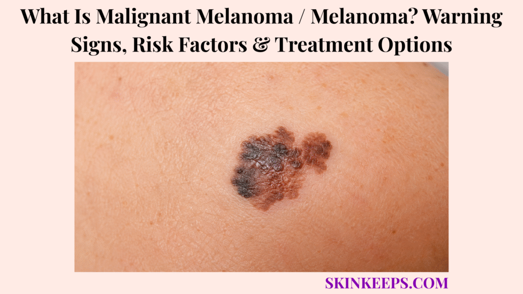
Melanoma is a malignant tumour of melanocytes that can invade deeper skin, spread to lymph nodes and metastasize to distant organs if it is not detected and treated early. Melanocytes are pigment-producing cells, and melanoma is distinct from the more common basal cell and squamous cell skin cancers because of its greater metastatic potential.
Early detection changes the treatment pathway substantially. Melanoma in situ and many thin localized melanomas can be treated surgically, while deeper, node-positive or metastatic disease may require nodal staging, immunotherapy, targeted treatment or other oncology-directed therapy according to stage and tumour biology.
Prompt dermatologic assessment is appropriate for a new or evolving pigmented lesion, an ugly-duckling spot, a rapidly enlarging firm nodule, unexplained bleeding or ulceration, a persistent non-healing growth, a new or changing longitudinal nail pigment band, or a changing lesion on a palm or sole. Melanoma can be smaller than 6 mm and does not need to be brown or black, so waiting for every ABCDE feature can delay assessment.
How Can You Recognize the Warning Signs of Melanoma?
Melanoma is suspected when a skin lesion is new, evolving, visually unusual or shows concerning asymmetry, border, colour or growth changes.
What Are the ABCDE Warning Signs of Melanoma?
The ABCDE rule identifies melanoma warning signs through asymmetry, border irregularity, colour variation, diameter and evolution.
A — Asymmetry: one half differs from the other. B — Border: the outline may be irregular, scalloped or poorly defined. C — Colour: a lesion can contain uneven tan, brown, black, white, red or blue areas.
D — Diameter: melanoma is often larger than 6 mm when diagnosed, but it can be smaller. E — Evolving: change in size, shape, colour, symptoms or structure is often more useful than waiting for a lesion to become large.
What Is the Ugly Duckling Sign?
The ugly-duckling sign describes a mole or spot that looks distinctly different from the person’s other lesions.
A lesion can be small and still deserve attention if it breaks the person’s usual mole pattern or continues to evolve. Looking different does not equal cancer, but difference plus change raises the level of suspicion.
What Does Nodular Melanoma Look Like?
Nodular melanoma often appears as a firm raised lesion that enlarges rapidly and may be dark, red, pink or skin-coloured.
It may look dome-shaped, continue growing vertically, crust, ulcerate or bleed. Because nodular melanoma can develop as a relatively symmetrical bump, ABCDE alone is less sensitive than the practical pattern of elevated or enlarging → firm → growing.
Can Melanoma Be Pink or Skin-Coloured?
Yes; amelanotic melanoma can appear pink, red or flesh-coloured because it contains little visible pigment.
These lesions can resemble a cyst, inflamed bump or non-healing sore. Persistent growth, recurrent bleeding or ulceration matters even when there is little or no dark pigment.
What Does Acral Melanoma Look Like?
Acral melanoma can appear as an irregular pigmented patch or changing lesion on the palm, sole, fingers, toes or nail unit.
It may resemble a bruise, callus, wart or wound and can occur on relatively sun-protected skin. Acral melanoma is particularly important to recognize across darker skin tones, but it is not exclusive to any one skin colour.
What Can Melanoma Beneath a Nail Look Like?
Nail melanoma may begin as a new or changing longitudinal brown or black band that progressively widens or becomes increasingly irregular.
Concern rises when a band changes, broadens, develops irregular pigment or extends onto surrounding nail-fold skin. Not every nail streak is melanoma, so the pattern requires clinical assessment rather than self-diagnosis from colour alone.
Can Melanoma Itch, Hurt or Bleed?
Melanoma can itch, hurt, bleed, ulcerate or crust, but many melanomas cause no symptoms.
Repeated bleeding, recurrent crusting, tenderness or a sore that does not heal can increase concern, but the absence of symptoms does not make an evolving lesion safe.
Figure 1. ABCDE is useful, but melanoma recognition also requires the ugly-duckling sign, rapidly growing nodules, non-pigmented lesions and palm, sole or nail warning signs.
Why Does Melanoma Develop, and Who Has a Higher Risk?
Melanoma develops when cancer-driving changes allow melanocytes to grow uncontrollably, with ultraviolet exposure, inherited susceptibility and other risk factors contributing differently across melanoma subtypes.
How Does Ultraviolet Radiation Contribute to Melanoma?
Ultraviolet radiation can damage melanocyte DNA and increase the chance that cancer-driving mutations accumulate.
Sunlight, blistering sunburns, tanning beds and tanning lamps all contribute ultraviolet exposure. UV is an important melanoma driver, but not every melanoma can be traced to one past sunburn and some melanomas arise where UV is not the dominant factor.
Who Has a Higher Risk of Developing Melanoma?
Melanoma risk rises with factors such as substantial UV exposure, previous melanoma, numerous or atypical moles, family history and immunosuppression.
Fair or burn-prone skin, freckles and red or blond hair increase UV susceptibility, but people without classic risk factors can still develop melanoma. Risk modifies surveillance; it does not decide whether a new lesion deserves attention.
Does Melanoma Always Develop From an Existing Mole?
No; melanoma can develop within a pre-existing melanocytic nevus or mole or arise de novo on previously normal-looking skin.
Self-examination therefore needs to track both change in established moles and completely new lesions.
Does Darker Skin Protect Against Melanoma?
No; melanoma can develop in every skin tone, including on palms, soles and nail units where ultraviolet exposure may not be the main driver.
Incidence differs between populations, but darker skin does not create immunity. Delayed recognition can be especially important when melanoma occurs on the sole or nail unit.
What Are the Main Types of Cutaneous Melanoma?
The major cutaneous melanoma patterns include superficial spreading, nodular, lentigo maligna and acral lentiginous melanoma.
Superficial spreading melanoma often begins as a flat or slightly raised irregularly pigmented lesion. Nodular melanoma grows more vertically as a firm expanding bump.
Lentigo maligna is an in-situ precursor on chronically sun-damaged skin; lentigo maligna melanoma refers to invasion developing from that lesion. Acral lentiginous melanoma arises on palms, soles or nail units.
Clinical appearance can suggest a subtype, but patients cannot reliably subtype melanoma from photographs alone.
What Is the Difference Between Melanoma In Situ and Invasive Melanoma?
Melanoma in situ remains confined to the epidermis, while invasive melanoma has crossed into the dermis and gained metastatic potential.
Stage 0 disease remains epidermal. Invasive melanoma has penetrated deeper tissue and can access lymphatic or blood vessels, but invasion does not mean spread has already occurred.
Figure 2. Melanoma in situ remains within the epidermis, while invasive melanoma crosses into the dermis and gains access to pathways that can support regional or distant spread.
How Is Melanoma Distinguished, Diagnosed and Staged?
Melanoma is confirmed by histopathology after biopsy, then staged using tumour depth, ulceration and evidence of regional or distant spread.
How Is Melanoma Different From an Ordinary Mole?
A benign mole is usually relatively stable and symmetrical, while melanoma becomes more suspicious when a lesion is new, evolving or unusually asymmetric, irregular or varied in colour.
A stable mole often has a predictable pattern, while melanoma concern rises with evolution, bleeding, ulceration or an ugly-duckling appearance. Visual inspection can identify suspicion but cannot confirm melanoma.
Which Other Skin Lesions Can Resemble Melanoma?
Dysplastic nevus can look irregular without being melanoma, which is why evolution and clinical context still matter.
Seborrhoeic keratosis can mimic a pigmented melanoma despite being a benign growth.
Basal cell carcinoma can occasionally be pigmented and enter the differential with melanoma.
Solar lentigines, blood beneath the nail, warts, calluses and persistent inflammatory lesions can also resemble melanoma. Persistent evolution should override reassurance from a familiar-looking label.
How Does Dermoscopy Help Assess Melanoma?
Dermoscopy helps dermatologists detect pigment, vascular and structural patterns that are not reliably visible to the naked eye and decide which lesions need biopsy.
It can reveal pigment-network irregularity, structural asymmetry and vascular clues, but dermoscopy improves selection and assessment rather than replacing histopathology.
Which Biopsy Is Used When Melanoma Is Suspected?
A suspicious melanoma should be biopsied in a way that preserves enough tissue for the pathologist to assess the tumour’s full invasive depth whenever technically feasible.
Adequate sampling is essential because diagnostic histology and Breslow depth determine staging and treatment planning. Cosmetic laser or superficial destructive treatment should not be used first on a suspicious pigmented lesion because it can remove or damage the tissue needed for diagnosis.
What Is Breslow Thickness?
Breslow thickness measures the vertical depth of an invasive melanoma in millimetres and is one of the most important determinants of stage and management.
Increasing depth generally raises metastatic risk and influences excision planning, sentinel-node discussion and stage. Ulceration is a separate staging feature that also changes risk classification.
What Does the Melanoma Pathology Report Need to Show?
A melanoma pathology report should establish whether the tumour is in situ or invasive and describe key features such as Breslow thickness, ulceration and margins.
Subtype, mitotic activity and other histologic features may also be reported, but the management-changing core for readers is in situ versus invasive disease, Breslow thickness, ulceration and evidence relevant to spread.
When Is a Sentinel Lymph-Node Biopsy Considered?
Sentinel lymph-node biopsy is considered for selected invasive melanomas when tumour thickness, ulceration or other risk features create a meaningful risk of occult regional spread.
The procedure samples the first draining lymph node or nodes for staging and prognosis. It is not required for every melanoma and is not the same as removing all regional lymph nodes.
When Are CT, PET or MRI Scans Needed for Melanoma?
Extensive body imaging is usually reserved for melanoma with higher-stage features, suspected nodal involvement, concerning systemic symptoms or established advanced disease.
Early localized melanoma does not automatically require whole-body PET, CT or MRI. Imaging intensity follows stage and clinical risk rather than the word “cancer” alone.
What Do Melanoma Stages 0–IV Mean?
Melanoma stages progress from Stage 0 disease confined to the epidermis to Stage IV disease with distant metastasis.
Stage 0 is melanoma in situ. Stages I–II describe invasive primary melanoma without established regional or distant metastasis, with thickness and ulceration contributing to stage.
Stage III includes regional nodal, satellite or in-transit spread. Stage IV means distant metastatic disease.
| Feature | Ordinary Mole | Dysplastic Nevus | Seborrhoeic Keratosis | Melanoma |
|---|---|---|---|---|
| Stability | Usually stable | Can look atypical but may remain stable | Usually benign and predictable | Evolution is an important warning sign |
| Symmetry | Often symmetrical | Can be irregular | Variable | Asymmetry is common |
| Colour | Often relatively uniform | May vary | Variable | Multiple or irregular colours possible |
| Growth / change | Limited | Needs clinical context | Usually predictable | New or evolving pattern is concerning |
| Definitive diagnosis | Clinical context | Clinical / biopsy when indicated | Clinical context | Histopathology |
Figure 3. Visual suspicion leads to dermoscopy and adequate biopsy; histopathology then provides Breslow thickness, ulceration and other findings that determine whether nodal or body-staging assessment is needed.
How Is Melanoma Treated?
Melanoma treatment is determined primarily by stage, resectability, regional spread and tumour molecular features.
How Is Melanoma In Situ Treated?
Stage 0 melanoma is treated with complete surgical excision because malignant cells remain confined to the epidermis.
Surgery removes the lesion with an appropriate surrounding margin and aims for microscopically clear edges. Completely removed melanoma in situ does not routinely require systemic melanoma therapy.
How Are Stage I and Stage II Melanomas Treated?
Wide local excision is the core treatment for Stage I and Stage II melanoma, with sentinel-node staging and adjuvant therapy considered for selected higher-risk tumours.
Surgical planning depends on pathological thickness. Selected higher-risk Stage II disease may be considered for adjuvant anti-PD-1 therapy, but this is individualized rather than automatic for every Stage II melanoma.
How Is Stage III Melanoma Treated?
Stage III melanoma is managed with a combination of surgery and systemic therapy according to resectability, regional disease pattern and tumour biology.
Adjuvant anti-PD-1 therapy can be used after appropriate local treatment, while BRAF V600-mutated melanoma may qualify for BRAF/MEK targeted therapy. The exact strategy is multidisciplinary and not identical for every Stage III patient.
Can Melanoma Treatment Be Given Before Surgery?
Yes; selected patients with resectable high-risk node-positive melanoma may receive neoadjuvant systemic therapy before surgery.
Neoadjuvant therapy means treatment before definitive surgery and can use checkpoint-inhibitor strategies in specialist settings. Evidence and approval status vary by regimen, so it should not be presented as a universal approach for all resectable melanoma.
How Is Unresectable or Metastatic Melanoma Treated?
Unresectable or metastatic melanoma is commonly treated with systemic immunotherapy, mutation-directed targeted therapy or other specialist oncologic approaches.
Options include anti-PD-1 therapy, combination checkpoint inhibition and PD-1/LAG-3 combinations. Combination immune therapy can increase toxicity, so a more intensive regimen is not automatically better for every patient.
When Is BRAF/MEK Targeted Therapy Used?
BRAF/MEK targeted therapy is used only when tumour testing confirms an actionable BRAF V600 mutation.
The treatment suppresses signalling through the BRAF and MEK pathway and can produce rapid tumour control in eligible disease. Melanoma diagnosis alone does not qualify a patient for BRAF-targeted treatment.
Are Other Treatments Used for Advanced Melanoma?
Yes; selected advanced melanomas may also be treated with tumour-infiltrating lymphocyte therapy, radiation, surgery, intralesional treatment or clinical-trial approaches.
The role of these therapies depends on disease distribution, previous treatment and specialist multidisciplinary planning rather than one universal sequence.
Figure 4. Stage 0 is surgery-only in most cases, Stages I–II remain surgery-led, Stage III combines regional and systemic planning, and advanced melanoma uses oncology-directed systemic therapy including mutation-directed treatment only when the tumour qualifies.
What Happens After Melanoma Treatment, and How Can Future Risk Be Reduced?
Melanoma follow-up is tailored to original stage and recurrence risk while also checking the entire skin for new primary melanomas.
What Determines the Outlook for Melanoma?
Melanoma prognosis depends strongly on how early the disease is detected and whether it has invaded deeply or spread beyond the primary site.
Breslow thickness, ulceration, lymph-node involvement, distant metastasis, the location and extent of metastatic disease and treatment response all influence prognosis.
Can Melanoma Come Back After Treatment?
Yes; melanoma can recur locally, in regional lymph nodes or at distant sites after treatment.
Recurrence risk varies with the original stage and pathology, so surveillance intensity is individualized rather than identical for everyone.
Can Someone Develop a Second New Melanoma?
Yes; a previous melanoma increases the risk of developing another primary melanoma elsewhere on the skin.
A second primary melanoma is a new tumour rather than a recurrence of the first one. Whole-skin surveillance matters because the original scar is not the only site at risk.
How Should Skin Self-Examination Be Performed After Melanoma?
Whole-body skin self-examination should compare current lesions with the person’s usual pattern and include easily missed sites such as the scalp, back, palms, soles and nails.
The back, buttocks, between the toes and nail units deserve deliberate inspection. New lesions, evolution, an ugly-duckling spot, a new firm nodule or a changing nail streak should be assessed rather than watched indefinitely.
Self-examination supports professional follow-up and does not replace stage-based dermatology or oncology surveillance.
How Can UV-Related Melanoma Risk Be Reduced?
Reducing unnecessary ultraviolet exposure lowers preventable melanoma risk but cannot eliminate melanoma risk completely.
Avoid tanning beds, seek shade, use sun-protective clothing and apply broad-spectrum sunscreen consistently. Acral melanoma and some other melanomas arise where UV is not the primary driver, so sun protection reduces risk rather than making melanoma impossible.
Which Changes Need Prompt Review After Previous Melanoma?
A melanoma survivor should seek prompt assessment for a new or evolving lesion, rapidly growing nodule, unexplained ulceration or bleeding, or a changing nail streak.
People with a higher-stage history should also report new enlarged nodes, persistent unexplained pain, neurological symptoms, significant shortness of breath or other concerning systemic changes.
Surveillance logic: complete initial treatment → stage-based follow-up → whole-body skin checks → UV-risk reduction → prompt review of new or evolving lesions.
What Should You Remember About Malignant Melanoma / Melanoma?
Melanoma is a malignant cancer of melanocytes whose treatment and prognosis depend heavily on how early it is detected, how deeply it has invaded and whether it has spread.
- Melanoma is malignant by definition; “malignant melanoma” is not a separate disease.
- ABCDE is useful but incomplete.
- Evolution and the ugly-duckling sign are high-value warning clues.
- Nodular melanoma can be a rapidly growing firm bump.
- Amelanotic melanoma can be pink or flesh-coloured.
- Acral and nail melanoma can occur on sun-protected sites.
- Melanoma can occur in every skin tone.
- Melanoma can arise without a previous mole.
- Biopsy and histopathology confirm diagnosis.
- Breslow thickness measures invasive depth.
- Ulceration and regional or distant spread influence stage.
- Early melanoma treatment is primarily surgical.
- Higher-stage disease may require immunotherapy or targeted therapy.
- BRAF-directed treatment requires an actionable BRAF V600 mutation.
- Recurrence and a second new primary melanoma remain possible.
- Whole-body surveillance remains important after treatment.
Recognize → biopsy → measure invasion → stage → treat by stage and biology → monitor recurrence and new melanoma.
Frequently Asked Questions About Malignant Melanoma / Melanoma
The most important melanoma questions concern its earliest warning signs, origin, non-classic appearances, diagnostic staging and curability when detected early.
What Are the Earliest Warning Signs of Melanoma?
Early melanoma may appear as a new or evolving lesion with asymmetry, irregular borders, colour variation or a pattern that looks different from the person’s other moles. Rapidly growing nodules and non-pigmented lesions are important exceptions to an ABCDE-only approach.
Can Melanoma Develop Without an Existing Mole?
Yes; melanoma can arise on previously normal-looking skin as well as within an existing melanocytic nevus. Both new lesions and changing established moles deserve attention.
Can Melanoma Be Pink or Smaller Than 6 mm?
Yes; melanoma can be pink or flesh-coloured and can be smaller than 6 mm, so colour and diameter alone cannot exclude it. Evolution remains one of the most useful warning signs.
How Is Melanoma Diagnosed and Staged?
Melanoma is diagnosed by biopsy and histopathology, then staged using invasion depth, ulceration and evidence of regional or distant spread. Breslow thickness and selected sentinel-node assessment help define management.
Can Melanoma Be Cured if It Is Found Early?
Early localized melanoma can often be treated successfully with complete surgery, while prognosis becomes less favourable as invasion and spread increase. This is why early recognition and adequate biopsy change the treatment pathway.
Which Sources Support This Melanoma Guidance?
American Academy of Dermatology — Melanoma Overview — Used for the melanoma definition, metastatic potential, early-detection importance, all-skin-tone relevance and surveillance context.
American Academy of Dermatology — ABCDEs of Melanoma — Used for asymmetry, border, colour, diameter caveat and evolution.
American Academy of Dermatology — Melanoma Signs and Symptoms — Used for new or changing lesions, unusual spots, firm dome-shaped growths, nail pigmentation, pink melanoma, itching, bleeding and ulceration.
American Academy of Dermatology — Melanoma Causes — Used for ultraviolet exposure, indoor tanning, sunburn, numerous or atypical moles, immunosuppression, family history, previous melanoma and all-skin-tone risk.
American Academy of Dermatology — Melanoma Diagnosis and Treatment — Used for dermatologic examination, dermoscopy, biopsy-based confirmation and treatment dependence on depth and spread.
National Cancer Institute — Melanoma Treatment (PDQ®) — Primary staging and treatment source used for Breslow-based risk, ulceration, sentinel-node concepts, Stage 0–IV treatment logic, immunotherapy, neoadjuvant strategies, BRAF/MEK targeted therapy and advanced-treatment options.
American Academy of Dermatology — Melanoma: Life After Treatment — Used for recurrence, second-primary melanoma risk, lifelong follow-up, skin self-examination and prevention after treatment.



