
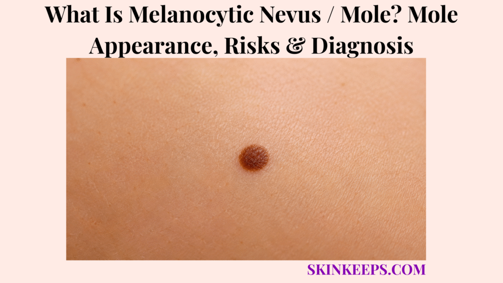
A melanocytic nevus, commonly called a mole, is a benign skin growth formed when melanocytes cluster together in a localized area. Melanocytes produce pigment, but a benign nevus can be skin-coloured, pink, tan, brown, very dark, flat, raised or dome-shaped depending on its type and stage of maturation.
Acquired moles commonly appear during childhood and adolescence and most remain harmless. Melanoma can sometimes resemble a mole, but the key safety question is not whether every mole might become cancer; it is whether a lesion meaningfully evolves or looks unlike the person’s usual mole pattern.
A new or changing mole deserves professional assessment when it develops new asymmetry, an increasingly irregular border, several changing colours, continued enlargement, a new lumpy surface, persistent itching, bleeding, crusting, oozing or an ugly-duckling appearance. A suspicious mole should be diagnosed before cosmetic removal or destruction.
How Can You Recognize a Typical Melanocytic Nevus?
A typical melanocytic nevus is usually a stable, symmetrical round or oval mole with a relatively clear border and fairly uniform internal colour.
What Does a Common Mole Usually Look Like?
A common acquired mole is typically round or oval, relatively symmetrical, clearly bordered and fairly uniform in colour.
Many ordinary moles are smaller than about 5 mm, but benign nevi can be larger. A nevus may begin flat and later become slightly elevated or dome-shaped as it matures.
Size is only one feature; a small lesion is not automatically safe, and a larger stable nevus is not automatically malignant. Stability and overall structure matter more.
What Colours Can Normal Moles Have?
Normal melanocytic nevi can be skin-coloured, pink, tan, light brown, dark brown or nearly black depending on nevus type and skin tone.
A benign mole often has relatively uniform colour within the lesion, but there is no single healthy mole colour for every person. Some benign blue nevi can appear blue-grey because pigment lies deeper in the skin.
Can Normal Moles Be Flat, Raised or Hairy?
Yes; benign moles can be flat, slightly raised, dome-shaped or hair-bearing.
Terminal hairs can grow through a nevus because hair follicles remain present beneath or within the lesion. Hair does not prove benignity; the lesion’s overall structure, stability and evolution are more important.
Where Can Melanocytic Nevi Develop?
Melanocytic nevi can develop on almost any skin surface, including both sun-exposed and covered areas.
Common locations include the trunk, limbs, face and scalp, but nevi can also occur on palms, soles and other less sun-exposed sites. A covered location is not automatically abnormal.
How Do Moles Normally Change With Age?
Moles commonly appear during childhood and adolescence and may enlarge proportionally, lighten, darken or become more raised while remaining benign.
As the body grows, a benign nevus can enlarge evenly and later mature into a more elevated lesion. Some nevi lighten or gradually involute later in life.
A genuinely new or changing lesion later in adulthood deserves more attention because ordinary acquired nevi become less common with age, but no single age cutoff diagnoses melanoma.
Figure 1. A common melanocytic nevus can mature from flat to slightly raised or dome-shaped while remaining benign when its structure stays stable and predictable.
Why Do Melanocytic Nevi Develop, and Which Mole Types Affect Risk?
Melanocytic nevi develop when melanocytes proliferate locally, with genetics, ultraviolet exposure and other host factors influencing how many and which types of moles a person develops.
Why Do Melanocytes Form a Mole?
A melanocytic nevus forms when melanocytes grow in a localized cluster rather than remaining evenly distributed through the skin.
The exact reason one particular nevus forms is not always known. Genetics, ultraviolet exposure and immune status influence nevus number and pattern, so no single cause explains every mole.
What Is an Acquired Melanocytic Nevus?
An acquired melanocytic nevus is a mole that develops after birth, most commonly during childhood or adolescence.
Junctional nevi have melanocytic nests near the epidermal–dermal junction, compound nevi extend into both junctional and dermal levels, and intradermal nevi are predominantly dermal. These pathology categories help describe maturation but are less important to patients than whether a mole is stable or evolving.
What Is a Congenital Melanocytic Nevus?
A congenital melanocytic nevus is present at birth or becomes evident very early in infancy and can range from small to very large.
Risk is not identical across congenital nevi. Small congenital lesions generally carry much lower melanoma risk than large or giant congenital nevi, so follow-up decisions depend on size, location and other characteristics rather than one universal percentage or one automatic removal rule.
What Is a Dysplastic or Atypical Nevus?
A dysplastic nevus is a benign melanocytic nevus with more atypical size, shape, border or colour features than an ordinary mole.
Dysplastic nevi may be larger, less symmetrical or more varied in pigmentation without being melanoma. Having several dysplastic nevi is more important as a marker of patient-level melanoma susceptibility than as proof that any one individual nevus will transform.
Does Having Many Moles Increase Melanoma Risk?
Yes; having numerous common nevi or multiple dysplastic nevi increases a person’s overall melanoma risk.
This is a patient-level risk concept. People with many or atypical nevi may benefit from closer skin examination, self-monitoring and selected photographic surveillance without treating each nevus as a high-risk lesion.
Can a Common Mole Turn Into Melanoma?
Yes, but transformation of an individual ordinary mole is uncommon, and most melanomas arise without a recognized pre-existing mole.
Most common and dysplastic nevi remain benign. Melanoma can arise de novo on previously normal-appearing skin, so surveillance must include both existing moles and new lesions.
This is why routine preventive removal of stable harmless nevi does not prevent most melanoma.
Figure 2. The chance that one ordinary mole transforms is low, while a high total nevus burden or multiple dysplastic nevi increases the person’s overall melanoma risk and surveillance need.
How Can You Tell a Benign Mole From a Possible Melanoma?
A benign mole is usually stable and consistent with the person’s other nevi, while melanoma becomes more concerning when a lesion evolves or behaves as an ugly duckling.
What Are the ABCDE Warning Signs?
The ABCDE rule flags suspicious mole changes through asymmetry, border irregularity, colour variation, diameter and evolution.
A — Asymmetry: one half differs from the other. B — Border: the edge becomes irregular, notched, blurred or poorly defined.
C — Colour: several colours or a changing internal colour pattern can increase concern. D — Diameter: melanoma is often larger than 6 mm at diagnosis, but it can be smaller.
E — Evolving: change in size, shape, colour, surface or symptoms is especially important. ABCDE raises suspicion; it does not confirm melanoma.
What Is the Ugly Duckling Sign?
The ugly-duckling sign is a mole or spot that looks distinctly different from the person’s usual mole pattern.
The outlier may be darker, lighter, larger, more irregular or structurally different. An ugly duckling deserves closer assessment but is not automatically melanoma.
Which Changes in an Existing Mole Matter Most?
Progressive enlargement, new asymmetry, changing colour, altered surface, persistent crusting, itching, oozing or bleeding are important reasons to reassess a mole.
A new raised or lumpy area also matters when it represents genuine evolution rather than the slow predictable maturation of a long-stable nevus. The practical decision is stable → observe; meaningfully evolving → assess.
How Is a Mole Different From a Solar Lentigo or Seborrhoeic Keratosis?
A melanocytic nevus is a melanocyte growth, whereas a solar lentigo or sunspot is a flat UV-related pigmented macule.
A seborrhoeic keratosis is a benign keratinocytic growth that often develops a waxy, keratotic or stuck-on surface.
Clinical overlap occurs, especially in pigmented lesions, so changing or uncertain lesions should be assessed rather than diagnosed from one surface feature.
Why Should a Suspicious Mole Not Be Treated With Laser First?
A suspicious mole should not be destroyed with laser first because intact tissue may be needed to determine whether melanoma is present and how deeply it has invaded.
Cosmetic destruction can remove pigment and tissue architecture needed for histopathologic diagnosis. A diagnosed benign lesion may have cosmetic options, but diagnostic uncertainty should be resolved before destructive treatment.
| Feature | Stable Benign Mole | Needs Closer Assessment |
|---|---|---|
| Symmetry | Relatively symmetrical | New asymmetry |
| Border | Stable and defined | Becoming irregular, notched or blurred |
| Colour | Relatively uniform for that lesion | Increasing or changing variation |
| Size | Stable or predictably proportional | Progressive enlargement |
| Pattern | Matches the person’s usual nevi | Ugly-duckling appearance |
| Symptoms | Usually unchanged | New bleeding, crusting, itching or oozing |
| Next step | Observe | Dermoscopy ± biopsy |
How Is a Melanocytic Nevus Diagnosed and Monitored?
Most ordinary melanocytic nevi can be diagnosed clinically, while dermoscopy, digital monitoring and biopsy are used when morphology or evolution creates uncertainty.
Can a Dermatologist Diagnose a Mole by Looking at It?
Yes; most typical melanocytic nevi can be identified clinically from their symmetry, colour, border, surface, location and stability.
A dermatologist also compares the lesion with the person’s other moles and reviews its history. Nevus assessment is stronger when the lesion is interpreted within the patient’s overall mole pattern rather than in complete isolation.
How Does Dermoscopy Examine a Mole?
Dermoscopy magnifies subsurface pigment and vascular structures that help distinguish typical benign nevus patterns from suspicious structural asymmetry.
Dermoscopy can show pigment networks, dots, globules, vessels, border structures and internal symmetry that are difficult to judge with the naked eye. It improves diagnostic confidence but does not replace histopathology when melanoma remains possible.
When Is Digital Mole Monitoring Useful?
Digital mole monitoring is useful when documenting baseline appearance can help determine whether an equivocal lesion or high-risk mole pattern genuinely changes over time.
Options include clinical photography, digital dermoscopy, selected short-term lesion monitoring and total-body mole mapping. These approaches are most useful for selected people with many or atypical nevi rather than everyone with one ordinary mole.
When Does a Mole Need a Biopsy?
A mole needs biopsy or excision when melanoma cannot be confidently excluded because of evolution, suspicious dermoscopic features, unusual appearance or persistent unexplained symptoms.
Triggers for tissue diagnosis can include an ugly-duckling lesion, progressive structural change, persistent bleeding or crusting, or an equivocal lesion that cannot be safely classified by examination and dermoscopy. Routine biopsy of every common or dysplastic nevus is unnecessary.
Why Is Histopathology Important?
Histopathology determines whether a sampled lesion is an ordinary nevus, dysplastic nevus, melanoma or another skin growth.
Microscopy evaluates tissue architecture and cellular features that visual inspection cannot confirm. If melanoma is seriously suspected, tissue sampling should preserve the information needed for diagnosis rather than simply destroying visible pigment.
Figure 3. Stable typical moles can be observed, equivocal lesions can be examined with dermoscopy or selected digital monitoring, and evolving or suspicious lesions move to biopsy and histopathology.
When Should a Melanocytic Nevus Be Removed?
A benign mole does not need routine preventive removal; removal is mainly appropriate for diagnostic uncertainty, repeated irritation or a person’s informed cosmetic preference after benignity is established.
Do Normal Moles Need Preventive Removal?
No; removing every ordinary or dysplastic mole does not prevent most melanomas.
Most individual nevi never transform, and many melanomas arise independently on previously normal-looking skin. Careful surveillance is usually more useful than blanket preventive removal.
When Is Excision Medically Appropriate?
Mole excision is medically appropriate when melanoma cannot be confidently excluded, histopathologic diagnosis is needed or the lesion repeatedly becomes irritated or traumatized.
A benign mole can also be removed for cosmetic reasons after diagnostic confidence is established, but cosmetic preference and medical necessity are different indications.
How Are Moles Removed?
Mole-removal technique depends on whether the lesion is diagnostically suspicious, its depth and its clinical appearance.
A suspicious flat melanocytic lesion generally needs surgical sampling that preserves tissue for pathology. A clearly benign protruding nevus may be suitable for shave removal in selected cases.
No one technique fits every mole, and a suspicious lesion should not be cosmetically destroyed before diagnosis.
Can a Mole Grow Back After Removal?
Yes; residual nevus cells can sometimes produce recurrent pigmentation or regrowth after incomplete or superficial mole removal.
This recurrent-nevus phenomenon can look irregular within a scar and may mimic melanoma clinically. Previous pathology and the pattern of new pigmentation matter, so recurrent pigment in a removal scar should be reassessed rather than automatically assumed harmless.
Should a Benign Mole Be Removed at Home?
No; cutting, burning or chemically removing a mole at home can cause injury and can destroy tissue needed for an accurate diagnosis.
Home removal can lead to bleeding, infection, scarring, incomplete removal and delay in diagnosing skin cancer. The safe sequence is diagnosis first → professional removal if justified.
What Should You Remember About Melanocytic Nevi / Moles?
Melanocytic nevi are benign melanocyte growths, and the safest strategy is to know the normal mole pattern and investigate meaningful evolution rather than remove every lesion preventively.
- A melanocytic nevus is benign.
- Normal moles vary in colour and elevation.
- Hair can grow through a benign mole but does not prove benignity.
- Stability is reassuring.
- Acquired nevi commonly develop during childhood and adolescence.
- Congenital nevi have size-dependent risk considerations.
- Dysplastic nevi are not melanoma.
- Numerous or atypical nevi raise patient-level melanoma risk.
- Most individual moles remain benign.
- Most melanomas do not require a pre-existing mole.
- ABCDE helps identify suspicious change.
- Evolution and the ugly-duckling sign are especially useful.
- Dermoscopy improves lesion assessment.
- Biopsy confirms genuinely suspicious or uncertain lesions.
- Routine preventive removal of all moles is unnecessary.
- Suspicious lesions should not be cosmetically destroyed before diagnosis.
Know baseline → watch evolution → compare with other moles → use dermoscopy when needed → biopsy genuinely suspicious lesions → remove only when justified.
Frequently Asked Questions About Melanocytic Nevi / Moles
The most important mole questions concern why nevi develop, melanoma transformation, suspicious changes, dysplastic-nevus management and when professional assessment is needed.
What Causes Melanocytic Nevi or Moles to Develop?
Melanocytic nevi develop when melanocytes grow in localized clusters, with genetics, ultraviolet exposure and other host factors influencing mole patterns. No single cause explains every mole.
Can a Normal Mole Turn Into Melanoma?
Yes, but transformation of an individual ordinary mole is uncommon, and most melanomas arise without a recognized pre-existing mole. Lesion-level transformation risk is different from the higher overall melanoma risk associated with having many or atypical nevi.
How Can You Tell a Benign Mole From a Suspicious Mole?
A benign mole is usually stable and consistent with the person’s normal mole pattern, while evolution, ABCDE changes or an ugly-duckling appearance justify closer assessment. Dermoscopy or biopsy is used when clinical uncertainty remains.
Do Atypical or Dysplastic Moles Need to Be Removed?
No; dysplastic nevi do not automatically require removal simply because they look atypical. Lesions that evolve or remain diagnostically suspicious may require biopsy, while stable lesions are often monitored.
When Should a Changing Mole Be Checked by a Dermatologist?
A mole should be checked when it develops persistent growth, new asymmetry, changing colour, an irregular surface, bleeding, crusting or an appearance unlike the person’s other moles. Evolution warrants assessment but does not itself prove melanoma.
Which Sources Support This Melanocytic Nevus / Mole Guidance?
National Cancer Institute — Common Moles, Dysplastic Nevi, and Risk of Melanoma — Primary evidence source for benign nevus definition, common-mole morphology, dysplastic nevi, many-nevi risk, low individual transformation risk, most melanomas arising independently, ABCDE-related changes, surveillance and selective removal.
DermNet — Melanocytic Naevus — Used for body distribution, acquired and congenital nevi, age-related changes, junctional/compound/intradermal context, genetics and UV influences, clinical diagnosis, digital monitoring and removal concepts.
American Cancer Society — Risk Factors for Melanoma Skin Cancer — Used specifically for patient-level risk from many or dysplastic nevi and for the size-dependent melanoma-risk context of congenital melanocytic nevi.
DermNet — Dermoscopy — Used for pigment-network, dots, globules, vascular and border assessment and for the role of digital image archiving and mole mapping.
American Academy of Dermatology — Moles: Signs and Symptoms — Used for normal mole colour and surface variation, hair-bearing moles, body locations and ABCDE features including the fact that melanoma can be smaller than 6 mm.
American Academy of Dermatology — Moles: Diagnosis and Treatment — Used for clinical diagnosis, medically appropriate removal, excision versus shave concepts, microscopy after removal and the warning against home mole removal.
The Recurrent Nevus Phenomenon — Review — Used for recurrent pigmentation after partial removal, the link with superficial/shave removal and the need to distinguish recurrent nevus from melanoma in a scar.



