
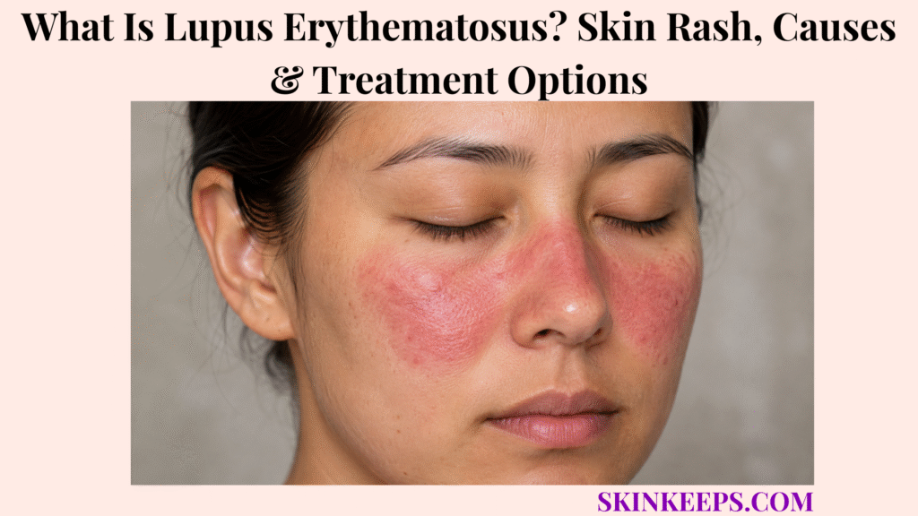
Lupus erythematosus is an autoimmune disease spectrum that can affect the skin alone or occur as part of systemic lupus involving multiple organs. Cutaneous lupus erythematosus, or CLE, describes lupus-specific skin disease, while systemic lupus erythematosus, or SLE, can involve skin together with joints, kidneys, blood, nervous system and other organs.
The main cutaneous patterns include the acute malar or butterfly rash, photosensitive subacute cutaneous lupus and persistent discoid plaques. Identifying the subtype matters because the association with systemic disease, the risk of scarring and the treatment priorities differ substantially.
Medical assessment is particularly important when a lupus-like rash occurs with chest pain, breathing difficulty, facial or leg swelling, abnormal urine, marked proteinuria, seizures, severe neurological symptoms, persistent fever or substantial systemic illness. These findings can indicate disease beyond the skin and require broader evaluation.
How Can You Recognize Lupus Erythematosus on the Skin?
Cutaneous lupus can appear as a butterfly-shaped facial rash, photosensitive ring-shaped or scaly lesions, or persistent discoid plaques that may scar.
What Does the Butterfly Rash Look Like?
The lupus butterfly or malar rash typically extends across both cheeks and the bridge of the nose and is especially associated with acute cutaneous lupus and SLE.
The eruption often worsens after ultraviolet exposure and may appear during an SLE flare. Not everyone with lupus develops a butterfly rash, and not every red facial rash represents lupus, so facial shape alone should never be treated as diagnostic proof.
What Do Discoid Lupus Lesions Look Like?
Discoid lupus erythematosus produces persistent raised or scaly plaques that can develop atrophy, pigment change and permanent scarring.
The face, scalp and ears are common sites. Lesions can have adherent scale and evolve from active inflammation into central atrophy, dyspigmentation and scar.
Scalp discoid lupus can permanently destroy hair follicles and cause scarring alopecia. Early treatment aims to stop further follicular loss rather than promise regrowth after a follicle has already been destroyed by scar.
What Does Subacute Cutaneous Lupus Look Like?
Subacute cutaneous lupus usually causes a widespread photosensitive rash made of ring-shaped or scaly lesions that generally heal without true scarring.
SCLE often appears on sun-exposed upper chest, upper back and arms as annular lesions or a papulosquamous eruption. The scale can resemble psoriasis or eczema, and pigment change may remain even when true scarring does not.
Can Lupus Affect the Mouth, Hair and Nails?
Yes; lupus can cause oral or nasal sores, hair loss and nail or periungual changes in addition to ordinary skin lesions.
Hair loss can be diffuse or patchy. Inflammatory non-scarring loss may recover as disease activity settles, while discoid scarring alopecia can be permanent because the follicle itself is destroyed.
How Can Lupus Rash Appear Across Different Skin Tones?
Lupus lesions can appear red-brown, violet, grey-brown or darker than surrounding skin rather than always appearing bright red.
Hyperpigmentation and hypopigmentation may be especially visible in deeper skin tones, and residual colour change can persist after inflammation becomes quiet. Skin colour alone cannot determine whether a lesion is active, scarred or lupus-related.
Figure 1. Acute, subacute and chronic/discoid cutaneous lupus have different morphology, systemic associations and scarring potential.
Why Does Lupus Erythematosus Develop, and Which Cutaneous Types Matter?
Lupus develops through autoimmune dysfunction influenced by genetic and environmental factors, while cutaneous disease is clinically organized into acute, subacute and chronic forms.
What Causes Lupus Erythematosus?
No single cause explains lupus; genetic susceptibility interacts with immune and environmental influences to produce autoimmune tissue inflammation.
Ultraviolet exposure, cigarette smoking, infections and selected medicines can influence disease in susceptible people, but none explains every case. Lupus should not be framed as the result of one food, one stressful event or a personal behaviour.
Why Does Sunlight Trigger Lupus Skin Flares?
Ultraviolet radiation can trigger or worsen cutaneous lupus inflammation in susceptible skin.
Sunlight and relevant artificial ultraviolet sources can provoke lesions even when other symptoms are relatively quiet. This is why photoprotection is part of disease treatment rather than optional cosmetic skincare.
What Is Acute Cutaneous Lupus?
Acute cutaneous lupus erythematosus includes the classic malar rash and is strongly associated with active systemic lupus.
ACLE may be localized to the cheeks and nasal bridge or appear as a more widespread acute eruption. It is generally non-scarring, but residual pigment change can remain after inflammation clears.
What Is Subacute Cutaneous Lupus?
Subacute cutaneous lupus erythematosus causes photosensitive annular or scaly lesions that generally heal without permanent scarring.
SCLE commonly affects sun-exposed skin and can be triggered by selected medicines. Medication start dates and rash onset therefore matter, but a prescribed drug should not be stopped independently without clinician review.
What Is Chronic Cutaneous or Discoid Lupus?
Chronic cutaneous lupus, especially discoid lupus, produces persistent inflammatory plaques that can permanently scar skin and destroy scalp hair follicles.
Atrophy, pigment alteration and scar distinguish the damage phase from active inflammation. Early disease control aims to prevent irreversible structural loss.
| CLE Type | Typical Pattern | Scarring Potential | Systemic Context |
|---|---|---|---|
| Acute CLE | Malar or broader acute eruption | Usually non-scarring | Strong systemic association |
| SCLE | Annular or papulosquamous photosensitive rash | Usually non-scarring | Variable systemic association |
| Chronic / discoid CLE | Persistent inflammatory plaques | Scarring and permanent alopecia possible | Often more skin-limited |
How Is Cutaneous Lupus Distinguished From Similar Rashes, and When Might It Indicate SLE?
Cutaneous lupus is distinguished by its morphology, photosensitive distribution, scarring behaviour and systemic context rather than by one visual feature alone.
How Is a Lupus Butterfly Rash Different From Rosacea?
Rosacea more often causes persistent facial flushing, visible vessels and papules or pustules, while a lupus malar rash occurs in an autoimmune and often photosensitive clinical context.
Rosacea has its own trigger pattern and may show central facial papules and pustules that are not defining features of a classic malar eruption. A butterfly-shaped distribution by itself does not establish lupus.
How Is Subacute Cutaneous Lupus Different From Psoriasis or Eczema?
SCLE is distinguished from psoriasis by its photosensitive distribution, annular or papulosquamous pattern and supporting clinical, laboratory or biopsy findings.
Dermatitis or eczema can also produce inflamed scaly skin, but its distribution, exposure pattern and barrier features differ from classic photosensitive SCLE.
When morphology overlaps, medication history, autoantibody context and biopsy can change the diagnostic decision.
How Is Discoid Lupus Different From Other Scalp or Facial Plaques?
Discoid lupus becomes especially concerning when persistent inflammatory plaques develop central scarring, pigment change and permanent follicular loss.
The pattern of an active inflammatory edge with a damaged or scarred centre and loss of follicular openings is different from uncomplicated dandruff, eczema or ordinary psoriasis. The presence of scar is a treatment urgency signal because established follicular destruction cannot simply be moisturized or scaled away.
Does Having Cutaneous Lupus Mean You Have Systemic Lupus?
No; cutaneous lupus can occur without systemic lupus, and the likelihood of SLE varies substantially between CLE subtypes.
ACLE has the strongest systemic association. SCLE can occur with systemic lupus in some patients, while localized discoid disease is more commonly primarily skin-limited.
Which Symptoms Suggest Lupus May Be Affecting More Than the Skin?
Joint inflammation, persistent fever, chest symptoms, swelling, abnormal urine findings or neurological symptoms can indicate lupus involvement beyond the skin.
Other clues can include fatigue, painful or swollen joints, oral or nasal ulcers, Raynaud phenomenon, chest pain with breathing, eyelid or leg swelling, urinary changes, headache, confusion or seizures. Nonspecific fatigue alone does not prove SLE, but these findings can move the assessment from CLE classification toward systemic-lupus evaluation.
Figure 2. A lupus-like skin pattern is classified first; systemic symptoms then determine whether the evaluation remains CLE-focused or expands to SLE and organ assessment.
| Feature | Lupus Malar / SCLE | Rosacea | Psoriasis / Eczema |
|---|---|---|---|
| Photosensitivity | Important clinical clue | Triggers differ | Variable |
| Papules / pustules | Not defining | Common in rosacea | Not defining for classic psoriasis |
| Scale | Can occur in SCLE | Usually not dominant | Common |
| Systemic autoimmune context | Possible | No lupus-specific context | Different disease pathway |
How Is Lupus Erythematosus Diagnosed?
Lupus is diagnosed by combining clinical findings with skin biopsy and targeted blood or urine testing rather than relying on one laboratory result.
How Does a Dermatologist Diagnose Cutaneous Lupus?
A dermatologist evaluates cutaneous lupus by examining lesion morphology, distribution, photosensitivity and other skin, scalp, nail, mucosal and systemic findings.
The history includes sun response, scalp or hair change, oral lesions, nail changes, medication exposure and symptoms outside the skin. A classic subtype may be suspected clinically, but testing is often needed when the pattern overlaps with another inflammatory disease.
When Is a Skin Biopsy Needed for Cutaneous Lupus?
A skin biopsy is useful when the suspected lupus eruption must be distinguished from another inflammatory skin disorder or its subtype needs confirmation.
Histopathology can show interface dermatitis and other subtype-related features. Direct immunofluorescence is useful in selected cases, but it is not mandatory for every patient with a clinically convincing pattern.
Which Blood Tests Are Used When Lupus Is Suspected?
Systemic lupus evaluation commonly begins with ANA and then uses more specific antibody and laboratory testing when the clinical picture supports further investigation.
Depending on the presentation, testing can include anti-double-stranded DNA, anti-Smith antibodies, complement levels and a complete blood count. A positive ANA alone does not diagnose SLE and must be interpreted with the clinical picture and additional testing.
Why Are Urine and Kidney Tests Important?
Urine and kidney tests are important because systemic lupus can affect the kidneys even when the most visible complaint is a skin rash.
Urinalysis can identify protein or other abnormalities, while kidney-function blood tests assess renal involvement. Selected patients require nephrology assessment or kidney biopsy, but this article does not replace a dedicated lupus-nephritis evaluation.
Why Must the Medication History Be Reviewed?
Medication history matters because some drugs can trigger lupus-like syndromes or subacute cutaneous lupus eruptions.
Clinicians compare medication start dates with eruption onset and consider whether the pattern is strongly photosensitive. Prescription medicines should not be stopped independently.
Figure 3. Cutaneous lupus diagnosis integrates morphology, photosensitivity, biopsy where useful, targeted serology and urine/kidney testing when systemic disease is suspected.
How Is Lupus Erythematosus Treated and Skin Flares Prevented?
Lupus skin disease is managed with rigorous photoprotection, site-appropriate anti-inflammatory therapy and systemic treatment when lesions are persistent, widespread or associated with SLE.
Why Is Sun Protection Part of Lupus Treatment?
Photoprotection is a core part of cutaneous lupus treatment because ultraviolet exposure can trigger new lesions and worsen existing inflammation.
Shade, protective clothing, sun-protective habits and consistent use of broad-spectrum sunscreen work together to reduce UV-provoked flares. Sunscreen is one part of a complete trigger-reduction strategy rather than the only measure.
Which Topical Treatments Are Used for Lupus Rash?
Localized cutaneous lupus lesions are commonly treated with topical corticosteroids, with calcineurin inhibitors used as steroid-sparing options on selected sensitive sites.
Treatment strength is matched to the body site, and selected thick discoid lesions can be treated with intralesional corticosteroid. Tacrolimus or pimecrolimus may be useful where steroid exposure is a concern.
For scalp DLE, preventing new scar formation is a more realistic goal than restoring follicles after permanent follicular destruction has already occurred.
When Is Hydroxychloroquine Used for Lupus?
Hydroxychloroquine is a major systemic treatment for persistent or widespread cutaneous lupus and is also foundational therapy for many patients with SLE unless contraindicated.
Antimalarial therapy can reduce lesions and flares but may take months to show full benefit. Clinician-directed monitoring includes attention to retinal toxicity and other treatment considerations; patient-specific doses are outside the scope of this article.
What If Cutaneous Lupus Does Not Respond to First-Line Treatment?
Treatment-resistant cutaneous lupus should first prompt reassessment of photoprotection, adherence, smoking exposure and diagnostic accuracy before stronger systemic therapy is added.
The escalation sequence is usually local therapy → antimalarial therapy → specialist systemic treatment. Methotrexate, mycophenolate or other immune-modifying therapies may be used in selected patients according to subtype, severity, pregnancy context and concurrent systemic disease.
How Is Systemic Lupus Treated When Internal Organs Are Involved?
Systemic lupus with internal-organ involvement requires treatment matched to the affected organ and disease severity rather than skin-directed therapy alone.
Treatment categories can include hydroxychloroquine, corticosteroids, conventional immunosuppressive therapy and targeted biologic treatments. The goals are to suppress active disease, prevent flares, reduce permanent organ damage and limit long-term glucocorticoid exposure.
Can Lupus Skin Damage Be Permanent?
Yes; discoid lupus can cause permanent skin scarring and irreversible scalp hair loss, while acute and subacute cutaneous lupus more often heal without true scars.
ACLE and SCLE can leave residual pigmentation even when scar is absent. DLE can produce atrophy, permanent scar and follicular destruction, so active inflammation, residual pigment and established scar should not be used interchangeably.
When Should Lupus Be Reassessed Promptly?
A lupus rash should be reassessed promptly when new skin activity occurs with signs suggesting increased systemic disease or organ involvement.
Substantial joint swelling, unexplained fever, chest pain, breathing difficulty, eyelid or leg swelling, urinary changes or neurological symptoms can represent more than an isolated cutaneous flare.
Figure 4. Photoprotection and anti-inflammatory treatment reduce flares, while persistent or widespread disease may need systemic therapy. Active scalp DLE should be treated early because established follicular scarring can be irreversible.
What Should You Remember About Lupus Erythematosus?
Lupus erythematosus is an autoimmune disease spectrum in which skin disease may remain cutaneous or occur as part of systemic lupus affecting internal organs.
- Lupus is not one single rash.
- CLE is the primary dermatologic lens of this article.
- ACLE includes the classic malar or butterfly rash and has a strong systemic association.
- SCLE produces photosensitive annular or scaly lesions that are usually non-scarring.
- Discoid lupus can permanently scar skin.
- Scalp DLE can cause irreversible scarring alopecia.
- Ultraviolet exposure can trigger cutaneous flares.
- CLE does not automatically mean SLE.
- Systemic symptoms require broader assessment.
- No single blood test establishes SLE.
- A positive ANA alone is insufficient for diagnosis.
- Skin biopsy can support subtype diagnosis.
- Photoprotection is part of treatment.
- Hydroxychloroquine is important for persistent or widespread CLE.
- Major-organ SLE requires a different treatment-intensity pathway.
Recognize CLE pattern → check systemic symptoms → confirm diagnosis → treat by subtype and severity → protect from UV → prevent permanent damage.
Frequently Asked Questions About Lupus Erythematosus
The most important lupus skin questions concern rash appearance, systemic-lupus risk, photosensitivity, diagnosis and permanent skin or hair damage.
What Does a Lupus Skin Rash Usually Look Like?
Lupus skin disease can appear as a butterfly-shaped facial rash, photosensitive ring-shaped or scaly lesions, or persistent discoid plaques. The appearance depends on whether the pattern is acute, subacute or chronic cutaneous lupus.
Does Having a Lupus Rash Mean You Have Systemic Lupus?
No; cutaneous lupus can occur without systemic lupus, although systemic association varies by CLE subtype. ACLE has a stronger SLE association than many localized discoid presentations.
Why Does Sunlight Make Lupus Rash Worse?
Ultraviolet radiation can activate cutaneous lupus inflammation and trigger new or worsening lesions in susceptible skin. This is why photoprotection is part of treatment rather than optional cosmetic care.
How Is Cutaneous Lupus Erythematosus Diagnosed?
Cutaneous lupus is diagnosed by combining the clinical skin pattern with biopsy and targeted laboratory evaluation when needed. Systemic testing is guided by subtype, symptoms and findings outside the skin.
Can Lupus Skin Rashes and Hair Loss Become Permanent?
Discoid lupus can cause permanent skin scars and irreversible scarring hair loss, while many acute and subacute lupus eruptions heal without true scarring. Residual pigmentation is different from a permanent scar.
Which Sources Support This Lupus Erythematosus Guidance?
American Academy of Dermatology — Lupus and Your Skin: Overview — Used for the CLE versus SLE distinction, cutaneous-lupus spectrum and skin-limited versus systemic context.
American Academy of Dermatology — Lupus and Your Skin: Signs and Symptoms — Used for malar rash, discoid morphology, scalp damage, SCLE, oral/hair/nail findings and skin-tone variation.
American Academy of Dermatology — Lupus and Your Skin: Causes — Used for autoimmune mechanism, multifactorial causation, ultraviolet triggers and environmental influences.
American Academy of Dermatology — Lupus and Your Skin: Diagnosis and Treatment — Used for dermatologist evaluation, biopsy, photoprotection, topical treatment, antimalarial therapy and prevention of scarring and irreversible hair loss.
DermNet — Cutaneous Lupus Erythematosus — Used for CLE classification, ACLE/SCLE/chronic CLE morphology, biopsy, laboratory context, treatment and scarring distinctions.
DermNet — Subacute Cutaneous Lupus Erythematosus — Used for annular and papulosquamous SCLE, photosensitivity, non-scarring course and drug-associated SCLE.
DermNet — Discoid Lupus Erythematosus — Used for persistent plaques, atrophy, dyspigmentation, scarring alopecia, biopsy and early treatment priorities.
NIAMS — Systemic Lupus Erythematosus: Diagnosis, Treatment, and Steps to Take — Used for the no-single-test principle, ANA context, targeted blood testing, urine/kidney evaluation and systemic-treatment concepts.
EULAR — Recommendations for the Management of Systemic Lupus Erythematosus: 2023 Update — Used specifically for hydroxychloroquine’s foundational SLE role, systemic treatment principles, flare prevention and glucocorticoid-reduction goals.



