
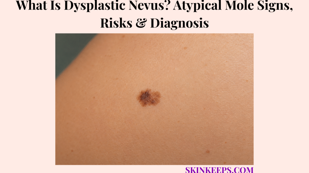
A dysplastic nevus is an atypical-looking mole that may have irregular color, shape, size, border, or microscopic features, but it is usually not melanoma.
Dysplastic nevus is also called an atypical mole, atypical nevus, dysplastic mole, or atypical melanocytic nevus. This page covers appearance, ABCDE signs, ugly-duckling warning, locations, causes, risk factors, melanoma-risk meaning, similar lesions, diagnosis, biopsy, pathology, monitoring, removal decisions, prevention, mistakes, and doctor-warning signs.
What Is a Dysplastic Nevus and Why Is It Called an Atypical Mole?
A dysplastic nevus is an atypical-looking mole that may have irregular color, shape, size, border, or microscopic features, but it is usually not melanoma.
Melanocytic means it comes from melanocytes, the pigment-producing cells that form many moles.
The word atypical means the mole does not look or read under the microscope exactly like a common mole, but atypical does not automatically mean cancer.
Why Is Dysplastic Nevus Not the Same as Melanoma?
A dysplastic nevus is not the same as melanoma because it is usually a benign mole with atypical features, not skin cancer.
It matters because it can resemble melanoma and because multiple atypical moles can signal higher melanoma-risk monitoring needs.
The safest wording is “atypical mole that needs monitoring or biopsy if suspicious,” not “cancer mole.”
Why Does the Word “Dysplastic” Appear in Pathology Reports?
The word “dysplastic” describes atypical cell and growth patterns that may be seen under a microscope.
Pathology reports may mention mild, moderate, or severe atypia.
The grade helps guide follow-up, re-excision, or monitoring discussions, but pathology wording should be interpreted with a dermatologist rather than searched alone online.
Practical rule: A dysplastic nevus is usually benign, but a changing, ugly-duckling, symptomatic, or ABCDE-positive mole should be checked because melanoma can mimic an atypical mole.
What Does a Dysplastic Nevus Look Like?
A dysplastic nevus may look larger, more irregular, more unevenly colored, or less symmetrical than a common mole.
Appearance alone cannot confirm the diagnosis, but it can help decide when a dermatologist should examine the spot.
What Atypical Mole Signs Are Common?
Common atypical mole signs include uneven shape, irregular border, mixed colors, larger size, or a mole that looks different from nearby moles.
Colors may include tan, brown, dark brown, black, pink, red, gray, or darker pigment in one lesion.
Some dysplastic nevi are flat, some are slightly raised, and some have a flat center with a raised area.
What Is the ABCDE Warning System?
The ABCDE system is a warning-sign checklist used to decide when a mole or new spot should be examined for possible melanoma.
A means asymmetry, B means border irregularity, C means color variation, D means diameter or growth, and E means evolving size, shape, color, elevation, or symptoms.
ABCDE signs are warning clues, not proof of melanoma, but evolving change is especially important.
What Is the Ugly Duckling Sign?
The ugly duckling sign means one mole looks clearly different from the person’s usual pattern of moles.
Many people have a personal mole pattern, so a new outlier in color, size, shape, pattern, or behavior deserves dermatologist review.
An ugly-duckling mole may need dermoscopy, photography, biopsy, or excision if melanoma cannot be ruled out.
| Sign | What User May Notice | Why It Matters |
|---|---|---|
| Asymmetry | One half unlike the other. | Melanoma warning clue. |
| Irregular border | Blurry, jagged, notched, or uneven edge. | Atypical pattern. |
| Color variation | Several colors in one mole. | Needs assessment. |
| Larger size | Bigger than nearby moles. | Risk clue, not diagnosis. |
| Evolving | Changing size, shape, color, elevation, or symptoms. | Strong warning sign. |
| Ugly duckling | Looks unlike the person’s other moles. | Dermatologist review / biopsy consideration. |
Where Can Dysplastic Nevi Appear?
Dysplastic nevi can appear anywhere on the skin, including sun-exposed and covered areas, so full-body skin checks matter.
They may appear on the back, chest, abdomen, arms, legs, scalp, buttocks, palms, soles, or less visible body sites.
New or changing pigment under a nail, on a palm, sole, scalp, genital area, or mucosal surface needs careful evaluation because hidden sites are easy to miss.
| Site | Why It Can Be Missed | Safer Checking Tip |
|---|---|---|
| Back | Hard to see alone. | Use mirror, helper, or mole map. |
| Scalp | Hidden by hair. | Ask barber, stylist, partner, or dermatologist. |
| Chest / abdomen | Easy to miss if not comparing over time. | Photograph if advised. |
| Arms / legs | Often noticed but may be dismissed. | Watch ABCDE and ugly-duckling signs. |
| Buttocks / covered areas | Not sun-exposed, so often ignored. | Include in full-body checks. |
| Palms / soles | Less common but important. | Check new pigment carefully. |
| Nails | Pigment line can be overlooked. | Dermatologist review for changing nail pigment. |
What Causes Dysplastic Nevi?
Dysplastic nevi develop from pigment-producing melanocytes, and risk is influenced by genetics, family history, number of moles, UV exposure, tanning-bed use, and skin type.
There is usually no single cause for one atypical mole.
How Do Genetics and Family History Affect Atypical Moles?
Genetics and family history can affect atypical moles because some people naturally develop many moles or inherit melanoma-risk patterns.
Family history of melanoma, multiple atypical moles, and high total mole count can change surveillance needs.
FAMMM syndrome and inherited melanoma-risk genes such as CDKN2A belong in selected high-risk family discussions, not routine testing for every single atypical mole.
How Does UV Exposure Affect Mole and Melanoma Risk?
UV exposure can increase melanoma risk by damaging skin-cell DNA over time.
Sunburn history, childhood sunburns, outdoor work, tanning beds, and intentional tanning can all shape risk.
Sun protection reduces future UV injury, but sunscreen does not remove or reverse existing dysplastic nevi.
Does a Dysplastic Nevus Happen Because of Poor Hygiene?
A dysplastic nevus does not happen because of poor hygiene.
It is not dirt, infection, poor washing, or something that can be scrubbed off.
Cutting, freezing, burning, tying, acid-treating, or irritating a mole at home increases harm and can delay proper diagnosis.
| Factor | How It Relates | Care Implication |
|---|---|---|
| Many moles | Higher mole burden. | More careful monitoring. |
| Family melanoma history | Inherited risk context. | Dermatology plan. |
| Multiple dysplastic nevi | Higher-risk marker. | Regular skin exams. |
| UV exposure | Skin DNA damage risk. | Sun protection. |
| Tanning beds | Avoidable UV risk. | Avoid completely. |
| Personal skin cancer history | Higher future risk. | Specialist follow-up. |
| Immune suppression | Higher caution context. | Lower threshold for review. |
How Much Does a Dysplastic Nevus Increase Melanoma Risk?
A single dysplastic nevus usually does not mean melanoma will develop, but having multiple dysplastic nevi increases a person’s overall melanoma-risk monitoring needs.
The key idea is risk-marker logic: the mole may be benign, while the person’s broader mole pattern may need closer monitoring.
Why Are Dysplastic Nevi Called Risk Markers?
Dysplastic nevi are called risk markers because they can show that a person’s mole pattern is linked with higher melanoma risk.
Most concern is about the person’s whole skin, not only one mole.
Melanoma can appear as a new lesion or arise elsewhere on the skin, so monitoring should include the whole body.
When Does Mole Count Change Follow-Up Needs?
Mole count changes follow-up needs because people with several dysplastic nevi or many total moles may need regular dermatologist surveillance.
People with more than five dysplastic nevi may be advised to have yearly skin exams, while prior melanoma or strong family history can lead to a more individualized schedule.
Follow the dermatologist’s plan rather than a universal online rule.
| Situation | Risk Meaning | Follow-Up Direction |
|---|---|---|
| One stable atypical mole | Usually low immediate concern. | Monitor and check if changing. |
| Several dysplastic nevi | Higher melanoma-risk marker. | Regular skin exams. |
| Many moles + family melanoma | Higher-risk profile. | Dermatology surveillance. |
| Prior melanoma | Higher future risk. | Specialist follow-up. |
| Changing mole | Possible melanoma. | Prompt evaluation/biopsy. |
| Ugly-duckling lesion | Does not fit personal pattern. | Dermatologist review. |
How Is a Dysplastic Nevus Different From Other Moles or Skin Spots?
Dysplastic nevi can resemble common moles, congenital moles, Spitz nevi, seborrheic keratoses, lentigines, blue nevi, and melanoma, so clinical exam and dermoscopy are important.
The goal is balanced: avoid panic about every atypical mole while not ignoring a mole that is changing or clearly different.
How Is a Dysplastic Nevus Different From a Common Mole?
A common mole is usually smaller, rounder, more symmetrical, and more evenly colored than a dysplastic nevus.
A dysplastic nevus may have irregular shape, irregular border, color variation, or a different pattern from nearby moles.
A common mole can still need review if it develops ABCDE changes.
How Is a Dysplastic Nevus Different From Melanoma?
Melanoma is skin cancer, while a dysplastic nevus is usually a benign atypical mole.
Melanoma becomes more concerning when a lesion is changing, asymmetric, multi-colored, symptomatic, fast-growing, bleeding, crusting, painful, or ugly-duckling.
Biopsy is needed when melanoma cannot be ruled out clinically.
How Is Dysplastic Nevus Different From Seborrheic Keratosis?
Seborrheic keratosis often looks stuck-on, waxy, rough, or wart-like, while a dysplastic nevus is a melanocytic mole.
Some seborrheic keratoses are dark and can mimic melanoma or an atypical mole.
Dermoscopy or biopsy may be needed when a dark rough lesion is changing or uncertain.
How Is Dysplastic Nevus Different From a Congenital Mole?
A congenital mole is present at birth or early infancy, while a dysplastic nevus is defined by atypical clinical or microscopic features.
Congenital nevi can also be monitored for change, but large congenital nevi have separate melanoma-risk discussions.
Do not treat congenital-nevus risk rules as the same as every dysplastic nevus.
How Is Dysplastic Nevus Different From Blue Nevus or Lentigo?
Blue nevus and lentigo can both be pigmented skin spots, but their color pattern, depth, border, and dermoscopic features differ from dysplastic nevus.
Blue nevus may look blue-gray or blue-black because pigment sits deeper in the skin. Lentigo is often a flat brown sunspot-like lesion.
Changing blue-black, irregular, or new adult lesions deserve dermatologist review.
| Lesion | Main Clue | Why Confusion Happens | Safer Next Step |
|---|---|---|---|
| Common mole | Symmetric, even color. | Pigment overlap. | Monitor for change. |
| Dysplastic nevus | Irregular but often benign. | Looks melanoma-like. | Dermoscopy/biopsy if suspicious. |
| Melanoma | Evolving, ugly duckling, ABCDE. | Can mimic atypical mole. | Prompt biopsy. |
| Seborrheic keratosis | Stuck-on/waxy/rough. | Dark rough spots mimic melanoma. | Dermoscopy or biopsy if uncertain. |
| Congenital nevus | Present from birth. | May be large/pigmented. | Separate risk plan. |
| Spitz nevus | Pink/brown changing bump, often younger patients. | Can mimic melanoma. | Dermatology diagnosis. |
| Blue nevus | Blue-gray deeper pigment. | Dark mole overlap. | Review if changing or atypical. |
| Lentigo | Flat brown sunspot-like lesion. | Pigment overlap. | Check if changing. |
A non-healing, shiny, dark, crusting, or bleeding lesion can sometimes resemble a pigmented basal cell carcinoma, so uncertain lesions should be examined instead of labeled as a harmless mole at home.
How Is a Dysplastic Nevus Diagnosed?
A dysplastic nevus is diagnosed through skin examination, dermoscopy, comparison with other moles, and biopsy when the mole looks suspicious or is changing.
Diagnosis is dermatologist-led because the decision to monitor or biopsy depends on both the single lesion and the person’s overall risk pattern.
What Does a Dermatologist Check?
A dermatologist checks the mole’s size, shape, border, color, symmetry, surface, symptoms, and whether it is new or changing.
The exam may also review whether the lesion is an ugly duckling, total mole count, family history of melanoma, personal skin cancer history, sunburn history, tanning-bed use, and immune suppression.
Hard-to-see areas such as the back, scalp, nails, palms, soles, and covered skin may need a full-body skin exam.
How Does Dermoscopy Help?
Dermoscopy helps by magnifying pigment structures that are not easily seen with the naked eye.
It helps separate reassuring patterns from suspicious patterns and can guide whether to monitor, photograph, or biopsy.
Dermoscopy does not replace biopsy when melanoma remains possible.
When Is Biopsy Recommended?
Biopsy is recommended when a mole is changing, suspicious, ugly-duckling, symptomatic, or cannot be confidently separated from melanoma.
Biopsy may be considered for ABCDE change, rapid growth, new adult mole, bleeding, crusting, ulceration, itching, pain, tenderness, irregular pigment, suspicious dermoscopic structures, high-risk history, or diagnostic uncertainty.
A new mole after age 30 should be examined by a dermatologist for melanoma signs.
- When the mole first appeared.
- Whether it is new after adulthood.
- Photos showing change over time.
- Size, shape, color, border, or elevation changes.
- Bleeding, crusting, itching, pain, tenderness, or ulceration.
- Whether it looks different from other moles.
- Personal history of melanoma or skin cancer.
- Family history of melanoma.
- Number of moles or atypical moles.
- Sunburn history and tanning-bed use.
- Immune suppression or transplant medicines.
- Prior biopsy or pathology report.
- Whether any mole is on scalp, nail, palm, sole, genital area, or mucosa.
What Does a Dysplastic Nevus Biopsy or Pathology Report Mean?
A biopsy report may describe a dysplastic nevus as having mild, moderate, or severe atypia, which helps the dermatologist decide whether monitoring or additional removal is needed.
Pathology language should be interpreted with the clinical exam, margin status, lesion appearance, and patient risk.
What Does Mild Atypia Usually Mean?
Mild atypia usually means the mole has slight atypical features under the microscope.
It is often lower concern after biopsy, but follow-up still depends on margins, clinical appearance, and personal risk.
Do not interpret mild atypia as “ignore forever” without the dermatologist’s guidance.
What Does Moderate Atypia Usually Mean?
Moderate atypia means the mole has more atypical features than mild atypia but is not the same as melanoma.
Management varies by margin status, clinical concern, pathologist interpretation, and dermatologist judgment.
Some cases are monitored, and some are re-excised; do not assume “moderate” means cancer.
What Does Severe Atypia Usually Mean?
Severe atypia means the mole has higher-level atypical features and may be managed more cautiously.
Severe atypia can overlap more closely with melanoma in situ in the differential diagnosis.
Re-excision or dermatopathology second opinion may be discussed when the report or clinical situation is uncertain.
| Report Wording | Simple Meaning | Typical Next Discussion |
|---|---|---|
| Mild atypia | Slightly atypical mole. | Often monitor after biopsy. |
| Moderate atypia | More atypical features. | Monitor vs re-excision depends. |
| Severe atypia | High-level atypia. | Often more cautious removal. |
| Positive margins | Atypical cells at edge. | Depends on grade/clinical concern. |
| Cannot exclude melanoma | Diagnostic uncertainty. | Further excision/specialist review. |
Does a Dysplastic Nevus Need to Be Removed?
A dysplastic nevus does not always need removal, but a suspicious, changing, symptomatic, or severely atypical lesion may need biopsy or complete excision.
Stable atypical moles may be monitored with photos and follow-up when the dermatologist feels the pattern is low concern.
Cosmetic removal should not replace diagnostic evaluation, and suspicious moles should not be shaved, cut, frozen, burned, tied, or acid-treated at home.
| Mole Situation | Best Direction | Why |
|---|---|---|
| Stable atypical mole | Monitor/photograph. | Avoid unnecessary procedures. |
| Ugly duckling | Dermatology review. | Rule out melanoma. |
| ABCDE change | Biopsy. | Change is concerning. |
| Severe atypia | Discuss excision. | Higher diagnostic concern. |
| Cosmetic concern only | Dermatology discussion. | Confirm benign first. |
| Home removal idea | Avoid. | Missed melanoma, scar, infection risk. |
How Should Dysplastic Nevi Be Monitored Over Time?
Dysplastic nevi are monitored by watching for change, comparing moles with each other, using photos or mole maps, and keeping dermatologist follow-up based on personal risk.
Monitoring should be steady and practical, not obsessive checking many times per day.
How Should Self-Skin Checks Be Done?
Self-skin checks should look for ABCDE changes, ugly-duckling lesions, new spots, and symptoms across the whole skin.
Include the back, scalp, palms, soles, nails, covered areas, and any hard-to-see sites.
Use a mirror, helper, body map, or photos when advised, and report changing, bleeding, itchy, painful, or crusting lesions early.
When Is Total-Body Photography Useful?
Total-body photography can be useful when someone has many moles, multiple dysplastic nevi, family history of melanoma, prior melanoma, or hard-to-monitor back lesions.
It helps detect new or changing lesions over time.
It is risk-based and dermatologist-guided, not mandatory for every single atypical mole.
How Often Should Dermatologist Exams Happen?
Dermatologist exam frequency depends on personal risk, mole count, family history, prior melanoma, and clinician judgment.
More than five dysplastic nevi may prompt yearly skin-exam discussion.
Prior melanoma, immune suppression, or high-risk family history may need a different dermatologist-led schedule.
- Know your usual mole pattern.
- Check ABCDE signs.
- Look for ugly-duckling lesions.
- Include back, scalp, palms, soles, nails, and covered areas.
- Use mole map or photos if advised.
- Keep dermatologist follow-up based on risk.
- Report changing, bleeding, itchy, painful, or crusting lesions early.
How Can Melanoma Risk Be Reduced If You Have Dysplastic Nevi?
Having dysplastic nevi does not mean melanoma is inevitable, but sun protection and regular skin monitoring help reduce risk and catch suspicious changes early.
Use broad-spectrum sunscreen, hats, sleeves, sunglasses, UPF clothing, and shade, and avoid tanning beds, intentional tanning, and sunburn.
Darker skin can still develop melanoma, and pigment changes on palms, soles, nails, scalp, genital areas, or mucosal surfaces should not be ignored.
- Use broad-spectrum sunscreen.
- Wear sun-protective clothing and hats.
- Seek shade.
- Avoid tanning beds.
- Avoid intentional sunburn or tanning.
- Protect children from sunburn.
- Know your family melanoma history.
- Use mole mapping or photos if advised.
- Keep dermatologist checks.
- Report ABCDE or ugly-duckling changes early.
What Dysplastic Nevus Mistakes Should You Avoid?
The biggest dysplastic nevus mistake is either panicking that every atypical mole is melanoma or ignoring a mole that is clearly changing.
Do not rely on phone apps alone, remove moles at home, skip follow-up if you have many atypical moles, forget family history, or assume darker skin cannot get melanoma.
Do not cosmetically remove a suspicious mole without diagnostic review and pathology when needed.
| Mistake | Why It Fails | Better Action |
|---|---|---|
| “Atypical = cancer” | Causes unnecessary panic. | Confirm with dermatologist/pathology. |
| Ignoring evolution | Misses melanoma warning. | Check changing lesions. |
| Home mole removal | No diagnosis + scar/infection risk. | Biopsy/excision by clinician. |
| Phone-app-only judgment | Can miss context and whole-skin pattern. | Use dermatologist exam when concerned. |
| No photos | Hard to track change. | Mole map or clinical photos. |
| Skipping exams | Risk marker missed. | Follow risk-based schedule. |
When Should an Atypical Mole Be Checked by a Dermatologist?
An atypical mole should be checked when it is new, changing, ugly-duckling, asymmetric, irregularly bordered, multi-colored, growing, bleeding, crusting, itching, painful, or different from your other moles.
Prompt review is also important for pigment under a nail, pigment on palms or soles, scalp lesions, genital-area pigment, mucosal pigment, or moles in hard-to-see places.
Which Mole Changes Need Prompt Review?
Prompt review is needed for a new adult mole, ABCDE changes, ugly-duckling appearance, rapid growth, bleeding, crusting, ulceration, itching, pain, or a new pigmented lesion in a hard-to-check area.
A new mole after age 30 should be checked.
A mole that has existed for years should not be dismissed if it is now changing.
Which People Should Be More Cautious?
People with many moles, more than five dysplastic nevi, family history of melanoma, prior melanoma, tanning-bed use, immune suppression, or hard-to-monitor moles should be more cautious.
History of blistering sunburns, personal skin cancer, FAMMM syndrome concern, or inherited melanoma-risk family should be discussed with a dermatologist.
High-risk people should follow a clinician-guided skin-check plan rather than guessing from a single mole photo.
Seek dermatologist review if a mole is:
- New after adulthood.
- Changing in size, shape, color, elevation, or symptoms.
- An ugly duckling compared with your other moles.
- Asymmetric.
- Jagged, blurred, or irregularly bordered.
- Multi-colored.
- Rapidly growing.
- Bleeding, crusting, ulcerated, itchy, painful, or tender.
- A dark line under a nail or changing nail pigment.
- On a palm, sole, scalp, genital area, or mucosal surface.
- Present in someone with many atypical moles, family melanoma history, prior melanoma, tanning-bed exposure, or immune suppression.
What Should You Remember About Dysplastic Nevus?
A dysplastic nevus is an atypical mole that is usually benign, but it matters because it can resemble melanoma and can signal increased melanoma-risk monitoring needs.
Safe care is not panic removal and not neglect; it is dermatologist-guided review of suspicious change, whole-skin monitoring, and sun protection.
- Dysplastic nevus means atypical mole.
- It is usually not melanoma.
- It may be larger, irregular, unevenly colored, or different from nearby moles.
- ABCDE and ugly-duckling signs guide when to seek care.
- Multiple dysplastic nevi increase melanoma-risk monitoring needs.
- Diagnosis may include dermoscopy, photos, biopsy, and pathology.
- Mild, moderate, and severe atypia guide management discussions.
- Stable lesions may be monitored.
- Suspicious or changing lesions should be biopsied.
- Do not remove moles at home.
- Sun protection and regular skin checks are essential.
Frequently Asked Questions About Dysplastic Nevus
Is a dysplastic nevus cancer?
No. A dysplastic nevus is usually not cancer; it is an atypical mole. Having atypical moles can increase melanoma-risk monitoring needs.
Can a dysplastic nevus turn into melanoma?
It can rarely happen, but most concern is that dysplastic nevi are risk markers and melanoma can also develop elsewhere on the skin. The more dysplastic nevi a person has, the more careful monitoring may become.
What does an atypical mole look like?
An atypical mole may have irregular shape, uneven border, multiple colors, larger size, or a pattern that looks different from the person’s other moles.
What is the ugly duckling sign?
The ugly duckling sign means one mole looks clearly different from the person’s usual mole pattern. It should be checked because melanoma can mimic an atypical mole.
How is a dysplastic nevus diagnosed?
Diagnosis may include skin exam, dermoscopy, comparison with other moles, clinical photography or mole mapping in selected people, and biopsy when melanoma cannot be ruled out.
Does every dysplastic nevus need removal?
No. Stable lesions may be monitored, while suspicious, changing, symptomatic, ugly-duckling, or severely atypical lesions may need biopsy or excision.
How often should I get skin checks if I have dysplastic nevi?
Frequency depends on risk factors such as number of atypical moles, total mole count, family history, prior melanoma, and dermatologist judgment. People with more than five dysplastic nevi may be advised to have yearly skin exams.
When should an atypical mole be checked urgently?
An atypical mole should be checked for ABCDE changes, ugly-duckling appearance, rapid growth, bleeding, crusting, ulceration, itching, pain, new mole after age 30, or any mole that looks clearly different from the rest.
Sources & Evidence About Dysplastic Nevus
National Cancer Institute — Common Moles, Dysplastic Nevi, and Risk of Melanoma was used for dysplastic nevi features, melanoma-risk framing, more-than-five dysplastic nevi and yearly skin-exam guidance, FAMMM/CDKN2A context, and full excisional biopsy recurrence framing.
DermNet — Atypical Melanocytic Naevus was used for atypical nevi as melanoma-risk markers, clinical/dermoscopic diagnosis, signature mole pattern, ugly-duckling sign, and biopsy/excision consideration for outlier lesions.
The Skin Cancer Foundation — Atypical Moles was used for the distinction that atypical moles are not skin cancer but are melanoma risk factors.
The Skin Cancer Foundation — Melanoma Risk Factors was used for UV exposure, indoor tanning, many moles, atypical mole features, immune-suppression context, and FAMMM context.
American Academy of Dermatology — Moles: Signs and Symptoms was used for ABCDE warning signs and immediate dermatologist appointment guidance for concerning moles or new spots.
American Academy of Dermatology — When Is a Mole a Problem? was used for new mole after age 30, bleeding or itchy mole warning, Body Mole Map, mole tracking, and checking for changes.
American Academy of Dermatology — Moles: Who Gets and Types was used for atypical mole terminology, mole types that increase melanoma risk, and FAMMM context.
DermNet — Dermoscopy of Atypical Naevi was used for dermoscopy/atypical-nevus context, serial photography support, and specialist-context caution for changing elevated atypical nevi.
Educational Disclaimer: This SkinKeeps article is for educational purposes only and does not diagnose or replace medical care. A new, changing, ugly-duckling, asymmetric, irregularly bordered, multi-colored, rapidly growing, bleeding, crusting, ulcerated, itchy, painful, tender, nail-related, palm-related, sole-related, scalp-related, genital-area-related, mucosal, or uncertain mole or pigmented spot should be checked by a qualified healthcare professional or dermatologist. People with many moles, multiple dysplastic nevi, family history of melanoma, personal history of melanoma or skin cancer, immune suppression, tanning-bed exposure, blistering sunburn history, or hard-to-monitor moles should follow a clinician-guided skin-check plan. Do not cut, shave, burn, freeze, acid-treat, tie off, scratch off, or cosmetically remove a suspicious mole at home or without appropriate diagnostic review.



