
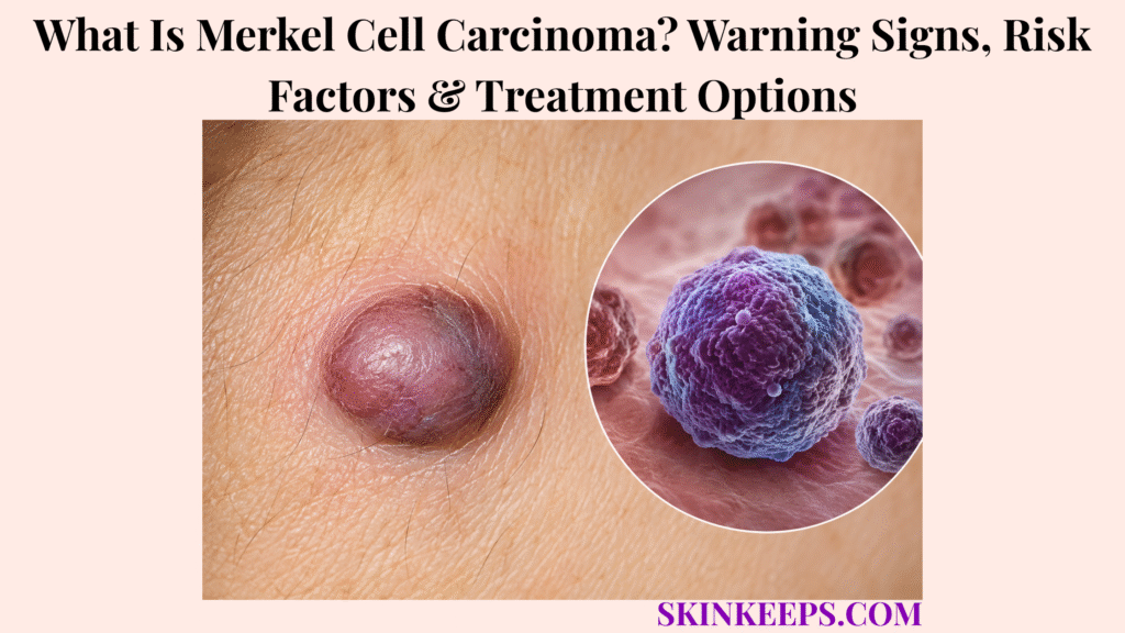
Merkel cell carcinoma is a rare aggressive neuroendocrine skin cancer that commonly appears as a rapidly enlarging, painless skin nodule. The lesion is often firm and smooth and may be pink, red, purple or skin-coloured; the head and neck are common locations, but MCC can develop on other body sites.
Its early appearance is nonspecific and can resemble a cyst, insect bite, pimple or sore. Because MCC can spread to regional lymph nodes and distant sites relatively early, a suspicious lesion needs biopsy confirmation followed by accurate staging rather than simple visual observation alone.
Prompt medical assessment is appropriate for a rapidly enlarging unexplained skin lump, a firm painless nodule, a changing red-purple lesion, bleeding or ulceration, a sore that appears to heal and then returns, or a new nearby lymph-node lump. A rapidly growing painless lesion should not be assumed harmless simply because it does not hurt.
How Can You Recognize Merkel Cell Carcinoma?
Merkel cell carcinoma most often appears as a firm, painless skin nodule that enlarges noticeably over weeks or months.
What Does Merkel Cell Carcinoma Usually Look Like?
Merkel cell carcinoma usually presents as a solitary firm nodule that may be skin-coloured, pink, red, blue-red or purple.
The surface can be smooth, shiny or dome-shaped. The lesion is often painless at first and may later crust, ulcerate or bleed, but ulceration is not required for MCC to be present.
How Quickly Does Merkel Cell Carcinoma Grow?
Merkel cell carcinoma often enlarges noticeably over weeks to months, making rapid growth one of its strongest practical warning signs.
Serial photographs, measurements or comparison between appointments can make short-term enlargement easier to recognize. A lesion that keeps growing over a few weeks behaves differently from a benign bump that has remained stable for years.
The practical rule is growth behaviour > harmless-looking surface.
Where Does Merkel Cell Carcinoma Usually Develop?
Merkel cell carcinoma often develops on chronically sun-exposed head and neck skin but can occur anywhere on the body.
Other sites include the arms, legs and trunk. Younger patients and people with darker skin can also develop MCC, so location or complexion alone cannot exclude the diagnosis.
What Is the AEIOU Warning Pattern for Merkel Cell Carcinoma?
The AEIOU mnemonic summarizes common MCC clues: asymptomatic, expanding rapidly, immune suppression, older age and ultraviolet-exposed fair skin.
A — Asymptomatic: MCC is often painless. E — Expanding rapidly: noticeable short-term growth is a particularly useful clue.
I — Immune suppression: reduced immune surveillance increases risk. O — Older than 50: incidence rises with age. U — UV-exposed fair skin: this is a common risk context.
AEIOU is a recognition aid rather than a diagnostic test, and a patient does not need all five features.
Which Skin-Lump Changes Make Merkel Cell Carcinoma More Concerning?
Continued rapid enlargement, firmness, unusual red-purple colour, bleeding, ulceration or nearby new lymph-node lumps make an unexplained lesion more concerning for MCC.
A sore that seems to heal and then returns also deserves reassessment. Lack of pain does not reduce concern when the lesion is changing quickly.
Figure 1. MCC can initially resemble a harmless painless bump; continued enlargement over weeks to months is the behaviour that should move the lesion toward biopsy.
Why Does Merkel Cell Carcinoma Develop, and Who Has a Higher Risk?
Merkel cell carcinoma can develop through a Merkel cell polyomavirus-associated pathway or a predominantly ultraviolet-damage pathway, with older age and immune suppression increasing risk.
What Role Does Merkel Cell Polyomavirus Play?
Merkel cell polyomavirus is associated with a large proportion of MCC cases and can contribute to tumour growth after viral material becomes integrated into cancer cells.
NCI reports that roughly 70–80% of U.S. MCC cases are associated with MCPyV. In virus-positive tumours, integrated viral material and viral oncoproteins can support malignant growth.
MCPyV exposure is common in adults, while MCC is rare. Ordinary viral exposure therefore does not mean a person is likely to develop Merkel cell carcinoma.
Can Merkel Cell Carcinoma Develop Without Polyomavirus?
Yes; virus-negative Merkel cell carcinoma forms through a separate pathway strongly associated with accumulated ultraviolet-induced DNA damage.
The two major biologic models are virus-associated MCC and UV-mutated virus-negative MCC. Clinical appearance alone cannot determine which pathway produced an individual tumour.
How Does Sun Exposure Affect Merkel Cell Carcinoma Risk?
Long-term ultraviolet exposure increases MCC risk and helps explain why the cancer often appears on chronically sun-exposed skin.
Relevant exposures include natural sunlight, tanning devices and substantial PUVA exposure. UV damage is important but does not explain every MCC.
Why Does Immune Suppression Increase MCC Risk?
Immune suppression increases MCC risk because weakened immune surveillance reduces the body’s ability to control abnormal or virus-associated cells.
Higher-risk contexts include solid-organ transplantation, chronic lymphocytic leukemia, HIV, lymphoma and long-term immunosuppressive treatment. Immune suppression can also worsen prognosis after MCC develops.
How Important Are Age and Skin Tone?
Merkel cell carcinoma occurs mainly in older adults, but younger people and people with darker skin can also develop the disease.
Age above 50 is part of the AEIOU risk pattern, with risk rising further in older age. Fair skin and chronic UV exposure are common contexts, but darker skin does not provide complete protection from MCC.
Figure 2. MCC can arise through an MCPyV-associated pathway or a virus-negative UV-damage pathway, while reduced immune surveillance increases susceptibility and can worsen disease behaviour.
How Is Merkel Cell Carcinoma Diagnosed and Staged?
Merkel cell carcinoma is confirmed by skin biopsy and then staged by evaluating regional lymph nodes and, when appropriate, imaging for further spread.
Why Is a Skin Biopsy Necessary for Merkel Cell Carcinoma?
Skin biopsy is necessary because Merkel cell carcinoma cannot usually be distinguished reliably from other benign or malignant skin lesions by appearance alone.
MCC can resemble melanoma, especially when a lesion is pigmented or nodular.
Squamous cell carcinoma can also enter the differential when a lesion is rapidly enlarging, crusted or ulcerated.
A harmless-looking epidermal cyst can be another clinical mimic when MCC presents as a smooth painless bump.
Biopsy provides tissue for histopathology and immunohistochemistry, allowing a dermatopathologist to distinguish these entities.
What Does the Pathologist Look for in Merkel Cell Carcinoma?
Histopathology identifies the malignant neuroendocrine tumour pattern, while immunohistochemistry helps confirm MCC and exclude important mimics.
The microscopic pattern is typically a malignant small-cell neuroendocrine carcinoma. CK20 is an important marker in the diagnostic panel, but one stain alone should not be presented as the entire diagnosis.
The marker panel is interpreted with morphology to distinguish MCC from melanoma, lymphoma and metastatic small-cell lung cancer.
Why Are Nearby Lymph Nodes Examined?
Regional lymph nodes are examined because Merkel cell carcinoma can spread through lymphatic pathways before enlarged nodes become obvious.
Initial staging therefore includes examination of the regional nodal basin even when no lymph-node lump is felt. Microscopic metastasis can change stage, prognosis and treatment planning.
Why Is Sentinel Lymph-Node Biopsy Important in MCC?
Sentinel lymph-node biopsy can detect hidden microscopic nodal metastasis in patients whose regional lymph nodes appear clinically negative.
SLNB samples the first draining lymph node or nodes for staging; it is not the same as removing all lymph nodes. When feasible, SLNB is planned before definitive wide excision because major surgery can alter lymphatic drainage.
When Are PET/CT, CT or MRI Used?
Imaging is used when tumour features, nodal findings, symptoms or stage create concern for regional or distant MCC spread.
PET/CT, CT or MRI may be selected according to the clinical question and suspected disease extent. There is no single identical scan protocol for every patient with MCC.
What Do the Main Merkel Cell Carcinoma Stages Mean?
MCC staging progresses conceptually from disease confined to the primary skin region to regional nodal spread and then distant metastatic disease.
Localized disease means no detected regional or distant spread. Regional disease includes nearby lymph-node involvement, satellite or in-transit disease or other regional extension.
Distant metastatic disease means spread to distant nodes or organs. Tumour size and whether nodal disease is microscopic or clinically apparent also influence formal staging.
Figure 3. Skin biopsy confirms MCC, but initial staging continues with regional-node assessment, sentinel-node biopsy when appropriate and imaging according to risk or suspected spread.
How Is Merkel Cell Carcinoma Treated?
Merkel cell carcinoma treatment depends on disease extent, with surgery forming the foundation for most localized disease and checkpoint immunotherapy preferred for most unresectable or metastatic disease.
How Is Localized Merkel Cell Carcinoma Treated?
Localized Merkel cell carcinoma is usually treated with complete surgical excision.
NCI describes excision margins of roughly 1–2 cm where anatomically feasible, with the goal of complete microscopic clearance while preserving function. Selected early lesions, particularly where tissue conservation matters, may be treated with Mohs or other margin-controlled approaches.
Why Is Radiation Therapy Often Added?
Radiation therapy is often considered because Merkel cell carcinoma is radiosensitive and has a meaningful risk of local or regional recurrence.
Postoperative radiation may target the primary tumour bed and, in selected cases, the draining nodal basin. Tumour size, margins, lymphovascular invasion, immune status, node findings and overall recurrence risk influence the decision.
Not every small low-risk localized MCC automatically needs radiation.
How Is Regional Lymph-Node Disease Treated?
Node-positive Merkel cell carcinoma may require combined surgery, regional radiation and multidisciplinary oncology management.
Microscopic disease detected by SLNB differs from clinically apparent nodal disease. Treatment varies with the number of involved nodes, distribution and total regional burden rather than following one universal node-management pathway.
How Is Metastatic or Unresectable Merkel Cell Carcinoma Treated?
Immune-checkpoint therapy is the preferred first systemic treatment for most metastatic or unresectable Merkel cell carcinoma.
Checkpoint inhibitors used in MCC include anti-PD-L1 therapy such as avelumab and anti-PD-1 therapies such as pembrolizumab and retifanlimab. These drugs restore anti-tumour immune activity and generally produce more durable responses than historical chemotherapy in advanced disease.
Is Chemotherapy Still Used for Merkel Cell Carcinoma?
Yes; chemotherapy can still be used in selected MCC cases, but immunotherapy is preferred for most metastatic patients because chemotherapy responses are often less durable.
Chemotherapy may remain an option when a patient cannot receive immunotherapy, when disease progresses despite immunotherapy or when another individualized systemic strategy is needed.
Is Immunotherapy Used Before or After Surgery for Every High-Risk MCC?
No; checkpoint immunotherapy is established in advanced MCC, while routine neoadjuvant or adjuvant immunotherapy for resectable disease remains under active investigation.
Trials continue to study checkpoint therapy before surgery and after definitive local treatment. This investigational boundary matters because every Stage I–III patient should not be assumed to require routine checkpoint treatment.
| Disease Extent | Core Treatment Logic | Surveillance Link |
|---|---|---|
| Localized MCC | Surgery ± selected radiation | Close early skin + node follow-up |
| Regional node-positive MCC | Surgery/radiation with multidisciplinary management | Regional-node surveillance is central |
| Unresectable or metastatic MCC | Checkpoint immunotherapy | Response and recurrence monitoring follow oncology plan |
| Selected immunotherapy-ineligible/progressive disease | Alternative systemic strategy including chemotherapy where appropriate | Follow-up remains individualized |
What Happens After Merkel Cell Carcinoma Treatment, and Which Signs Need Prompt Follow-Up?
Merkel cell carcinoma requires close follow-up after treatment because recurrence can develop locally, in regional lymph nodes or at distant sites, especially during the first few years.
Can Merkel Cell Carcinoma Return After Treatment?
Yes; Merkel cell carcinoma can recur at the original site, in regional lymph nodes or in distant organs after treatment.
Recurrence is not inevitable, but the possibility remains after the surgical wound has healed. Surveillance therefore extends beyond the scar itself.
When Is Merkel Cell Carcinoma Recurrence Risk Greatest?
Close surveillance is particularly important during the first two to three years after MCC treatment because many recurrences occur relatively early.
AAD notes that follow-up visits are often approximately every three to four months during the first two to three years, but the schedule is individualized by stage, immune status and treatment history rather than used as a universal rule.
What Happens During Merkel Cell Carcinoma Follow-Up?
MCC follow-up examines the original tumour site, surrounding skin, regional lymph nodes and the rest of the skin for recurrence or another skin cancer.
The visit can include the scar, surrounding tissue, draining nodal basin and whole-skin examination. Selected patients may also need follow-up imaging according to stage and clinical risk.
Which New Changes Need Earlier Review After MCC?
A new lump near the scar, enlarging lymph node, another rapidly growing skin lesion or unexplained persistent systemic symptom warrants earlier reassessment after MCC.
For someone with a history of advanced disease, persistent bone pain, respiratory symptoms, neurological symptoms or marked unexplained weight loss can also justify earlier review.
Does Sun Protection Still Matter After MCC Treatment?
Yes; continued ultraviolet protection reduces further UV damage and helps lower the risk of additional UV-associated skin cancers.
Sun protection should accompany ongoing skin surveillance. It reduces further UV injury but does not eliminate recurrence risk and should not be described as preventing virus-associated MCC recurrence.
What Determines the Outlook for Merkel Cell Carcinoma?
Merkel cell carcinoma prognosis depends strongly on stage at diagnosis, with localized disease generally having a better outlook than nodal or distant metastatic disease.
Immune suppression, tumour characteristics and treatment response also influence outcome. Older survival data may not fully reflect outcomes achievable with modern checkpoint immunotherapy.
What Should You Remember About Merkel Cell Carcinoma?
Merkel cell carcinoma is a rare aggressive skin cancer in which rapid painless growth, early biopsy, accurate lymph-node staging and extent-directed treatment are central to improving outcomes.
- MCC is a neuroendocrine skin cancer.
- It often appears as a painless rapidly growing nodule.
- Pink, red or purple colour is common but not required.
- Rapid growth matters more than the absence of pain.
- AEIOU is a recognition aid, not a diagnostic test.
- MCPyV and UV damage represent major biologic pathways.
- Age and immune suppression raise risk.
- Biopsy confirms MCC.
- Regional lymph nodes must be assessed.
- SLNB detects occult nodal spread when appropriate.
- Imaging follows clinical and stage risk.
- Localized disease is surgery-led.
- Radiation is individualized.
- Advanced disease is primarily immunotherapy-led.
- Routine adjuvant or neoadjuvant checkpoint treatment in resectable MCC remains an evolving evidence area.
- Early recurrence surveillance is important.
Recognize rapid growth → biopsy → stage nodes/spread → treat by extent → monitor recurrence closely.
Frequently Asked Questions About Merkel Cell Carcinoma
The most important Merkel cell carcinoma questions concern its early appearance, rapid growth, causes, diagnostic staging and whether early disease can be treated successfully.
What Does Merkel Cell Carcinoma Look Like When It First Appears?
Merkel cell carcinoma often begins as a firm, painless, rapidly enlarging pink, red, purple or skin-coloured bump. It can resemble a cyst, insect bite, pimple or sore.
How Quickly Does Merkel Cell Carcinoma Grow?
Merkel cell carcinoma can become noticeably larger over weeks to months, making rapid growth an important warning feature. Absence of pain does not make rapid enlargement reassuring.
What Causes Merkel Cell Carcinoma?
Merkel cell carcinoma can develop through a Merkel cell polyomavirus-associated pathway or through accumulated ultraviolet-related DNA damage. Older age and immune suppression increase risk but do not determine the diagnosis.
How Is Merkel Cell Carcinoma Diagnosed and Staged?
Merkel cell carcinoma is diagnosed by skin biopsy and staged by assessing regional lymph nodes and, when appropriate, imaging for further spread. Sentinel lymph-node biopsy is particularly important for selected clinically node-negative disease.
Can Merkel Cell Carcinoma Be Cured When Found Early?
Localized Merkel cell carcinoma has a better prognosis than node-positive or metastatic disease and is generally treated with complete surgical removal, often with radiation considered according to recurrence risk. Early diagnosis and accurate nodal staging therefore matter.
Which Sources Support This Merkel Cell Carcinoma Guidance?
American Academy of Dermatology — Merkel Cell Carcinoma: Signs and Symptoms — Used for rapid growth over weeks to months, painless appearance, pink/red/purple colour, head-and-neck tendency, harmless-looking mimics and skin-tone inclusivity.
American Academy of Dermatology — Merkel Cell Carcinoma: Causes — Used for age, UV exposure, immune suppression, transplant/HIV context and the distinction between common MCPyV exposure and rare MCC development.
American Academy of Dermatology — Merkel Cell Carcinoma: Diagnosis and Treatment — Used for specialist biopsy diagnosis, dermatopathology, imaging and selected margin-controlled/Mohs surgery context.
American Academy of Dermatology — Merkel Cell Carcinoma: Self-Care and Follow-Up — Used for early intensive follow-up, skin and lymph-node surveillance and ongoing UV protection after treatment.
National Cancer Institute — Merkel Cell Carcinoma Treatment (PDQ®), Health Professional Version — Primary staging and treatment source used for AEIOU, MCPyV/UV biology, immune suppression, SLNB, surgery, radiation, regional treatment, advanced checkpoint immunotherapy, chemotherapy and the investigational neoadjuvant/adjuvant boundary.
National Cancer Institute — Risk Factors: Infectious Agents — Used specifically to show that most adults have been infected with MCPyV while MCC remains rare, preventing ordinary viral exposure from being equated with cancer.
U.S. FDA — Avelumab (Bavencio) Merkel Cell Carcinoma Approval Record — Used specifically to support the approved role of anti-PD-L1 therapy in metastatic MCC.
U.S. FDA — Retifanlimab-dlwr Approval for Metastatic or Recurrent Locally Advanced MCC — Used specifically for the current approved anti-PD-1 treatment context in advanced MCC.



