
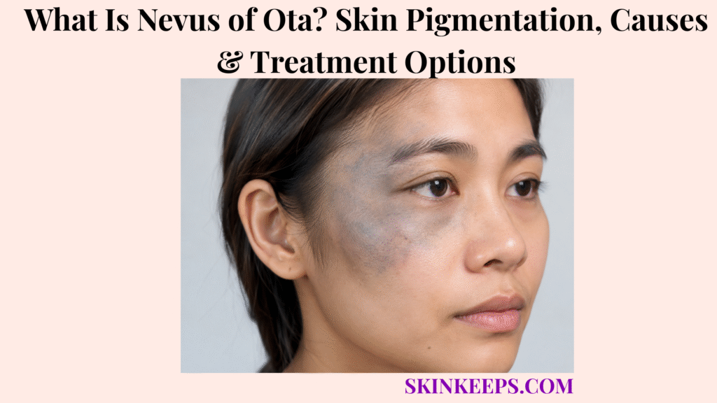
Nevus of Ota is a usually unilateral form of dermal melanocytosis that causes flat blue-grey, slate-brown or brown-violet pigmentation around the eye and may also pigment structures inside the eye. The colour comes from melanocytes and melanin located within the dermis rather than from ordinary superficial epidermal pigmentation.
Some lesions are visible from birth, while others become apparent during childhood, adolescence or later hormonal periods. The facial pigmentation is usually benign, but ocular melanocytosis changes the medical follow-up because glaucoma and uveal melanoma become surveillance concerns that remain separate from any cosmetic skin treatment.
This article is for educational purposes only and does not constitute medical advice, diagnosis, or treatment. New eye pain, changes or loss of vision, unexplained symptoms suggesting increased eye pressure, a new raised papule or nodule within previously flat pigmentation, rapid focal growth, ulceration, bleeding, or a major unexpected change in colour or shape should be evaluated by a licensed dermatologist, ophthalmologist, or other qualified healthcare professional.
How Can You Recognize Nevus of Ota?
Nevus of Ota usually appears as flat, poorly defined blue-grey, slate-blue or brown-violet pigmentation around one eye, sometimes accompanied by same-side pigmentation of the sclera or other ocular structures.
What Does Nevus of Ota Look Like, and Where Does It Appear?
Nevus of Ota produces patchy or confluent blue-grey to brown-violet pigmentation over the periocular skin, temple, forehead, cheekbone, upper cheek and nearby nose.
The pigment is usually flat and poorly defined rather than raised. Colour can range from blue-grey and slate-blue to brown-violet or grey-black depending on pigment depth, skin tone and lighting.
The distribution follows areas supplied mainly by the ophthalmic and maxillary divisions of the trigeminal nerve, but this anatomical pattern is a dermatologic localization clue rather than a neurologic disorder.
Is Nevus of Ota Usually on One Side of the Face?
Yes; Nevus of Ota is predominantly unilateral, although bilateral oculodermal melanocytosis can occur.
Unilateral involvement is therefore a strong recognition feature but not an absolute diagnostic requirement. A bilateral pattern broadens the differential rather than automatically excluding the diagnosis.
Can Nevus of Ota Affect the Eye or Mouth?
Yes; Nevus of Ota can pigment the conjunctiva, sclera, iris, choroid and other ocular structures, and oral mucosal pigmentation occasionally occurs.
Scleral pigmentation is especially visible and can be the first clue that the condition is truly oculodermal rather than skin-only. Pigment can also involve the iris and choroid, which is why eye assessment matters medically.
Oral involvement is less common and may affect areas such as the palate or buccal mucosa.
Does Nevus of Ota Change With Age?
Nevus of Ota can become darker or more extensive during growth, puberty, pregnancy or other hormonal periods, while spontaneous complete disappearance is uncommon.
Some lesions are present at birth, while others become clinically obvious in childhood, adolescence or adulthood. Hormonal change can modify pigment intensity without being the original developmental cause.
Figure 1. Nevus of Ota usually produces periocular facial pigmentation on one side, with possible same-side scleral, iris or choroidal melanocytosis.
Why Does Nevus of Ota Develop?
Nevus of Ota is thought to develop when melanocytes remain within the dermis during development, where their deeper melanin produces the characteristic blue-grey appearance.
Why Are Melanocytes Located in the Dermis?
The leading developmental explanation is that some melanocytes remain within the dermis during embryologic migration rather than reaching their usual more superficial location.
Melanocytes arise from neural-crest precursors that migrate during development. In dermal melanocytosis, some pigment-producing cells persist deeper in the skin.
Somatic alterations involving genes such as GNAQ or GNA11 have been identified in some lesions, but these findings do not mean every case has one identical genetic cause.
Why Does Deep Melanin Look Blue or Grey?
Melanin within the dermis appears blue or grey because light passes through overlying skin before reflecting from the deeper pigment.
Superficial melanin tends to look more brown, whereas deeper dermal pigment can appear blue-grey, slate or blue-brown. This optical effect explains why Nevus of Ota should not be described as ordinary surface hyperpigmentation.
Is Nevus of Ota Inherited or Caused by Sun Exposure?
Typical Nevus of Ota is considered a developmental, generally non-hereditary melanocytosis rather than a pigmentation disorder primarily caused by ultraviolet exposure.
Family inheritance is not the usual pattern, although genetic changes within melanocytes can contribute biologically. Ultraviolet exposure is not considered the main cause because the relevant melanocytes reside deeper in the dermis.
Who Develops Nevus of Ota, and Why Can Hormones Affect Its Colour?
Nevus of Ota occurs across populations but is reported more often in people of Asian ancestry and in females, while hormonal periods may increase melanocyte activity and darken existing pigmentation.
Cases also occur in people of African ancestry and other skin tones. Demographic frequency supports epidemiology but is never a diagnostic requirement.
Figure 2. The characteristic blue-grey appearance comes from melanocytes and melanin located within the dermis, with developmental persistence as the leading mechanistic model.
How Is Nevus of Ota Distinguished and Diagnosed?
Nevus of Ota is usually diagnosed clinically from its colour, unilateral periocular distribution and possible ocular pigmentation, with biopsy reserved for uncertain or structurally changing lesions.
How Is Nevus of Ota Different From Melasma, Hori Nevus and Nevus of Ito?
Nevus of Ota is typically unilateral, blue-grey and periocular with possible ocular pigmentation, while melasma is usually bilateral brown facial pigmentation, Hori nevus is acquired and bilateral without typical ocular involvement, and Nevus of Ito affects the shoulder region.
Melasma is primarily a facial hypermelanosis rather than oculodermal melanocytosis. Hori nevus can produce bilateral blue-grey facial pigment but usually spares ocular and oral structures.
Nevus of Ito represents dermal melanocytosis of the shoulder or upper-arm region rather than the periocular trigeminal distribution.
How Is Nevus of Ota Different From a Blue Nevus or General Hyperpigmentation?
Nevus of Ota usually forms a broad flat periocular field of dermal pigment, whereas a blue nevus is generally a more localized melanocytic lesion and hyperpigmentation is a much broader descriptive category.
The distinction is primarily one of morphology, distribution and tissue pattern. A localized papule or small nodule is not the classic presentation of diffuse oculodermal melanocytosis.
When Are Dermoscopy or Skin Biopsy Used?
Dermoscopy can document typical homogeneous blue-grey pigmentation, while skin biopsy is usually reserved for diagnostic uncertainty or a lesion that develops new structural changes.
Biopsy becomes more appropriate when a previously flat field develops a new papule, nodule, ulceration, major focal pigment change or another feature that no longer fits stable classic disease.
Histology can demonstrate melanocytes scattered within the dermis. Routine biopsy of a stable classic lesion is unnecessary.
Why Does Nevus of Ota Need an Ophthalmologic Examination?
Nevus of Ota requires eye assessment because ocular melanocytosis can involve structures that influence intraocular pressure and carries an increased risk of glaucoma and uveal melanoma.
Baseline ophthalmologic assessment can include visual evaluation, intraocular-pressure measurement, slit-lamp examination, gonioscopy and dilated examination of the retina and choroid.
Dermatologic recognition therefore does not complete the evaluation when ocular involvement is possible.
Figure 3. A typical stable facial lesion can usually be diagnosed clinically, while atypical structural change prompts biopsy and ocular pigment prompts baseline ophthalmology plus long-term surveillance.
How Is Nevus of Ota Treated?
Nevus of Ota does not usually require removal for medical reasons, but pigment-targeting laser treatment can substantially lighten facial pigmentation when cosmetic treatment is desired.
Does Nevus of Ota Have to Be Removed?
No; stable cutaneous Nevus of Ota is usually benign and can be observed or cosmetically camouflaged if the person does not want procedural treatment.
Skin treatment is primarily cosmetic or psychosocial rather than prophylactic. Observation and camouflage are legitimate choices when the pigmentation is stable.
Which Lasers Are Used for Nevus of Ota?
Q-switched pigment lasers are well-established treatments for Nevus of Ota because they target dermal melanin while limiting surrounding tissue damage.
Commonly used systems include 1064-nm Q-switched Nd:YAG, Q-switched alexandrite and Q-switched ruby lasers. Several sessions are often needed because melanocytes lie at variable depths and one treatment does not reliably clear all pigment.
Are Picosecond Lasers Better Than Q-Switched Lasers?
Picosecond lasers are increasingly supported alternatives for Nevus of Ota, but current evidence does not justify describing them as universally superior to established Q-switched systems.
A 2021 systematic review found the largest evidence base for Q-switched Nd:YAG while describing picosecond alexandrite as promising but still needing more safety data. A 2026 meta-analysis added newer supportive evidence for 755-nm picosecond alexandrite, but the literature still does not establish one universally best platform for every skin type or lesion.
What Side Effects or Recurrence Can Follow Laser Treatment?
Laser treatment can cause temporary inflammation, hyperpigmentation, hypopigmentation or uneven clearance, and pigmentation can recur after substantial initial lightening.
Redness and swelling are common early reactions, while post-inflammatory hyperpigmentation or hypopigmentation are more important pigmentary complications. Darker skin requires careful device and parameter selection by an experienced clinician because pigmentary adverse effects can be more consequential.
Repigmentation can occur even after excellent cosmetic improvement. Facial laser treatment does not remove melanocytes inside the eye.
Figure 4. Facial pigmentation can be observed, camouflaged or treated with pigment-targeting lasers, but ocular melanocytosis requires its own surveillance pathway even after excellent skin clearance.
What Long-Term Problems Can Nevus of Ota Cause, and How Should It Be Monitored?
The main long-term medical concerns are glaucoma and uveal melanoma when ocular melanocytosis is present, so eye monitoring must continue independently of facial pigment treatment.
How Can Nevus of Ota Increase Glaucoma Risk?
Ocular melanocytosis can increase glaucoma risk when pigment affects structures involved in aqueous-humour drainage and contributes to elevated intraocular pressure.
The pressure abnormality usually concerns the same-side eye. Glaucoma can remain silent until optic-nerve damage develops, so pressure and optic-nerve surveillance matter even when vision seems normal.
A 2025 ophthalmology study found that glaucoma itself was uncommon in its Thai cohort, while ocular hypertension and glaucoma suspicion were more frequent among eyes with ocular or oculodermal melanocytosis than among skin-only cases.
Can Nevus of Ota Be Associated With Uveal Melanoma?
Yes; ocular or oculodermal melanocytosis is a recognized risk factor for uveal melanoma, although this complication remains uncommon.
The choroid is an important site of concern. Risk varies with ocular involvement and population context, so surveillance should not be converted into a universal numerical prediction for every person.
Nevus of Ota itself is not melanoma, and uveal melanoma is biologically and clinically distinct from ordinary cutaneous melanoma.
How Is Ocular Melanocytosis Monitored Over Time?
Long-term ophthalmologic surveillance can include intraocular-pressure measurement, slit-lamp examination and dilated evaluation of the retina and choroid.
Selected suspicious findings can lead an ophthalmologist to use fundus photography, optical coherence tomography, autofluorescence or ultrasound. These tests are chosen according to the ocular findings rather than automatically performed at every visit.
Which Skin or Eye Changes Need Reassessment?
A new raised nodule, rapid focal growth, ulceration, bleeding, major pigment change, new eye pain or visual change should prompt reassessment rather than routine cosmetic treatment.
A suspicious new cutaneous melanocytic lesion may require evaluation in the context of melanoma, while uveal melanoma remains a separate ocular malignancy and should not be conflated with the cutaneous disease.
Successful facial laser treatment does not end ophthalmologic follow-up because skin clearance does not eliminate ocular melanocytosis.
What Should You Remember About Nevus of Ota?
Nevus of Ota is usually benign dermal melanocytosis around one eye, but ocular involvement makes glaucoma and uveal-melanoma surveillance an important part of care even after successful facial laser treatment.
- Nevus of Ota is a form of dermal melanocytosis.
- It usually produces blue-grey, slate or brown-violet facial pigmentation.
- Periocular and trigeminal distribution are characteristic.
- Most cases are unilateral, but bilateral disease can occur.
- Sclera, iris and choroid can also be pigmented.
- Oral mucosal pigmentation can occasionally occur.
- Deep dermal melanocytes explain the blue-grey colour.
- Developmental melanocyte persistence is the leading mechanistic model.
- Sun exposure is not the primary cause.
- Hormonal periods can darken existing pigment.
- Melasma, Hori nevus and Nevus of Ito differ in distribution and ocular involvement.
- Diagnosis is usually clinical.
- Dermoscopy is supportive.
- Biopsy is selective for uncertain or changing lesions.
- Baseline ophthalmologic assessment matters when ocular involvement is possible.
- Stable skin pigment does not require removal.
- Q-switched lasers are established cosmetic treatments.
- Picosecond systems are increasingly supported but are not universally superior.
- Hyperpigmentation, hypopigmentation and repigmentation can occur after laser treatment.
- Laser treatment does not remove ocular melanocytes.
- Glaucoma and uveal melanoma are long-term ocular concerns.
- Ocular follow-up continues after cosmetic skin clearance.
Recognize → Confirm distribution → Check eye involvement → Differentiate mimics → Treat skin pigment if desired → Continue ocular surveillance.
Frequently Asked Questions About Nevus of Ota
The most important Nevus of Ota questions concern its blue-grey colour, ocular involvement, melanoma association, laser treatment and recurrence after lightening.
What Causes the Blue-Grey Pigmentation in Nevus of Ota?
The blue-grey colour comes from melanin produced by melanocytes located within the dermis rather than only near the skin surface. Developmental persistence of these deeper melanocytes is the leading explanation for the appearance.
Can Nevus of Ota Affect the Eye and Vision?
Yes; Nevus of Ota can involve the sclera, iris, choroid and other ocular structures, which is why ophthalmologic evaluation is important. Glaucoma and uveal melanoma are surveillance concerns, but vision problems do not occur in everyone.
Can Nevus of Ota Turn Into Melanoma?
Nevus of Ota is usually benign, but ocular melanocytosis is associated with an increased risk of uveal melanoma and rare malignant changes can also occur elsewhere. Increased risk does not mean melanoma is inevitable.
Which Laser Treatments Are Used for Nevus of Ota?
Q-switched Nd:YAG, alexandrite and ruby lasers are established pigment-targeting options, while picosecond technologies are increasingly used. Multiple sessions are often needed, and darker skin requires careful management of pigmentary side effects.
Can Nevus of Ota Return After Laser Treatment?
Yes; substantial lightening can occur, but repigmentation or recurrence is possible after laser treatment. Even complete-looking skin clearance does not eliminate ocular surveillance requirements.
Which Sources Support This Nevus of Ota Guidance?
DermNet — Naevus of Ota, Hori and Ito — Used for dermal-melanocytosis terminology, blue-grey colour, facial/ocular distribution, Hori and Ito distinctions, demographics, hormonal darkening, GNAQ/GNA11 context, diagnosis, Q-switched treatment, pigmentary adverse effects and recurrence.
Ophthalmology Review — Oculodermal Melanocytosis / Nevus of Ota — Used for ocular anatomy, unilateral predominance, glaucoma and uveal-melanoma risk, and multimodal ophthalmic surveillance.
BMJ Open Ophthalmology 2025 — Glaucoma and Ocular Manifestations in Nevus of Ota — Used for current ocular-hypertension and glaucoma-risk context comparing ocular/oculodermal melanocytosis with skin-only disease.
Systematic Review — Q-Switched and Picosecond Lasers for Nevus of Ota — Used for the relative evidence base of Q-switched Nd:YAG, Q-switched ruby, Q-switched alexandrite and picosecond alexandrite systems.
2026 Systematic Review — 755-nm Picosecond Alexandrite for Nevus of Ota — Used specifically for newer evidence supporting picosecond alexandrite while preserving the boundary that increasing evidence does not prove universal superiority.
Meta-Analysis — Q-Switched Alexandrite vs Nd:YAG Laser — Used for established Q-switched laser treatment context and the need to individualize treatment rather than declare one platform universally best.



