
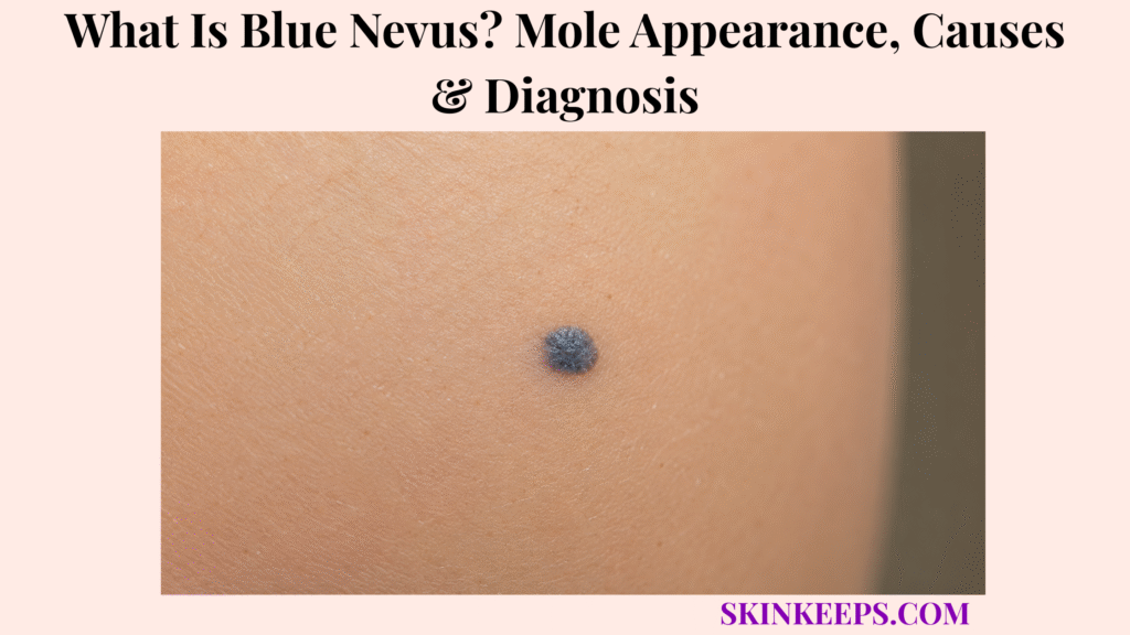
A blue nevus is a usually benign mole made of pigment-producing melanocytes located deeper in the skin, which can make it look blue, blue-gray, slate, or blue-black.
This page explains appearance, causes, body locations, diagnosis, dermoscopy, biopsy logic, monitoring, removal, mistakes, and warning signs. Blue nevus is usually benign, but a new, changing, painful, bleeding, crusting, irregular, or melanoma-like blue-black lesion should be checked.
What Is Blue Nevus and Why Does a Mole Look Blue?
Blue nevus is a usually benign melanocytic mole in which pigment-producing cells sit deeper in the skin, creating a blue, gray-blue, slate, or blue-black appearance.
It is also spelled blue naevus. A melanocytic nevus is a mole made from melanocytes, the pigment-producing cells that make melanin.
Many ordinary brown moles have pigment closer to the surface. In blue nevus, the pigment is deeper in the dermis, so the same pigment can be perceived as blue, gray, steel-blue, slate, or blue-black.
Why Can Deeper Pigment Create a Blue Color?
Deeper pigment can create a blue color because melanocytes sit in the dermis, where skin optics can make pigment appear blue, gray, slate, or blue-black rather than brown.
The dermis is the skin layer below the epidermis. When pigment is deeper, light and color perception change the way the lesion appears from the surface.
This is why a blue color alone does not automatically mean melanoma. The safer question is whether the lesion is stable, uniform, clinically typical, and confirmed when needed.
Why Is Blue Nevus Usually Benign but Still Needs Correct Identification?
Blue nevus is usually benign, but correct identification matters because melanoma and other dark lesions can sometimes look blue-black.
Common blue nevi typically stay stable over time. A classic, stable lesion may be monitored after a clinician confirms the pattern.
The main risk is false reassurance. A new adult-onset, changing, symptomatic, irregular, rapidly growing, or different-looking blue-black lesion should not be treated as harmless without evaluation.
Practical rule: A stable blue nevus is often benign, but a new, changing, symptomatic, or unusual blue-black lesion needs professional confirmation.
What Does a Blue Nevus Look Like on Skin?
A typical blue nevus may appear as a small round or oval blue, gray-blue, blue-black, slate, or dark papule, nodule, plaque, or flat spot that is usually well-defined and stable.
The surface is often smooth. It may be flat like a macule, slightly raised like a papule, plaque-like, or nodular and firm.
What Color Can a Blue Nevus Have?
A blue nevus can look steel-blue, blue-gray, slate-gray, blue-black, or occasionally dark brown, brown-yellow, or nearly black.
Color may appear different across skin tones and lighting. A classic lesion often has a fairly uniform blue or gray tone rather than many different colors.
A very dark lesion still needs review if it is new, changing, growing, bleeding, crusting, painful, irregular, or different from the person’s other moles.
What Shape and Texture Can a Blue Nevus Have?
A blue nevus is often round or oval with a smooth surface and may be flat, dome-shaped, papular, plaque-like, or nodular.
A common blue nevus is often small and stable. Clinical references commonly describe common blue nevus as about 0.5–1 cm, while cellular blue nevus can be more nodular and larger.
Size is only a clue, not a home-diagnosis rule. A large, new, growing, or nodular blue-black lesion should be evaluated.
What Symptoms Are Not Typical for a Stable Blue Nevus?
Rapid growth, bleeding, crusting, ulceration, pain, persistent itching, multiple color zones, or an irregular spreading border are not typical features of a stable blue nevus.
These signs do not prove melanoma, but they raise the need for dermatology review. A lesion that looks unlike the person’s other moles, known as an ugly-duckling pattern, also deserves attention.
| Feature | Typical Blue Nevus Clue | When It Becomes Concerning |
|---|---|---|
| Color | Homogeneous blue, gray-blue, slate, or blue-black | Multiple colors or new dark change |
| Shape | Round or oval | Asymmetry or spreading irregular border |
| Surface | Smooth macule, papule, plaque, or nodule | Crusting, ulceration, or bleeding |
| Size | Often small if common blue nevus | Rapid enlargement or large new adult lesion |
| Symptoms | Usually painless and stable | Pain, persistent itch, tenderness, bleeding |
| Pattern | Looks similar over time | Ugly-duckling lesion unlike other moles |
Where Does Blue Nevus Usually Appear?
Blue nevus can appear on many body sites, including the hands, feet, scalp, face, arms, legs, buttocks, and sacrococcygeal area.
It can occur on the back of the hands or feet, face, scalp, buttocks, arms, legs, or lower back near the sacrococcygeal area. Location can support the pattern, but it cannot confirm diagnosis alone.
Scalp lesions and other hard-to-monitor lesions may be removed more often as a precaution because change history can be hard to track. Rare nail-adjacent or mucosal locations need specialist judgment when uncertain.
| Location | Common Clue | Diagnosis Caution |
|---|---|---|
| Scalp | Blue-gray or blue-black spot or nodule | Hard to monitor; history may be unclear |
| Face | Small blue-gray papule or macule | Cosmetic and melanoma-mimic concern |
| Back of hands | Distal extremity pattern | Separate from traumatic tattoo or vascular lesion |
| Feet | Blue-gray lesion on dorsal foot or toe area | Check changing or acral pigmented lesions |
| Arms / legs | Stable blue papule or spot | Track growth and symptoms |
| Buttocks | Cellular blue nevus can occur here | Larger or nodular lesions may need closer review |
| Sacrococcygeal area | Reported cellular-site pattern | Hard-to-see area; monitor carefully |
| Nail / mucosal area | Rare context | Specialist evaluation if uncertain |
What Types of Blue Nevus Are Important to Know?
Blue nevus has several forms, but the most useful patient-facing distinction is between common blue nevus, cellular blue nevus, and atypical or melanoma-like blue lesions that require closer evaluation.
Most readers do not need pathology-level subtyping. The practical question is whether the lesion is classic and stable or whether it is new, changing, symptomatic, large, nodular, or uncertain.
What Is a Common Blue Nevus?
A common blue nevus is usually a small, stable, blue-gray or blue-black benign mole that may be monitored if it has classic features.
It is often smooth, round or oval, and fairly uniform in color. Many common blue nevi remain stable for years.
A small blue lesion is not automatically benign. The full pattern, change history, dermoscopy, and clinician judgment matter.
What Is a Cellular Blue Nevus?
A cellular blue nevus can be larger or more nodular than a common blue nevus and may need closer evaluation if it is growing, atypical, symptomatic, or uncertain.
Cellular blue nevus can occur on the scalp, buttocks, or sacrococcygeal area. It may be harder to distinguish clinically from concerning lesions when it is large, deep, nodular, or changing.
Rare malignant transformation is mainly discussed in cellular blue nevus contexts. This should not be exaggerated, but it supports biopsy or excision when the lesion is atypical or changing.
What Is Blue Nevus-Like Melanoma?
Blue nevus-like melanoma is a rare but important melanoma pattern that can resemble a blue nevus, especially when a blue-black lesion grows, changes, ulcerates, bleeds, hurts, or shows multiple colors.
Diagnosis requires biopsy and pathology when melanoma cannot be confidently ruled out. Appearance alone should not be used to reassure a changing blue-black nodule.
Stable common blue nevus should not be framed as commonly turning into melanoma. The key is identifying lesions that do not behave like a stable blue nevus.
| Type / Pattern | Typical Clue | Diagnosis Concern | Likely Next Step |
|---|---|---|---|
| Common blue nevus | Small, stable, homogeneous blue-gray lesion | Usually low if classic and stable | Monitor after confirmation |
| Cellular blue nevus | Larger or nodular blue-black lesion | Can be harder to distinguish | Dermatology review; biopsy if atypical |
| Combined blue nevus | Blue nevus with another nevus pattern | Mixed appearance may confuse diagnosis | Dermoscopy or biopsy if uncertain |
| Subungual blue nevus | Blue macule near or under nail unit | Nail pigmentation can be complex | Specialist assessment if uncertain |
| Blue nevus-like melanoma | Changing, irregular, multi-color, symptomatic lesion | Melanoma rule-out | Prompt biopsy/pathology |
What Causes Blue Nevus?
Blue nevus forms when pigment-producing melanocytes are located deeper in the skin, creating a blue or gray visual effect rather than the more typical brown color of many surface moles.
Melanocytes are pigment-producing cells. In blue nevus, the cells are located in the dermis, below the more superficial epidermis.
Some blue nevi appear in childhood or adolescence, while some appear later. Most are sporadic or acquired. Rare congenital blue nevi can occur.
Blue nevus is not caused by dirt, bruising, or poor hygiene. A bruise, graphite mark, or traumatic tattoo can mimic a blue lesion, so history matters.
How Is Blue Nevus Different From Other Dark or Blue Skin Lesions?
Blue nevus can resemble melanoma, an ordinary mole, a bruise, graphite tattoo, vascular lesion, seborrheic keratosis, pigmented basal cell carcinoma, or other dark skin lesions, so change history and dermatologist assessment matter.
Color overlap is common. A stable, homogeneous blue lesion is different from a lesion that changes, bleeds, ulcerates, grows, becomes painful, or develops several colors.
How Is Blue Nevus Different From Melanoma?
Blue nevus is usually stable and homogeneous, while melanoma concern rises when a lesion changes, grows, bleeds, ulcerates, hurts, itches persistently, or develops multiple colors or irregular borders.
Melanoma can sometimes look blue-black. A changing mole, jagged border, multiple colors, growth, bleeding, sore-like behavior, or ugly-duckling pattern should be checked.
Biopsy is needed when melanoma cannot be ruled out clinically.
How Is Blue Nevus Different From a Bruise or Graphite Mark?
Blue nevus is different from a bruise or graphite mark because a bruise usually changes color and fades, while a graphite mark often follows a pencil injury history.
A blue nevus usually has a mole-like structure and stable pigment. A graphite or traumatic tattoo may stay gray-blue but often has a clear injury story.
History matters, especially in children. A persistent or uncertain blue-gray mark should be examined rather than guessed from appearance alone.
How Is Blue Nevus Different From a Vascular Lesion?
Blue nevus is pigment-based, while vascular lesions are blood-vessel-based and may look red, purple, blue, blanch, bleed, or change with pressure.
A hemangioma, venous lake, angioma, or other vascular lesion follows blood-vessel logic rather than dermal pigment logic.
Dermoscopy and clinical exam can help separate pigment patterns from vascular patterns.
| Condition | Common Clue | Why Confusion Happens | Safer Next Step |
|---|---|---|---|
| Blue nevus | Stable homogeneous blue/gray lesion | Dark color can worry users | Dermoscopy if uncertain |
| Melanoma | Changing, irregular, multi-color, bleeding, painful, or ulcerated lesion | Can look blue-black | Prompt biopsy if suspicious |
| Bruise | Color changes and fades | Blue/purple color overlap | Track healing timeline; review if persistent |
| Graphite tattoo | History of pencil or graphite injury | Gray-blue mark | Clinical history and exam |
| Vascular lesion | Red-purple/blue, may blanch or bleed | Dark vascular color | Dermoscopy or clinical exam |
| Basal cell carcinoma | Dark, shiny, pearly, bleeding, or non-healing growth | Pigmented tumors may mimic moles | Dermatology review |
| Benign keratosis | Waxy or stuck-on pigmented growth | Dark raised lesion overlap | Dermoscopy/biopsy if uncertain |
How Is Blue Nevus Diagnosed?
Blue nevus is often diagnosed by clinical appearance and dermoscopy, but biopsy is recommended when the lesion is new in adulthood, changing, symptomatic, atypical, or difficult to distinguish from melanoma.
Diagnosis is not based on color alone. A clinician reviews the lesion’s history, location, size, shape, color pattern, symptoms, and whether it looks different from the person’s other moles.
What Does Dermoscopy Show in a Blue Nevus?
Dermoscopy of a typical blue nevus often shows homogeneous blue pigmentation with a structureless, uniform pattern that supports the diagnosis.
The pattern may look steel-blue, hazy, or ground-glass under dermoscopy. A well-defined border that fades into surrounding skin can also support the diagnosis.
Dermoscopy supports clinical diagnosis, but it does not replace biopsy when the lesion is suspicious. Network, dots, clods, streaks, vessels, extra colors, or other atypical structures may trigger further investigation.
When Is Biopsy Considered for Blue Nevus?
Biopsy is considered when a blue nevus-like lesion appears newly in adulthood, grows, changes, becomes symptomatic, looks irregular, has multiple colors, or cannot be confidently separated from melanoma.
Biopsy may also be considered for a large or nodular lesion, a cellular-blue-nevus concern, an ugly-duckling lesion, a scalp or hard-to-monitor lesion, or a lesion with bleeding, crusting, ulceration, pain, or persistent itch.
Stable classic lesions may be monitored clinically after appropriate confirmation. Rapidly changing lesions should not be watched without evaluation.
What Does Pathology Confirm?
Pathology confirms whether the lesion is a blue nevus type or another pigmented lesion, including melanoma when that concern cannot be ruled out clinically.
Pathology can separate benign blue nevus from blue nevus-like melanoma, pigmented basal cell carcinoma, vascular lesions, traumatic pigment, and other mimics.
It also helps decide whether complete removal or follow-up is needed.
Does Blue Nevus Need Treatment or Removal?
A stable, classic blue nevus often does not need treatment, but removal may be considered for diagnosis, symptoms, cosmetic reasons, repeated trauma, or melanoma-like features.
The decision depends on confidence in the diagnosis, change history, location, size, symptoms, cosmetic goals, and scar tradeoff.
When Is Monitoring Enough for Blue Nevus?
Monitoring may be enough when a dermatologist confirms a classic blue nevus that is stable, symptom-free, and not changing in size, color, shape, or elevation.
Photos can help track whether the lesion changes. Monitoring should include clear instructions about what changes require re-checking.
Monitoring is not the same as ignoring. A new adult lesion or changing lesion should be evaluated before being monitored.
When Is Removal Recommended or Considered?
Removal or biopsy may be considered when diagnosis is uncertain, the lesion is new in adulthood, changing, symptomatic, repeatedly traumatized, cosmetically bothersome, or difficult to monitor.
Bleeding, crusting, ulceration, pain, persistent itch, change in elevation, growth, multiple colors, irregular border, large nodular shape, or scalp location can increase the reason for biopsy or removal.
Patient preference can matter after counseling, especially if the lesion is cosmetically bothersome or repeatedly irritated.
What Should Users Expect From Blue Nevus Removal?
Blue nevus removal is usually done by a clinician when tissue diagnosis or cosmetic removal is needed, and it can leave a scar.
Excision is often used when tissue diagnosis is needed. Because the pigment sits deeper, removal may need to go deeper than some superficial brown lesions.
Pathology may be sent if diagnosis is uncertain. A recurrent blue lesion at a prior removal site should be checked rather than assumed to be harmless regrowth.
| Situation | Likely Approach | Main Goal | Caution |
|---|---|---|---|
| Classic stable lesion | Monitor after confirmation | Avoid unnecessary scar | Track changes |
| New adult lesion | Biopsy or excision consideration | Rule out melanoma | Do not delay review |
| Changing lesion | Biopsy | Diagnosis | Prompt evaluation |
| Large/nodular lesion | Dermatology assessment | Assess cellular blue nevus or mimic | Pathology may be needed |
| Scalp/hard-to-monitor lesion | Removal may be considered | Safer monitoring or diagnosis | Scar tradeoff |
| Cosmetic concern | Elective removal discussion | Appearance | Scarring possible |
| Symptomatic lesion | Exam, biopsy, or removal | Rule out concern | Symptoms matter |
What Blue Nevus Mistakes Should You Avoid?
The biggest blue nevus mistake is either panicking that every blue mole is melanoma or ignoring a changing blue-black lesion because blue nevus is usually benign.
Do not self-diagnose a new blue-black mole. Do not rely only on color. Pattern, history, symptoms, dermoscopy, and biopsy logic matter.
Do not remove, burn, freeze, cut, scrape, or apply mole-removal creams at home. DIY treatment can scar the skin and delay diagnosis if the lesion is not a benign blue nevus.
| Mistake | Why It Fails | Better Action |
|---|---|---|
| Calling every blue mole cancer | Creates unnecessary fear | Get dermatologist assessment if uncertain |
| Ignoring change | Melanoma can mimic blue nevus | Check changing lesions |
| DIY mole removal | Scarring and missed diagnosis risk | Diagnose first |
| Watching symptoms too long | Pain, bleeding, crusting, or ulceration can matter | Dermatology review |
| Avoiding biopsy when advised | Delays diagnosis | Follow clinician plan |
| Relying on color alone | Blue-black color has many causes | Assess pattern, history, dermoscopy |
| Ignoring ugly-duckling sign | One different lesion can be important | Compare with other moles and seek care |
When Should a Blue Nevus Be Checked by a Dermatologist?
A blue nevus or blue-black mole should be checked by a dermatologist when it is new in adulthood, growing, changing color or shape, bleeding, crusting, painful, itchy, ulcerated, irregular, unusually large, or different from your other moles.
Hard-to-monitor areas such as the scalp, nail unit, back, buttocks, and sacrococcygeal region may need extra caution because changes may be missed.
Which Blue Nevus Warning Signs Need Faster Review?
Faster review is needed for a new adult-onset blue lesion, rapid growth, color or shape change, bleeding, crusting, ulceration, pain, persistent itch, firm enlarging nodule, multiple colors, or ugly-duckling pattern.
Do not wait and see for a changing blue-black lesion. A dermatologist can decide whether dermoscopy, photography, biopsy, excision, or monitoring is safest.
What Should You Bring to a Blue Nevus Appointment?
A useful blue nevus appointment starts with photos, timing, change history, symptoms, injury history, skin cancer history, family melanoma history, immune status, and previous biopsy records.
Bring details about when the lesion appeared, whether it changed, whether it hurts or itches, and whether there was trauma, pencil injury, previous removal, or prior biopsy.
Seek dermatology review if a blue or blue-black lesion is:
What Should You Remember About Blue Nevus?
The most important thing to remember about blue nevus is that it is usually a benign blue or gray mole, but diagnosis should be confirmed when the lesion is new, changing, symptomatic, or melanoma-like.
Stable classic lesions may be monitored after confirmation. New, changing, symptomatic, large, nodular, irregular, or uncertain lesions need biopsy logic.
Frequently Asked Questions About Blue Nevus
Is blue nevus cancer?
Usually no. Blue nevus is usually benign, but blue-black melanoma-like lesions can mimic it, so new, changing, or symptomatic lesions should be checked.
Why is a blue nevus blue?
Pigment-producing melanocytes sit deeper in the skin, and the way light interacts with deeper pigment makes the mole appear blue, gray, slate, or blue-black.
What does blue nevus look like?
A blue nevus may look like a small, usually well-defined blue, blue-gray, slate, or blue-black spot, papule, plaque, or nodule that is often smooth and stable. Dermoscopy may show homogeneous blue pigmentation.
Does blue nevus need biopsy?
Not always. A classic, stable lesion may be monitored, but a new adult lesion, changing lesion, symptomatic lesion, irregular lesion, large or nodular lesion, or uncertain lesion may need biopsy.
Can blue nevus turn into melanoma?
Malignant transformation is rare, but melanoma can mimic blue nevus or rarely arise in association with blue nevus, especially in cellular blue nevus contexts. Changes should be evaluated promptly.
Can blue nevus be removed?
Yes. A blue nevus can be removed for diagnosis, symptoms, repeated irritation, hard-to-monitor location, or cosmetic reasons, but removal may leave a scar and should be clinician-performed.
When should a blue mole be checked?
A blue mole should be checked when it is new in adulthood, rapidly growing, changing color or shape, bleeding, crusting, ulcerated, painful, persistently itchy, multi-colored, irregular, large, nodular, or different from the person’s other moles.
Sources & Evidence About Blue Nevus
DermNet — Blue Naevus was used for blue nevus definition, dermal melanocyte location, blue/gray color mechanism, clinical features, common sites, diagnosis, excisional biopsy if uncertain, treatment, outcome, and scalp-monitoring caution.
DermNet — Blue Naevus Dermoscopy was used for homogeneous blue pigmentation, structureless or uniform pattern, well-defined fading border, steel-blue/hazy/ground-glass pattern, and dermoscopic features that warrant melanoma investigation.
StatPearls / NCBI Bookshelf — Blue Nevus was used for blue nevi as melanotic dermal lesions, common versus cellular types, melanoma confusion, stable lesion monitoring, biopsy for new or changing lesions, excision context, and recurrence-at-excision-site caution.
American Academy of Dermatology — Melanoma Signs and Symptoms was used for changing mole, jagged border, more than one color, growing lesion, bleeding sore-like growth, nail-line warning, and ABCDE warning-sign support.
American Academy of Dermatology — When Is a Mole a Problem? was used for dermatologist evaluation when a mole looks different from others, itches, bleeds, or changes, and for removal reasons such as irritation or cosmetic concern.
Educational Disclaimer: This SkinKeeps article is for educational purposes only and does not diagnose or replace medical care. A new, changing, rapidly growing, bleeding, crusting, ulcerated, painful, persistently itchy, irregular, multi-colored, large, nodular, adult-onset, ugly-duckling, hard-to-monitor, or melanoma-like blue-black lesion should be checked by a qualified healthcare professional or dermatologist.



