
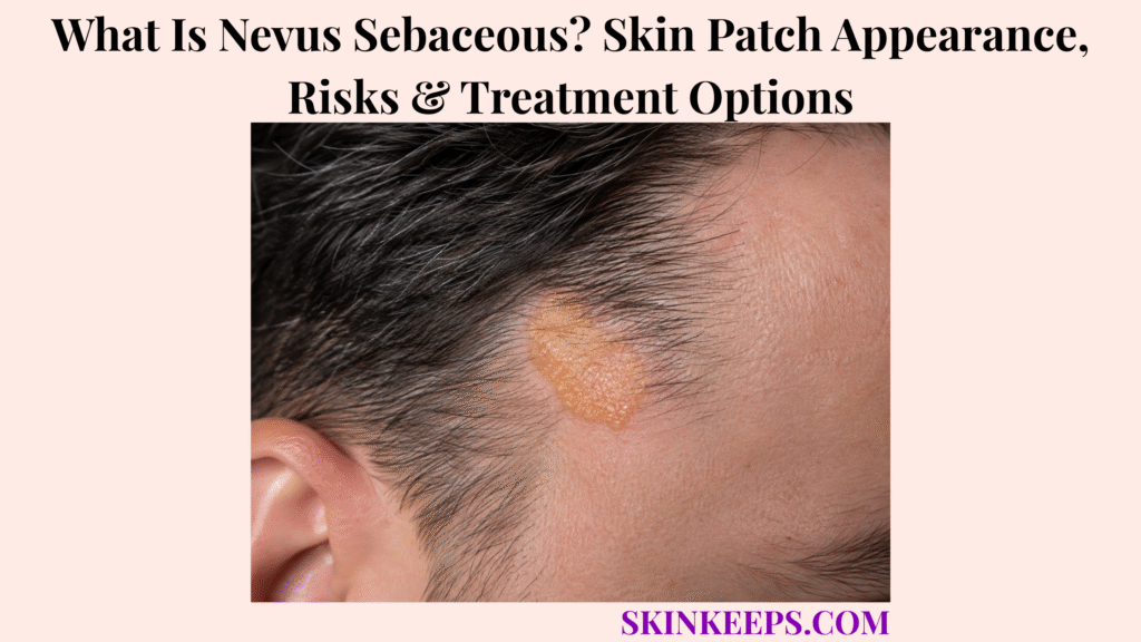
Nevus sebaceous is a congenital organoid hamartoma that usually appears as a smooth, hair-poor or hairless patch on the scalp or face and becomes thicker or more verrucous around puberty. It contains abnormal development of several skin structures, including the epidermis, sebaceous glands, hair follicles and sometimes other adnexal components.
The lesion usually persists throughout life, and puberty-related thickening is an expected maturation change rather than automatic evidence of cancer. Secondary tumours can develop later, but most are benign, so modern management balances observation against elective excision instead of requiring preventive childhood surgery for every lesion.
This article is for educational purposes only and does not constitute medical advice, diagnosis, or treatment. A nevus sebaceous that develops a new discrete lump or nodule, rapid focal enlargement, persistent crusting, a non-healing sore, ulceration, repeated bleeding, significant new pain, or another unexpected change from its usual appearance should be evaluated by a licensed dermatologist or qualified healthcare professional.
How Can You Recognize a Nevus Sebaceous?
Nevus sebaceous usually begins as a well-defined smooth or slightly raised, hair-poor plaque on the scalp or face and becomes thicker and more textured during adolescence.
What Does Nevus Sebaceous Look Like in Childhood?
In a baby or young child, nevus sebaceous usually appears as a smooth or slightly raised yellow-orange, yellow-tan or skin-toned plaque with little or no terminal hair.
The plaque is often oval or linear and sharply defined. Yellow colouring can be subtle, so congenital onset, texture and localized hair loss are often more useful recognition features than colour alone.
Where Does Nevus Sebaceous Usually Develop, and Why Is the Scalp Area Hairless?
Nevus sebaceous most often affects the scalp, followed by the preauricular region, face and neck, and scalp lesions are usually hair-poor because their follicles are abnormally developed.
Trunk or limb lesions occur less often and can follow Blaschko lines. Within scalp lesions, immature or abnormal follicles fail to produce normal terminal hair, creating localized alopecia.
What Happens to Nevus Sebaceous During Puberty?
Puberty can naturally make nevus sebaceous thicker, more raised, pebbled, scaly, papillomatous or wart-like as sebaceous and other adnexal structures mature.
The key distinction is pattern: gradual change affecting most of the plaque during adolescence is expected maturation, whereas a new discrete nodule, ulcer or persistently bleeding focus is a separate event that should be assessed.
Does Nevus Sebaceous Look the Same on Every Skin Tone or Cause Symptoms?
No; yellow colouring may be less obvious in darker skin, and most uncomplicated lesions are painless and non-itchy.
Across skin tones, congenital onset, localized alopecia, texture, location and age-related maturation remain useful recognition clues. New pain, bleeding, crusting or a focal nodule should prompt reassessment rather than being attributed automatically to normal maturation.
Figure 1. Nevus sebaceous usually remains relatively smooth through childhood and becomes more textured during puberty; a new discrete focal growth is the change that deserves separate assessment.
Why Does Nevus Sebaceous Develop, and What Is Its Tumour Risk?
Nevus sebaceous develops from postzygotic mosaic abnormalities in skin development, commonly involving the RAS/MAPK pathway, and can later develop secondary tumours that are much more often benign than malignant.
What Causes Nevus Sebaceous, and Is It Inherited?
Nevus sebaceous usually results from a postzygotic mosaic genetic alteration rather than a mutation inherited through every cell from a parent.
The genetic change arises after fertilization and is therefore limited to a subset of cells. HRAS and KRAS alterations within the RAS/MAPK pathway are well-established molecular findings, while ordinary isolated nevus sebaceous is usually sporadic and does not require routine genetic testing.
Why Does the Lesion Become More Prominent With Age?
Hormonal maturation during adolescence stimulates sebaceous and other adnexal structures within the nevus, making the lesion thicker and more papillomatous.
This predictable developmental response links a congenital lesion to its classic adolescent texture. Hormonal maturation itself is not a secondary tumour.
Which Secondary Tumours Can Develop Inside Nevus Sebaceous?
Secondary tumours can develop within nevus sebaceous, especially in adulthood, but the majority are benign adnexal neoplasms.
Trichoblastoma and syringocystadenoma papilliferum are among the classic benign secondary tumours. Trichilemmoma, sebaceoma and poroma can also occur, but a long tumour catalogue is less useful than recognizing that most secondary growths are benign.
Can Nevus Sebaceous Become Cancerous?
Yes, but malignant secondary tumours are uncommon, and contemporary evidence indicates that older teaching substantially exaggerated the cancer risk.
Basal cell carcinoma is the best-known malignant association, but major contemporary clinical references estimate BCC in less than 1% of nevus-sebaceous lesions.
Other malignancies, including squamous cell carcinoma, are rarer. A new focal growth still deserves assessment because appearance alone may not reliably distinguish a benign secondary tumour from a malignant one.
Figure 2. Postzygotic RAS-pathway mosaicism creates the congenital organoid nevus; puberty changes the lesion naturally, while secondary tumours form a later branch in which benign neoplasms predominate.
How Is Nevus Sebaceous Distinguished and Diagnosed?
A typical nevus sebaceous is usually diagnosed clinically from congenital onset, scalp or facial location, localized alopecia and predictable age-related maturation, while biopsy is reserved for uncertainty or new focal changes.
Which Conditions Can Resemble Nevus Sebaceous?
The differential diagnosis changes with age and can include aplasia cutis congenita, other epidermal nevi, congenital melanocytic nevi and later wart-like or adnexal growths.
In infancy, congenital skin defects and other nevi can resemble a smooth hair-poor plaque. Later, an epidermal nevus, wart, seborrhoeic keratosis or secondary adnexal tumour can resemble the more verrucous stage.
How Is Nevus Sebaceous Different From a Congenital Melanocytic Nevus?
Nevus sebaceous is an epidermal-adnexal organoid hamartoma that is often hair-poor and yellowish, whereas a melanocytic nevus / mole arises from melanocytes and is usually more pigmented.
Nevus sebaceous is defined by abnormal epidermal and adnexal structures and characteristically becomes more verrucous during puberty. Congenital melanocytic nevi belong to a different melanocytic disease category.
When Is Skin Biopsy Needed?
Skin biopsy is most useful when the diagnosis is uncertain or a nevus sebaceous develops a new focal lump, persistent crust, ulceration, bleeding or another unexpected structural change.
A new discrete tumour, unexpected childhood change or suspected secondary neoplasm should be sampled or excised according to clinical context rather than being labelled automatically as either normal puberty maturation or cancer.
Histology changes with age: childhood lesions show immature pilosebaceous structures, while post-pubertal lesions can show more prominent sebaceous glands, acanthosis, papillomatosis and hyperkeratosis.
When Should Nevus Sebaceous Syndrome Be Considered?
Nevus sebaceous syndrome should be considered mainly when lesions are large, extensive or multifocal and occur with neurological, ocular or skeletal abnormalities.
Seizures, developmental abnormalities, ocular findings or skeletal asymmetry can justify targeted neurology, ophthalmology or orthopedic assessment. One small isolated scalp nevus does not automatically imply Schimmelpenning syndrome.
Figure 3. Stable classic lesions can be followed clinically, focal structural change prompts tissue assessment, and broader syndromic evaluation belongs mainly to extensive lesions with extracutaneous abnormalities.
Should Nevus Sebaceous Be Removed?
Not every nevus sebaceous requires preventive removal; observation and complete surgical excision are both reasonable options depending on lesion change, symptoms, cosmetic impact, reconstruction needs and patient preference.
Does Every Nevus Sebaceous Need Preventive Surgery?
No; routine prophylactic excision of every childhood nevus sebaceous is not mandatory because malignant transformation is uncommon and usually occurs later in life.
A stable uncomplicated lesion can be monitored. Modern risk estimates and newer cohorts have moved management away from the older assumption that every lesion should be removed early solely to prevent cancer.
When Does Surgical Excision Become More Reasonable?
Excision becomes more reasonable when a suspicious secondary growth develops, the lesion repeatedly bleeds or ulcerates, cosmetic impact is substantial, or the patient prefers definitive removal after discussing scarring.
Lesion size, anatomical site, recurring irritation, future reconstruction complexity and patient or parent preference all affect the decision. A suspicious focal change can be biopsied first or removed completely depending on the clinical situation.
What Does Complete Excision Involve, and What Is the Best Age?
Full-thickness surgical excision is the definitive removal method, but there is no single best age because timing depends on lesion size, location, anesthesia, scarring and patient preference.
Complete excision removes the full lesion, provides tissue for histopathology and inevitably leaves a surgical scar. Childhood surgery is not universally necessary; later childhood, adolescence or adulthood can also be reasonable depending on anatomy and practical considerations.
Can Laser, Shave Removal or Topical Medicines Replace Complete Excision?
Superficial laser, shave or dermabrasion procedures may improve surface appearance but generally do not remove the full-depth nevus and therefore are not equivalent to complete excision.
Residual abnormal tissue can remain and surface recurrence can occur, so continued monitoring may still be needed. Limited emerging reports suggest topical sirolimus may flatten selected lesions, but it is not an established definitive replacement for full-thickness excision.
| Clinical Context | Main Management Direction | Key Trade-Off |
|---|---|---|
| Stable uncomplicated lesion | Observation | Monitor rather than create an unnecessary surgical scar |
| Expected pubertal maturation | Continue monitoring | Whole-plaque thickening is not automatically malignant |
| Cosmetic / reconstructive concern | Elective excision discussion | Definitive removal leaves a scar |
| New focal tumour-like change | Biopsy and/or excision | Benign and malignant secondary tumours can look similar |
| Definitive removal desired | Full-thickness surgical excision | Pathology obtained; reconstruction may be needed |
| Superficial laser / shave / dermabrasion | Explain limitations | Residual tissue and recurrence are possible |
Which Changes in Nevus Sebaceous Need Long-Term Follow-Up?
Long-term monitoring should focus on new focal changes within the nevus rather than treating every predictable age-related surface change as dangerous.
Does Nevus Sebaceous Go Away, and Which Puberty Changes Are Expected?
Nevus sebaceous usually persists throughout life, and gradual thickening, scaling or verrucous change across the plaque can occur normally during puberty.
A thicker, pebbled, wart-like or more yellow-orange plaque can therefore represent expected maturation. The important comparison is general maturation of the whole plaque versus a new focal lesion arising inside it.
Which Changes Need Earlier Dermatology Review?
A new discrete nodule, rapid focal enlargement, persistent crusting, non-healing sore, ulceration, recurrent bleeding or major unexpected texture change deserves earlier dermatology assessment.
These features do not prove malignancy, but they change the lesion from predictable maturation to a structural event that may require biopsy.
Does a New Lump Automatically Mean Cancer?
No; secondary tumours arising within nevus sebaceous are more often benign than malignant, but a new lump still requires evaluation because appearance alone may not reliably determine the tumour type.
Trichoblastoma and syringocystadenoma papilliferum are classic benign examples. Reassurance about the overall risk should not be used as a reason to ignore a genuinely new focal growth.
What Happens After Complete Excision?
After complete excision, follow-up mainly concerns wound healing, scar outcome and pathology findings from the removed tissue.
If pathology shows only nevus sebaceous or a benign secondary tumour, routine surgical follow-up is usually sufficient. If a malignancy is identified, further management follows that tumour type and margin status rather than assuming every excised nevus needs oncology surveillance.
What Should You Remember About Nevus Sebaceous?
Nevus sebaceous is a congenital organoid hamartoma that usually begins as a smooth hair-poor scalp or facial plaque, naturally thickens around puberty and only uncommonly develops a malignant secondary tumour.
- Nevus sebaceous is congenital.
- It is an organoid hamartoma involving multiple skin structures.
- The scalp and face are common sites.
- Localized alopecia is characteristic.
- Yellow colour may be less obvious in darker skin.
- Puberty naturally changes lesion texture.
- Pubertal thickening does not automatically mean cancer.
- Whole-plaque maturation differs from a new focal growth.
- Postzygotic HRAS/KRAS mosaicism is an important developmental mechanism.
- Typical isolated disease is usually not inherited.
- Secondary tumours can develop later.
- Most secondary tumours are benign.
- Trichoblastoma and syringocystadenoma papilliferum are classic benign examples.
- Malignant transformation is uncommon.
- BCC risk is far lower than older teaching suggested.
- Diagnosis is usually clinical.
- New focal change can require biopsy.
- Extensive lesions with extracutaneous findings can justify syndromic evaluation.
- Automatic childhood excision is not mandatory.
- Observation is a valid management option.
- Complete full-thickness excision is definitive when removal is chosen.
- Surgery leaves a scar.
- Superficial destructive treatment may leave residual nevus tissue.
- Long-term monitoring focuses on new focal change.
Recognize congenital plaque → Expect puberty maturation → Monitor focal change → Biopsy when needed → Observe or excise individually.
Frequently Asked Questions About Nevus Sebaceous
The most important nevus-sebaceous questions concern childhood appearance, puberty-related thickening, cancer risk, preventive removal and changes that require biopsy.
What Does Nevus Sebaceous Look Like in a Baby or Child?
Nevus sebaceous usually appears as a smooth or slightly raised yellow-orange, yellow-tan or skin-toned hair-poor plaque on the scalp or face. It is often well-defined and oval or linear, with localized alopecia when the scalp is involved.
Why Does Nevus Sebaceous Become Thicker During Puberty?
Hormonal maturation stimulates sebaceous and other adnexal structures within the nevus, making the plaque thicker, bumpier or more verrucous. This expected maturation does not automatically indicate cancer.
Can Nevus Sebaceous Become Cancerous?
Yes, but malignant secondary tumours are uncommon, while benign tumours developing within nevus sebaceous are substantially more common. Basal cell carcinoma is the best-known malignant association, but modern risk estimates are much lower than older teaching suggested.
Does Every Nevus Sebaceous Need to Be Surgically Removed?
No; a stable uncomplicated lesion can be observed, while excision can be chosen for suspicious change, symptoms, cosmetic concerns or patient preference. There is no universal mandatory age for surgery, and complete excision leaves a scar.
Which Changes in Nevus Sebaceous Should Be Biopsied?
A new focal lump, persistent crust, ulceration, recurrent bleeding, non-healing sore or other unexpected structural change can justify biopsy or excision. A new growth is not automatically malignant but should not be assumed harmless without assessment.
Which Sources Support This Nevus Sebaceous Guidance?
DermNet — Nevus Sebaceous — Primary source for organoid-hamartoma definition, congenital morphology, scalp/facial distribution, skin-tone variation, pubertal maturation, RAS-pathway mosaicism, benign secondary tumours, BCC risk, biopsy, observation, excision and superficial-treatment limitations.
StatPearls / NCBI Bookshelf — Nevus Sebaceus — Used for localized alopecia, abnormal pilosebaceous development, pubertal histology, clinical diagnosis, differential diagnosis, treatment context and the modern low-malignancy framing.
2024 Systematic Review and Meta-Analysis — Secondary Tumours Associated With Nevus Sebaceous — Used specifically for contemporary benign-versus-malignant secondary-neoplasm risk interpretation while avoiding universalizing pooled estimates.
2025 Cohort of 953 Patients With Nevus Sebaceous — Used for current individualized surgical-timing context and the lack of one universally mandatory prophylactic-excision approach.
American Academy of Dermatology — Birthmarks: Signs and Symptoms — Used for the patient-facing description of childhood hairless scalp lesions, adolescent thickening and lifelong persistence.
DermNet — Epidermal Naevus Syndromes — Used only for nevus sebaceous syndrome / Schimmelpenning context and associated neurologic, ocular and skeletal abnormalities.



