
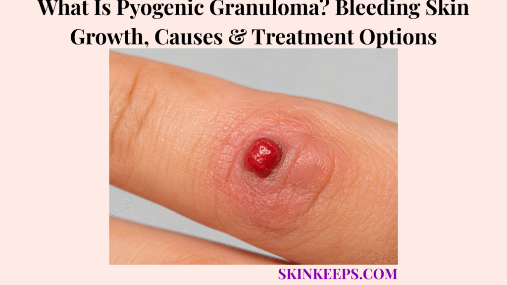
Pyogenic granuloma is a benign vascular growth made of proliferating capillaries that often grows quickly and bleeds easily after minor contact. It is more accurately described as a lobular capillary hemangioma because the lesion is made of fragile blood-vessel tissue rather than pus or a conventional granulomatous process.
The name can therefore be misleading. Trauma, irritation, pregnancy-related hormonal change and selected medicines are associated with some cases, but many lesions have no clear trigger; diagnosis may require histology when the appearance is atypical, removal options vary by site and patient factors, and recurrence can occur after treatment.
This article is for educational purposes only. A rapidly growing, repeatedly bleeding or uncertain skin growth should be professionally evaluated.
What Is Pyogenic Granuloma and What Does It Look and Feel Like?
Pyogenic granuloma is a benign capillary-rich vascular growth that typically enlarges rapidly and forms a fragile red or reddish-purple lesion that bleeds easily.
Why Is Pyogenic Granuloma Considered a Vascular Growth?
PG is considered vascular because it consists of proliferating capillary blood vessels that create a soft, friable lesion with a strong tendency to bleed.
The basic mechanism is vascular proliferation → raised growth → fragile capillaries → easy bleeding. That vascular architecture explains why a relatively small lesion can bleed disproportionately after rubbing, shaving, clothing friction or another minor contact.
Why Is the Name “Pyogenic Granuloma” Misleading?
The name is misleading because PG is neither primarily a pus-forming infection nor a classic granuloma; it is better classified as a lobular capillary hemangioma.
The older name remains widely used, but it should not lead readers to assume that the lesion is a bacterial abscess or that antibiotics are the defining treatment.
How Does a Pyogenic Granuloma Usually Begin and Grow?
PG often starts as a small papule and enlarges rapidly into a raised vascular growth that may ulcerate or repeatedly bleed after minor trauma.
A typical sequence is small papule → rapid enlargement → raised or fleshy vascular lesion → surface ulceration or crusting → recurrent bleeding. Growth over a short period is characteristic, but rapid growth alone does not establish the diagnosis.
What Color, Surface and Locations Are Typical?
PG commonly appears bright red to reddish-purple with a smooth, shiny, lobulated, crusted or ulcerated surface and can occur on the hands, fingers, face, lips, gingiva or other skin and mucosal sites.
The lesion may be raised on a stalk or sit more broadly on the skin, and a pale or keratinized collarette can surround its base. The pattern is useful for recognition, but colour or bleeding should never be treated as proof by themselves.
Figure 1. Pyogenic granuloma is best recognized as a rapidly enlarging, friable vascular growth that bleeds easily, often with surface ulceration, crusting, a stalk-like shape or a collarette—but appearance alone does not confirm the diagnosis.
What Causes Pyogenic Granuloma and Which Factors Can Trigger It?
Pyogenic granuloma has a multifactorial origin, with local trauma, irritation, pregnancy-related hormonal changes and selected medicines associated with some lesions, while many cases have no obvious trigger.
Can Skin Injury or Irritation Trigger PG?
Minor injury or repeated irritation can precede some pyogenic granulomas, although many affected people do not recall any specific trauma.
Trauma is therefore a reported association rather than a universal explanation. Repeated friction or local irritation may contribute in some sites, but the absence of remembered injury does not make PG unlikely.
Why Can PG Develop During Pregnancy?
Pregnancy-associated hormonal changes can promote PG, particularly on gingival or other oral mucosal tissue.
These pregnancy-associated lesions may be called granuloma gravidarum or pregnancy tumour. The word “tumour” in this context describes a growth and does not automatically imply malignancy.
Can Medicines Contribute to Pyogenic Granuloma?
Yes; medication-associated PG is recognized, but drug exposure is only one possible contributor and should not be assumed in every case.
A medication history can therefore be useful when a new lesion appears, especially if lesions are multiple or occur around nails, but the page should not reduce PG to one medicine-specific reaction.
Does Every Pyogenic Granuloma Have an Identifiable Cause?
No; PG can develop without a clear trauma, hormonal or medication trigger, so its pathogenesis should be presented as multifactorial rather than explained by one universal cause.
The most defensible model is susceptibility to abnormal capillary proliferation plus possible modifying factors, not “one event caused every lesion.”
Figure 2. Trauma, irritation, pregnancy-related hormonal change and selected medicines are possible modifiers, not mandatory causes; many pyogenic granulomas develop without one clearly identifiable trigger.
How Is Pyogenic Granuloma Diagnosed and Distinguished From Other Growths?
PG is often suspected clinically from its rapid growth and friable bleeding surface, but atypical or uncertain lesions may require biopsy or removal for histologic confirmation because other growths can mimic it.
How Does a Dermatologist Identify PG?
A dermatologist evaluates PG through the growth history, speed of enlargement, bleeding pattern, surface appearance and dermoscopy when useful.
The practical sequence is history → rapid growth and bleeding pattern → clinical examination → dermoscopy when helpful → biopsy or removal when diagnostic certainty is insufficient. A white collarette and vascular dermoscopic structures can support the impression, but they do not remove the need for judgment.
What Skin Growths Can Resemble Pyogenic Granuloma?
Benign vascular lesions, irritated growths, warts and some skin cancers can resemble PG, particularly when a lesion is red, ulcerated or repeatedly bleeding.
Keratinized or irritated lesions such as warts belong to a different disease process but can enter the visual differential when the surface is rough or traumatized.
A repeatedly bleeding or atypical lesion may also require assessment for skin cancer rather than being labeled from appearance alone.
Why Is Amelanotic Melanoma an Important Mimic?
Amelanotic melanoma can sometimes appear as a pink, red or bleeding growth rather than a dark mole, which is why rapidly growing atypical lesions should not automatically be labeled PG.
The purpose of this comparison is not to imply that most PG-like lesions are malignant; it is to preserve the safety rule that an unusual bleeding growth may need tissue diagnosis. For melanoma-specific recognition, see the dedicated melanoma page.
When Is Biopsy or Histology Needed?
Biopsy or excision is especially useful when the diagnosis is uncertain because histology can confirm lobular capillary architecture and exclude another lesion.
Under the microscope, PG typically shows lobules of capillary-sized vessels in a vascular stroma, often with surface ulceration or a collarette. A classic lesion may be removed and submitted for histology as part of treatment, while atypical lesions deserve a lower threshold for diagnostic confirmation.
Figure 3. A rapidly growing bleeding lesion may fit PG, but atypical or uncertain lesions should move toward tissue diagnosis so benign and malignant mimics are not missed.
How Is Pyogenic Granuloma Treated or Removed?
PG treatment removes or destroys the abnormal vascular tissue while controlling bleeding, with excision, shave or curettage with cautery, cryotherapy, laser or selected conservative treatments chosen according to lesion site and patient factors.
When Is Surgical Excision Used?
Complete surgical excision can remove a PG and provide tissue for histopathologic diagnosis, making it useful when definitive removal or diagnostic certainty is especially important.
The tradeoff is straightforward: excision can give both treatment and a complete tissue specimen, but it is a procedure and can leave a scar. The decision depends on location, lesion size, recurrence history and the need for diagnostic certainty.
How Are Shave Removal, Curettage and Cautery Used?
Shave removal or curettage can remove visible PG tissue, while cautery helps control bleeding and treat the remaining lesion base.
These are clinician-performed procedural approaches, not home techniques. The goal is to remove the vascular growth while managing hemostasis and reducing residual tissue that could contribute to recurrence.
When Are Cryotherapy or Laser Considered?
Cryotherapy or laser can be selected for certain PG lesions depending on size, location, cosmetic priorities, patient age and expected recurrence or scarring risk.
Neither method should be presented as universally superior. Treatment choice depends on the lesion and the clinical setting, and repeat treatment may sometimes be needed.
Are Non-Surgical Treatments Available?
Some PG lesions can be managed with clinician-directed non-surgical approaches when procedural removal is undesirable, but evidence and effectiveness vary by method and clinical setting.
Conservative options should remain professionally selected because the first problem is still diagnostic certainty: a lesion that has not been correctly identified should not be treated at home as though it were confirmed PG.
Figure 4. Treatment choice is individualized: excision offers complete tissue removal and histology, while other procedural or selected non-surgical approaches trade different levels of invasiveness, scarring, repeat treatment and recurrence risk.
How Should Bleeding Be Managed, and Can Pyogenic Granuloma Return After Treatment?
Bleeding from PG should be managed with firm direct pressure and protection of the lesion, while repeated bleeding, recurrence or diagnostic uncertainty should prompt professional reassessment rather than repeated home treatment.
What Should Be Done When a Pyogenic Granuloma Bleeds?
Firm direct pressure is the safest first response to PG bleeding, followed by protection of the lesion and medical assessment if bleeding does not stop or keeps recurring.
Use a clean dressing or cloth and maintain steady pressure rather than repeatedly lifting it to check the site. Bleeding that is difficult to control, unusually heavy or associated with dizziness or weakness warrants urgent medical attention.
Can Pyogenic Granuloma Grow Back After Treatment?
Yes; PG can recur, particularly when residual vascular tissue remains or a local contributing factor continues.
Recurrence risk varies with lesion site, treatment method and whether an ongoing local trigger remains, so no removal method should be described as guaranteeing permanent cure.
Can PG Removal Leave a Scar or Pigment Change?
Yes; excision and other destructive treatments can leave a scar or pigment change, with cosmetic outcome depending on lesion size, location, treatment method and individual healing.
This tradeoff matters most on visible or functionally sensitive sites, where treatment selection may balance diagnostic certainty, bleeding control, recurrence risk and cosmetic outcome.
When Should a Suspected PG Be Evaluated Promptly?
Professional evaluation is appropriate when a growth enlarges rapidly, bleeds repeatedly or spontaneously, remains ulcerated or crusted, recurs after treatment, or has an uncertain diagnosis.
Assessment is also appropriate when bleeding is difficult to stop, clothing or shaving repeatedly traumatizes the lesion, or pain, swelling or discharge raises concern for a complication. A rapidly growing bleeding lesion should not be cut, tied off or burned at home because the diagnosis may be wrong and significant bleeding can occur.
What Should You Remember About Pyogenic Granuloma?
Pyogenic granuloma is a benign capillary vascular growth that grows quickly and bleeds easily, but diagnosis should be confirmed when the appearance is atypical because other benign and malignant lesions can mimic it.
- Pyogenic granuloma is benign.
- It is a vascular proliferation.
- Lobular capillary hemangioma is the more descriptive term.
- PG is not primarily a bacterial infection.
- PG is not a classic granuloma.
- Rapid growth is common.
- Easy bleeding is characteristic.
- Surface ulceration or crusting can occur.
- Hands, fingers, face and oral mucosa can be affected.
- Trauma may contribute but is not universal.
- Pregnancy-related hormonal change can contribute.
- Medicines can be associated with some lesions.
- Many cases have no identifiable trigger.
- Diagnosis often begins clinically.
- Dermoscopy can support recognition.
- Other benign growths can mimic PG.
- Warts can enter the differential.
- Skin cancer occasionally needs exclusion.
- Amelanotic melanoma is an important atypical mimic.
- Histology can confirm lobular capillary architecture.
- Excision is an effective removal option.
- Shave removal, curettage and cautery are procedural options.
- Cryotherapy and laser can be selected in appropriate cases.
- Selected non-surgical approaches exist.
- Firm direct pressure helps control bleeding.
- Recurrence is possible.
- Scarring or pigment change can occur.
- Home removal is unsafe.
- Persistent or recurrent bleeding deserves evaluation.
Rapid vascular growth → recognize easy bleeding → assess possible contributors → confirm diagnosis when needed → select professional treatment → control bleeding → monitor recurrence.
Frequently Asked Questions About Pyogenic Granuloma
The main PG questions concern cancer risk, infection, easy bleeding, spontaneous resolution and recurrence after removal.
Is Pyogenic Granuloma Cancerous?
No; pyogenic granuloma is a benign vascular growth, but some malignant lesions can resemble it and may require biopsy when the diagnosis is uncertain.
Is Pyogenic Granuloma Caused by an Infection?
No; PG is not primarily an infection despite the misleading word “pyogenic” in its name.
Why Does Pyogenic Granuloma Bleed So Easily?
PG bleeds easily because it contains many fragile capillary blood vessels close to its surface.
Can Pyogenic Granuloma Go Away Without Treatment?
Some PG lesions may regress in selected circumstances, particularly some pregnancy-associated lesions after delivery, but persistent, repeatedly bleeding or diagnostically uncertain lesions often require professional assessment and treatment.
Can Pyogenic Granuloma Come Back After Removal?
Yes; recurrence can occur after treatment, particularly when residual lesion tissue or a persistent local contributing factor remains.
Which Sources Support This Pyogenic Granuloma Guidance?
DermNet — Pyogenic Granuloma — Used for benign vascular classification, misleading terminology, clinical appearance, dermoscopy, bleeding, common sites, pregnancy/trauma associations, histology, differential diagnosis, treatment categories and recurrence.
NCBI Bookshelf / StatPearls — Pyogenic Granuloma — Used for lobular capillary hemangioma terminology, rapid friable growth, multifactorial pathogenesis, pregnancy and medication associations, histopathology and clinical evaluation.
Kaleeny & Janis — Pyogenic Granuloma Diagnosis and Management: A Practical Review — Used for treatment-comparison framing, surgical excision effectiveness, alternative procedural approaches and evidence limitations.
NHS — First Aid: Bleeding — Used for direct-pressure first aid and escalation when bleeding remains difficult to control.
Mayo Clinic — Severe Bleeding: First Aid — Used as additional support for firm direct-pressure bleeding control and seeking urgent help for severe bleeding.



