
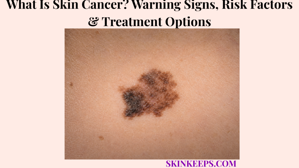
Skin cancer develops when abnormal skin cells acquire changes that allow them to grow without normal control and form a malignant lesion. The three major types readers need to distinguish are basal cell carcinoma (BCC), squamous cell carcinoma (SCC) and melanoma, because they arise from different skin cells and do not share the same pattern of invasion or spread.
Recognition should focus on abnormal behavior—such as a new, changing, growing, bleeding or non-healing lesion—rather than one color or shape. Examination can raise suspicion, but biopsy and pathology establish the diagnosis; treatment is then selected according to the cancer type, location, depth or other high-risk features, and whether disease remains local or has spread.
This article is for educational purposes only. New, changing, repeatedly bleeding, ulcerated or non-healing skin lesions should be professionally evaluated rather than self-treated.
What Is Skin Cancer and Which Main Types Develop in the Skin?
Skin cancer is malignant growth of abnormal skin cells, with basal cell carcinoma, squamous cell carcinoma and melanoma representing the three major types that differ in cell of origin, usual behavior and treatment priorities.
How Does Normal Skin Become Cancerous?
Skin cancer develops when DNA damage or other cancer-promoting cellular changes allow abnormal cells to escape normal growth control and form a malignant lesion.
Ultraviolet radiation from sunlight and tanning devices is an important source of DNA damage for several major skin cancers. Cancer development is not a single-step event: cellular damage accumulates, repair or growth-control mechanisms fail, abnormal cells expand, and some tumors acquire the ability to invade nearby tissue or spread beyond the skin.
What Is Basal Cell Carcinoma?
Basal cell carcinoma begins in basal cells of the epidermis and is usually more likely to cause progressive local tissue destruction than distant metastasis.
Basal cell carcinoma is the better-owner topic for BCC-specific morphology, risk stratification, treatment choices and recurrence. On this parent page, the key point is that BCC can damage nearby structures if neglected even though distant spread is uncommon compared with melanoma.
What Is Squamous Cell Carcinoma?
Squamous cell carcinoma develops from epidermal squamous keratinocytes and can invade surrounding tissue, with meaningful metastatic potential in selected higher-risk tumors.
Squamous cell carcinoma has its own risk features and management pathway. A routine localized SCC and a high-risk SCC are not biologically equivalent, so prognosis and treatment should not be generalized from the word “skin cancer” alone.
What Is Melanoma and Why Is It More Dangerous?
Melanoma begins in melanocytes and is less common than BCC or SCC but has a substantially greater tendency to invade and metastasize.
Melanoma requires melanoma-specific assessment of tumor thickness, ulceration, regional nodes and distant spread when indicated. Its biology is the reason melanoma warning signs and staging cannot simply be substituted for the management of all skin cancers.
What Warning Signs and Risk Factors Should Raise Concern for Skin Cancer?
Skin cancer can present as a new, growing, changing, bleeding, crusting or non-healing lesion, while ultraviolet exposure, previous skin cancer, immune suppression and selected personal or family factors increase risk without proving that a lesion is malignant.
What General Skin Changes Can Be Warning Signs?
A new lesion, enlarging spot, non-healing sore, repeated bleeding, persistent crusting or visible change in shape, color, texture or symptoms deserves evaluation because abnormal behavior is often more informative than one fixed appearance.
Skin cancer is not always a dark mole. BCC and SCC may be skin-colored, pink, red, brown, scaly, crusted, ulcerated or sore-like, while melanoma may be pigmented or less obviously pigmented. A lesion that behaves differently from the surrounding skin deserves attention even when it does not match a textbook photograph.
What Can BCC and SCC Look Like?
BCC and SCC can appear as persistent bumps, rough or scaly patches, ulcers, non-healing sores or lesions that repeatedly bleed, crust or enlarge.
BCC often has a shiny, pearly, scar-like, ulcerated or recurrent-sore pattern, but not every BCC is pearly. SCC may be rough, crusted, thickened, ulcerated or tender and can grow progressively. Morphology should raise suspicion, not replace biopsy when the diagnosis is uncertain.
What Are the ABCDE Warning Signs of Melanoma?
ABCDE highlights melanoma warning features: Asymmetry, irregular Border, varied Color, Diameter as one clue, and especially Evolving change over time.
Diameter is not a rule-out test because melanomas can be smaller than 6 mm. “Evolving” is particularly useful because change in size, shape, color or symptoms can identify a lesion that no longer behaves like a person’s baseline moles.
What Is the “Ugly Duckling” Sign?
The ugly-duckling sign describes one mole or spot that looks distinctly different from a person’s usual lesion pattern and therefore deserves closer assessment.
The concept depends on personal baseline rather than a universal color chart: if most lesions share a similar pattern and one becomes a clear outlier, that difference can be clinically meaningful even when the outlier does not satisfy every ABCDE feature.
Can Melanoma Appear Under a Nail or on Less Sun-Exposed Skin?
Yes; melanoma can occur beneath or around nails and on skin that receives little obvious sun exposure, so surveillance should not focus only on sun-exposed moles.
This is especially important across darker skin tones, where suspicious changes may appear on palms, soles, nails or other less sun-exposed sites. A persistent new nail streak, changing pigmented area, non-healing spot or bleeding lesion deserves evaluation rather than reassurance based on skin tone or body location.
How Does UV Radiation Increase Skin Cancer Risk?
Ultraviolet radiation from sunlight and indoor tanning devices can damage skin-cell DNA and increase the probability that cancer-promoting changes accumulate over time.
Risk is influenced by exposure pattern and cancer type. Cumulative UV exposure is strongly associated with keratinocyte cancers such as BCC and SCC, while intermittent intense exposure and sunburn are important in melanoma risk. UV exposure changes probability; it does not diagnose an individual lesion.
Can People With Darker Skin Develop Skin Cancer?
Yes; people of every skin tone can develop skin cancer, and suspicious new, changing, bleeding or non-healing lesions remain clinically important regardless of complexion.
Darker skin tone does not eliminate cancer risk. Delayed recognition can be dangerous, so assessment should be driven by lesion behavior and clinical context rather than the assumption that skin color rules skin cancer out.
How Is Skin Cancer Diagnosed and How Is Its Extent Determined?
Skin cancer diagnosis requires professional lesion assessment and tissue biopsy when cancer is suspected, while pathology and selected staging tests determine the cancer type, relevant high-risk features and extent.
How Does a Dermatologist Examine a Suspicious Lesion?
A dermatologist evaluates the lesion’s history of change, surface, border, color, symptoms and relationship to a person’s other spots and may use dermoscopy before deciding whether biopsy is needed.
Dermoscopy can reveal structures not easily seen with the unaided eye, but it does not convert visual inspection into definitive tissue diagnosis. The purpose of examination is to decide which lesions can be observed and which need sampling.
Why Is a Skin Biopsy Needed?
Biopsy provides tissue for microscopic examination so a pathologist can determine whether cancer is present and identify the specific cancer type.
This is the central boundary between suspicion and diagnosis: ABCDE, an ugly-duckling pattern, dermoscopy or a non-healing sore can justify biopsy, but none of them alone proves cancer. Histopathology is what turns a suspicious lesion into a defined diagnosis.
What Information From Pathology Guides Treatment?
Pathology provides the exact diagnosis and type-specific features—such as depth of invasion, ulceration, margin status or other high-risk findings—that influence staging and treatment.
The important details differ among BCC, SCC and melanoma. For melanoma, tumor thickness is a major staging factor; for keratinocyte cancers, location, histologic risk features, depth and other factors may affect local-risk assessment. One universal staging rule should not be imposed across all three cancers.
When Are Lymph-Node Tests or Imaging Needed?
Lymph-node assessment or imaging becomes more relevant when pathology or clinical findings suggest deeper invasion, high-risk SCC, melanoma with regional or metastatic potential, or possible spread beyond the primary lesion.
Small routine BCCs and many localized SCCs do not automatically require extensive scans. Staging tests are selected because of the cancer type and risk profile, not simply because the word “cancer” appears on a pathology report.
How Can Actinic Keratosis Enter the Differential?
Actinic keratosis is not the same as invasive skin cancer but is a UV-damage-associated precancerous lesion that can overlap visually with rough or scaly lesions requiring assessment.
Actinic keratosis has its own progression and treatment pathway. On a parent skin-cancer page, its role is to explain why a persistent rough sun-damaged patch may deserve evaluation even before invasive SCC is confirmed.
How Is Skin Cancer Treated?
Skin cancer treatment is selected according to cancer type, location, size, depth or other high-risk features, recurrence risk and spread rather than through one universal removal method.
When Is Surgical Excision Used?
Surgical excision is a major treatment for localized BCC, SCC and melanoma and aims to remove the cancer with an appropriate margin of surrounding tissue while preserving function whenever possible.
The size of the margin and the need for additional procedures depend on the diagnosis and risk profile. This article therefore avoids universal margin numbers: melanoma, low-risk BCC and high-risk SCC do not share one surgical rule.
When Is Mohs Surgery Used?
Mohs surgery is particularly useful for selected high-risk, recurrent or anatomically sensitive BCCs and SCCs because tissue is removed in stages while the surgical margins are examined during the procedure.
Mohs is not the default treatment for every skin cancer and is not a universal melanoma treatment. Its value is greatest when complete margin control and tissue preservation are especially important.
When Are Radiation or Other Local Treatments Used?
Radiation and selected topical, photodynamic or lesion-directed treatments may be appropriate for certain nonmelanoma skin cancers when cancer type, site, extent and patient factors support them.
These methods are not interchangeable. Some superficial or low-risk lesions can be managed with non-surgical local approaches, while invasive or higher-risk tumors usually require a different strategy. Diagnostic certainty must come before destructive treatment.
How Is Advanced Melanoma Treated?
Regional or metastatic melanoma may require systemic treatment such as immunotherapy or targeted therapy, with surgery or radiation used in selected situations.
Systemic therapy depends on stage, prior treatment, tumor biology and patient factors. The parent skin-cancer page should therefore explain the treatment category without reproducing melanoma drug regimens or mutation-specific algorithms.
Can Advanced BCC or SCC Require Systemic Treatment?
Yes; selected advanced BCCs and SCCs that cannot be adequately controlled with routine local treatment may require systemic targeted or immune-based therapy.
This is uncommon compared with ordinary localized disease but clinically important. “Nonmelanoma skin cancer” should not be interpreted as a guarantee that advanced disease never occurs.
How Can Skin Cancer Risk Be Reduced, Detected Earlier, and Know When to Seek Evaluation?
Skin-cancer risk reduction combines lowering avoidable ultraviolet exposure with ongoing awareness of a person’s baseline skin and prompt assessment of new, changing, bleeding or non-healing lesions.
How Does UV Protection Reduce Skin Cancer Risk?
Reducing avoidable ultraviolet exposure lowers an important source of DNA damage associated with BCC, SCC and melanoma risk.
A combined strategy is more realistic than relying on one product: seek shade when practical, use protective clothing and hats, protect exposed skin with broad-spectrum sunscreen, and avoid unnecessary intense UV exposure. These measures reduce risk; they do not guarantee that skin cancer will never occur.
Why Should Indoor Tanning Be Avoided?
Indoor tanning exposes skin to carcinogenic ultraviolet radiation and is not a safe alternative to outdoor sun exposure.
Tanning devices contribute additional UV dose and are associated with increased melanoma, SCC and BCC risk. A “base tan” does not convert UV exposure into a protective or cancer-free exposure.
How Can Regular Skin Self-Checks Help?
Regular self-checks help a person learn their baseline lesion pattern and notice new, changing, unusual, bleeding or non-healing spots earlier.
Self-checks are a detection aid, not a diagnostic test. Their value is recognizing change and seeking professional assessment rather than deciding at home that a lesion is benign or malignant.
Which Areas Should Be Included in a Skin Self-Exam?
A skin self-exam should include less visible sites such as the scalp, ears, back, buttocks, between the toes, soles and nails rather than only the face and arms.
Palms, soles and nails deserve particular attention in darker skin tones because melanoma and other skin cancers can occur in areas with little sun exposure. Whole-skin surveillance reduces the chance that an important lesion is ignored simply because it is hidden or not sun-exposed.
How Is Seborrheic Keratosis Different From Skin Cancer?
Seborrheic keratosis is a benign epidermal growth, but dark, irritated or changing SK-like lesions can resemble malignancy and should not be self-diagnosed when atypical.
Seborrheic keratosis is the better-owner page for benign “stuck-on” growths. The safety point here is that a familiar benign appearance does not justify home freezing, scraping or chemical destruction when a lesion is new, changing or diagnostically uncertain.
When Should a Spot or Growth Be Checked by a Dermatologist?
A lesion should be professionally evaluated when it is new and enlarging, changing, repeatedly bleeding, non-healing, ulcerated, markedly different from other spots or suspicious beneath or around a nail.
Persistent crusting, recurrence at a previous cancer site, ABCDE change, unexplained pain or itching and diagnostic uncertainty also justify assessment. New + changing + bleeding + non-healing + different = assessment, not DIY removal.
What Should You Remember About Skin Cancer?
Skin cancer is a parent category that includes biologically different malignancies, so recognition should lead to biopsy and type-specific assessment before prognosis or treatment is discussed.
- BCC, SCC and melanoma arise from different skin-cell populations and do not behave identically.
- UV radiation is an important risk factor and source of DNA damage, but risk factors do not diagnose a lesion.
- New, changing, growing, repeatedly bleeding, crusting or non-healing lesions deserve attention.
- ABCDE and the ugly-duckling sign help identify suspicious melanoma patterns but do not confirm melanoma.
- Melanoma can occur beneath nails, on soles and in other less sun-exposed sites.
- People of every skin tone can develop skin cancer.
- Clinical examination creates suspicion; biopsy and pathology establish the diagnosis.
- Pathology provides the cancer type and type-specific risk features that guide management.
- Not every skin cancer requires lymph-node testing or imaging.
- Surgical excision is common for localized skin cancers, while Mohs is selected for appropriate BCC/SCC cases rather than used universally.
- Advanced melanoma may need immunotherapy or targeted therapy.
- Selected advanced BCC or SCC may also require systemic therapy.
- UV protection is a combined risk-reduction strategy; sunscreen alone is not a guarantee.
- Indoor tanning adds carcinogenic UV exposure and should be avoided.
- Whole-skin self-awareness can help detect meaningful changes earlier.
- Suspicious lesions should not be cut, frozen, burned or chemically destroyed at home.
Core pathway: Notice abnormal lesion behavior → professional examination → biopsy when suspicious → pathology establishes type → assess extent when indicated → choose type/stage-specific treatment → reduce UV risk and continue surveillance.
Frequently Asked Questions About Skin Cancer
The main skin-cancer questions concern the major cancer types, first warning signs, risk across skin tones, early treatment outcomes and recurrence.
What Are the Three Main Types of Skin Cancer?
The three main types are basal cell carcinoma, squamous cell carcinoma and melanoma.
What Are the First Warning Signs of Skin Cancer?
Early warning signs include a new or changing growth, a non-healing sore, repeated bleeding, persistent crusting or a spot that looks different from a person’s other lesions.
Can People With Darker Skin Develop Skin Cancer?
Yes; people of every skin tone can develop skin cancer, so suspicious new or changing lesions should be evaluated regardless of complexion.
Can Skin Cancer Be Completely Cured When Found Early?
Many localized skin cancers can be treated successfully when detected early, but outcome depends on the cancer type, depth, location and whether it has spread.
Can Skin Cancer Come Back After Treatment?
Yes; recurrence can occur, and people who have had one skin cancer may also remain at increased risk of developing another skin cancer later.
National Cancer Institute — Skin Cancer (Including Melanoma) — Used for the parent skin-cancer framework and distinction among basal cell carcinoma, squamous cell carcinoma and melanoma.
National Cancer Institute — Skin Cancer Treatment (PDQ) — Used for BCC/SCC diagnosis, local treatment categories, surgery, Mohs, radiation and systemic treatment context for advanced disease.
National Cancer Institute — Melanoma Treatment (PDQ) — Used for biopsy, melanoma staging, tumor thickness, nodal assessment and stage-specific surgery/systemic treatment.
National Cancer Institute — Skin Cancer Prevention (PDQ) — Used for UV exposure as a risk factor and for the risk-reduction-versus-guaranteed-prevention distinction.
American Academy of Dermatology — How to Examine Your Skin for Skin Cancer — Used for ABCDE, BCC/SCC warning patterns and whole-skin self-examination.
American Academy of Dermatology — Finding Skin Cancer in Darker Skin Tones — Used for inclusive recognition, palms/soles/nails and warning signs across darker skin tones.
American Academy of Dermatology Association — Indoor Tanning and UV Risk — Used for the current association between indoor tanning and melanoma, SCC and BCC risk.



