
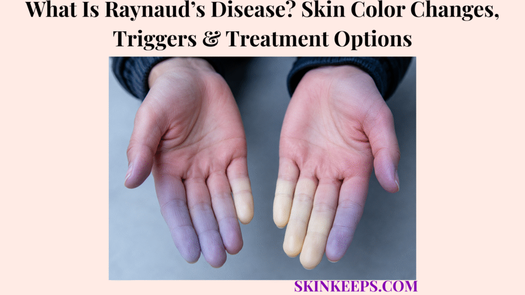
Raynaud phenomenon is an episodic blood-vessel spasm that temporarily reduces circulation to areas such as the fingers and toes, causing changes in color, temperature and sensation. Cold and emotional stress are common triggers because susceptible small arteries constrict more strongly than needed, then reopen as the attack resolves.
Primary Raynaud’s occurs without an identifiable underlying disease, while secondary Raynaud phenomenon is linked to another condition or exposure and can carry greater tissue risk. Diagnosis is mainly clinical, and treatment ranges from warming and trigger reduction to vasodilating medication or specialist care when attacks are severe, persistent or complicated by ulcers.
This article is for educational purposes only. Persistent severe pain, ulcers or circulation that does not return normally after warming should be medically evaluated.
What Is Raynaud’s Disease and What Does an Attack Look and Feel Like?
Raynaud phenomenon causes temporary spasms of small peripheral blood vessels, producing episodic color changes, coldness, numbness or pain—most often in the fingers and toes.
How Does Blood-Vessel Spasm Cause a Raynaud Attack?
A Raynaud attack occurs when cold, stress or another trigger causes excessive narrowing of small blood vessels, temporarily reducing blood flow until the vessels reopen.
The core sequence is trigger → vasospasm → reduced peripheral blood flow → color and sensation change → rewarming → reperfusion. This is more precise than calling Raynaud’s generic “poor circulation,” because the circulation change is episodic and driven by abnormal vasoconstriction during attacks.
Why Can Skin Turn White, Blue and Then Red?
Pale or white skin reflects reduced blood flow, blue or dusky change reflects reduced oxygenation, and red coloration can appear as circulation returns during rewarming.
Not every attack shows all three phases, and the order or intensity can vary. Some attacks are biphasic, while others may be dominated by one obvious change, so a strict white-blue-red sequence is not required.
What Sensations Occur During a Raynaud Attack?
Raynaud attacks can cause coldness, numbness, tingling, pins-and-needles, burning or pain, with throbbing sometimes occurring as blood flow returns.
Sensation can change across the attack, with numbness during reduced flow and tingling or throbbing during reperfusion. Symptom intensity ranges from mild inconvenience to substantial pain, especially in secondary disease.
Where Does Raynaud Occur, and How Long Can Attacks Last?
Raynaud most often affects fingers and toes, although other peripheral sites can be involved, and individual attacks can last from minutes to hours.
The nose, ears, tongue and other acral areas can occasionally be affected. Across different skin tones, the best clues may be contrast from baseline color, pallor or duskiness, temperature change and sensory symptoms rather than expecting textbook “blue fingers.”
Figure 1. A Raynaud attack is a reversible vasospastic sequence: a trigger narrows peripheral vessels, color and sensation change while flow is reduced, and rewarming allows reperfusion. Not every attack shows every color phase.
What Triggers Raynaud’s, and What Causes Secondary Raynaud Phenomenon?
Cold and emotional stress are common attack triggers, while secondary Raynaud phenomenon occurs when another disease or exposure contributes to the abnormal vascular response.
How Does Cold Trigger Raynaud’s?
Cold is the most common Raynaud trigger because susceptible vessels constrict excessively when the body responds to lower temperature.
Attacks can follow outdoor cold, contact with refrigerated or frozen objects, cold water, abrupt temperature changes or strong air conditioning. Cold provokes the vasospasm; it should not be described as the root cause of the disorder itself.
Can Stress, Smoking, Vaping or Medicines Trigger Attacks?
Emotional stress, nicotine and selected medicines or exposures can increase vasoconstriction and provoke or worsen Raynaud attacks in susceptible people.
Smoking, vaping or other nicotine exposure can worsen vessel narrowing. Some stimulant, decongestant, migraine, blood-pressure or other medicines may also matter, while vibration and occupational cold can be relevant exposures. Prescribed medication should not be stopped without clinician guidance.
What Is the Difference Between Primary and Secondary Raynaud’s?
Primary Raynaud’s occurs without an identifiable underlying disease, while secondary Raynaud phenomenon is associated with another medical condition or exposure and is more likely to cause severe ischemic complications.
Primary Raynaud’s is often milder and tissue injury is uncommon. Secondary Raynaud’s deserves a broader evaluation because ulcers, persistent ischemia or more severe attacks are more concerning when another vascular, rheumatic or systemic process is present.
Which Conditions Are Commonly Associated With Secondary Raynaud’s?
Secondary Raynaud phenomenon is particularly associated with connective-tissue diseases such as systemic sclerosis and lupus, although several other rheumatic, vascular and systemic disorders can also be involved.
Scleroderma is an important association because vascular abnormalities can be clinically significant. Lupus erythematosus is another connective-tissue context in which Raynaud phenomenon may occur, while Sjögren disease, inflammatory arthritis and other conditions can also be considered when the history suggests secondary disease.
Figure 2. Cold, stress, nicotine and other exposures are attack triggers. Primary Raynaud’s has no identified underlying disease, whereas secondary Raynaud phenomenon is linked to another condition or exposure and carries a higher tissue-risk profile.
How Is Raynaud’s Disease Diagnosed and Secondary Raynaud’s Identified?
Raynaud phenomenon is diagnosed mainly from the attack history and characteristic cold- or stress-related color changes, while nailfold examination and targeted blood tests help identify people who may have secondary disease.
How Does a Doctor Diagnose Raynaud’s From the Attack Pattern?
Diagnosis is primarily clinical and combines the color-change pattern, cold or stress relationship, sensory symptoms and physical examination.
Clinicians consider attack duration, symmetry, triggers, recovery with warming and whether ulcers or tissue injury are present. There is no single test that alone proves Raynaud phenomenon, so the full pattern matters more than one isolated color change.
What Is Nailfold Capillaroscopy?
Nailfold capillaroscopy examines the tiny vessels at the base of the fingernails for abnormalities that may suggest secondary Raynaud phenomenon or connective-tissue disease.
This examination is particularly useful when the history raises concern for an autoimmune or connective-tissue process. It supports the primary-versus-secondary distinction but should be interpreted alongside the rest of the clinical picture.
When Are Blood Tests Needed?
Blood tests are more relevant when secondary Raynaud’s is suspected and may include autoimmune testing such as ANA plus selected disease-specific tests based on the clinical picture.
Testing should be history-driven and clinician-selected rather than a universal panel for every person with cold fingers. The purpose is to investigate a suspected underlying condition, not to confirm every Raynaud attack with laboratory testing.
Which Features Make Secondary Raynaud’s More Concerning?
More severe pain, marked asymmetry, fingertip or toe ulcers, persistent tissue injury or signs of systemic disease make secondary Raynaud phenomenon more concerning.
Attacks that fail to recover normally, wounds that heal poorly, skin breakdown or systemic connective-tissue features deserve closer assessment because they change the task from simple attack management to tissue protection and underlying-disease evaluation.
Figure 3. Primary Raynaud’s is usually lower-risk and has no identified underlying disease, while secondary Raynaud phenomenon changes both investigation and urgency because ulcers and tissue injury are more plausible.
How Is Raynaud’s Disease Treated?
Raynaud treatment aims to reduce attack frequency and severity, improve peripheral blood flow and prevent ulcers or tissue injury, with medication reserved for attacks that remain troublesome or high risk despite trigger control.
What Is the Main Goal of Raynaud Treatment?
The treatment goal is to reduce vasospastic attacks, maintain adequate blood flow and prevent ischemic skin damage.
Management usually starts with trigger reduction and whole-body warming, then adds medication if attacks remain troublesome. Secondary disease also requires attention to the associated condition, while severe ischemia may need specialist vascular management.
When Are Calcium-Channel Blockers Used?
Vasodilating calcium-channel blockers such as nifedipine may be used when attacks are frequent, painful or inadequately controlled by warming and trigger avoidance.
Medication is clinician-directed and is not required for everyone with Raynaud phenomenon. The decision depends on attack burden, severity, blood-pressure tolerance, tissue risk and whether disease is primary or secondary.
What Other Medicines May Be Used for Difficult Raynaud’s?
Other vasodilating treatments may be considered when first-line therapy is ineffective, poorly tolerated or insufficient for severe secondary Raynaud’s.
The exact choice depends on the clinical situation, so second-line treatment is better presented by category than as one universal drug list. The treatment objective remains improved perfusion and tissue protection rather than simply changing skin color.
How Is Secondary or Severe Raynaud Phenomenon Treated?
Secondary Raynaud’s requires both attack control and treatment of the underlying condition, while severe ischemic disease may need specialist vascular therapy or, rarely, a procedure such as sympathectomy.
Sympathectomy is a selected specialist option rather than routine surgery, and its benefit may not be permanent. Severe disease with ulcers, persistent ischemia or tissue breakdown should be managed according to the underlying cause and vascular risk.
Figure 4. Raynaud treatment follows a severity-based path: start with warmth and trigger control, add medication when attacks remain troublesome, treat secondary disease when present, and escalate persistent ischemia to specialist care.
How Can Raynaud Attacks Be Prevented, and When Is Urgent Evaluation Needed?
Raynaud attacks can often be reduced by keeping the whole body warm, avoiding personal vasoconstrictive triggers and gently rewarming during attacks, while ulcers or persistent ischemia require prompt medical assessment.
How Can Keeping the Whole Body Warm Reduce Attacks?
Keeping the core, hands and feet warm can reduce the body’s cold-triggered vasoconstrictive response and help prevent Raynaud attacks.
Layered clothing, gloves, warm socks and head or core warmth can all matter. Gloves can also be useful when handling refrigerated or frozen items, and people who are sensitive to air conditioning may benefit from planning for sudden indoor temperature changes.
What Should Someone Do During a Raynaud Attack?
During an attack, move to a warmer environment and gently rewarm the affected area so circulation can return gradually.
Warm—not excessively hot—water can be used, and gentle movement may help restore comfort. Very hot water or direct high heat should be avoided because numb skin is easier to injure.
Why Should Smoking, Vaping and Personal Triggers Be Reduced?
Nicotine constricts blood vessels and can worsen Raynaud attacks, while identifying repeat stress, cold or exposure patterns can help reduce avoidable episodes.
A practical approach is attack → identify the recent exposure → ask whether the same pattern repeats → reduce avoidable triggers where possible. This keeps prevention individualized rather than assuming every person has the same trigger profile.
When Does Raynaud’s Need Prompt or Urgent Medical Evaluation?
Prompt assessment is needed when Raynaud attacks become markedly more severe or asymmetric, ulcers develop, wounds fail to heal, severe pain persists or circulation does not return normally after warming.
Persistent ischemia, infected ulcers, tissue breakdown or systemic illness need urgent assessment. Rare severe secondary disease can progress to tissue death such as gangrene, which is a different medical state from a typical reversible Raynaud attack. A typical attack should recover; persistent tissue-threatening ischemia requires urgent care.
What Should You Remember About Raynaud’s Disease?
Raynaud phenomenon is an episodic vasospastic disorder that temporarily reduces peripheral blood flow, while the key clinical task is distinguishing uncomplicated primary Raynaud’s from secondary disease that can threaten tissue.
- Raynaud is driven by episodic vasospasm.
- It is not simply generic poor circulation.
- Fingers and toes are the most common sites.
- Other acral sites can be affected.
- White or pale change can reflect reduced blood flow.
- Blue or dusky change can reflect reduced oxygen delivery.
- Red change can appear during reperfusion.
- Not every attack has all three colors.
- Numbness, tingling, burning or pain can occur.
- Attacks can last minutes to hours.
- Cold is a common trigger.
- Stress can trigger attacks.
- Nicotine can worsen vasoconstriction.
- Selected medicines and exposures can contribute.
- Primary Raynaud’s has no identified underlying disease.
- Secondary Raynaud’s has an associated condition or exposure.
- Secondary disease is more likely to cause tissue damage.
- Scleroderma is an important association.
- Lupus is an important association.
- Diagnosis is primarily clinical.
- Nailfold capillaroscopy can help investigate secondary disease.
- Blood tests are targeted rather than universal.
- Severe asymmetry or ulcers are concerning.
- Treatment starts with trigger reduction and warming.
- Calcium-channel blockers may be used when needed.
- Other vasodilators are selected for difficult disease.
- Secondary disease also needs underlying-cause treatment.
- Sympathectomy is reserved for severe selected cases.
- Whole-body warmth helps prevention.
- Gentle rewarming helps during attacks.
- Persistent ischemia or ulcers require medical evaluation.
Trigger → vasospasm → temporary ischemia → recognize the attack → determine primary versus secondary Raynaud’s → prevent and treat attacks → escalate tissue injury.
Frequently Asked Questions About Raynaud’s Disease
The main Raynaud questions concern danger, autoimmune disease, color changes, long-term persistence and the difference between primary and secondary forms.
Is Raynaud’s Disease Dangerous?
Primary Raynaud’s is usually mild and does not commonly cause tissue damage, while secondary Raynaud phenomenon can be more serious and may cause ulcers or ischemic injury.
Is Raynaud’s Disease an Autoimmune Disease?
Primary Raynaud’s is not itself defined as an autoimmune disease, but secondary Raynaud phenomenon can occur with autoimmune and connective-tissue disorders.
Why Do Fingers Turn White or Blue During a Raynaud Attack?
Fingers may turn pale when blood flow drops and blue or dusky when oxygen delivery remains reduced during vasospasm.
Can Raynaud’s Disease Go Away Completely?
Raynaud symptoms can improve or become easier to control, but the long-term course varies and persistent secondary disease depends partly on its underlying cause.
What Is the Difference Between Primary and Secondary Raynaud’s?
Primary Raynaud’s occurs without an identified underlying disease, while secondary Raynaud phenomenon is associated with another condition or exposure and has a greater risk of tissue complications.
Which Sources Support This Raynaud Guidance?
NIAMS — Raynaud’s Phenomenon — Used for terminology, vasospasm mechanism, primary versus secondary disease, common triggers, color and sensory changes, diagnosis, nailfold examination, treatment and tissue-risk framing.
MSD Manual Professional Edition — Raynaud Syndrome — Used for primary versus secondary management, vasodilator treatment, skin-color variation and severe ischemic complications.



