
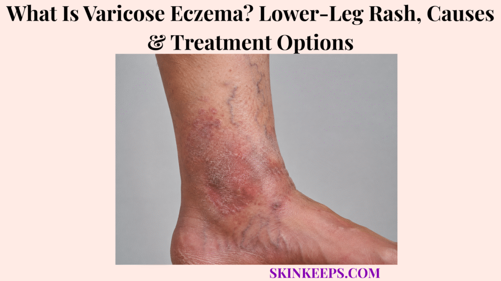
Varicose eczema is a chronic inflammatory skin condition of the lower legs caused by impaired venous return and sustained venous hypertension. Also called venous eczema, gravitational eczema or stasis dermatitis, it commonly produces ankle swelling, itching, scaling and persistent brown, red, purple or darker skin changes.
The visible rash is only one part of the problem. Effective care requires controlling eczema and repairing the skin barrier while also reducing edema and venous pressure, distinguishing chronic dermatitis from cellulitis or contact allergy, and watching for lipodermatosclerosis, open wounds or sudden vascular changes that need further assessment.
Medical note: This article is for educational purposes only. Rapidly worsening warmth, pain or redness, fever, a new open wound, or sudden one-sided painful leg swelling should be medically assessed promptly.
What Is Varicose Eczema and What Does It Look Like on the Lower Legs?
Varicose eczema is a lower-leg dermatitis driven by chronic venous hypertension and commonly causes itching, scaling, edema and red, brown, purple or otherwise darker discoloration around the ankles and lower legs.
How Does Poor Venous Return Cause Varicose Eczema?
When venous valves and the calf-muscle pump do not return blood efficiently toward the heart, pressure rises in the lower-leg venous system. Fluid can accumulate in surrounding tissues, and the resulting edema and inflammatory response damage the skin barrier and promote eczema.
Why Is Varicose Eczema Also Called Stasis Dermatitis?
Varicose eczema, venous eczema, gravitational eczema and stasis dermatitis are overlapping names for the same venous-pressure–related dermatitis. The different terms emphasize venous disease, blood and fluid pooling, or the effect of gravity rather than describing separate disorders.
Do Visible Varicose Veins Have to Be Present?
No. Prominent varicose veins can coexist with stasis dermatitis, but they are not required. Chronic venous insufficiency and venous hypertension can be present even when no large bulging surface vein is obvious.
Where Does Varicose Eczema Usually Begin?
Varicose eczema commonly starts around the lower leg and inner ankle, where dependent venous pressure and edema are often greatest. Early changes may be subtle and include ankle swelling near the end of the day, itching, dryness and discolored skin.
What Does Early Varicose Eczema Look Like?
Early disease often causes dry or scaly skin, itch, soreness, ankle swelling and patches that may look red, brown, purple, gray or darker than the surrounding skin. General barrier dryness is discussed separately in Dry Skin / Xerosis, but dryness here is a consequence of venous dermatitis rather than the root cause.
Can Varicose Eczema Become Wet, Blistered or Crusted?
Yes. During an active flare, the skin may develop small blisters, oozing, crusting and fissures. Chronic disease can instead look increasingly dry, scaly and thickened. Oozing alone does not prove bacterial infection; the surrounding clinical pattern matters.
Why Does the Skin Become Brown or Darker?
Long-standing venous disease can produce persistent orange-brown, brown, purple or darker discoloration. One contributor is hemosiderin, an iron-containing pigment left after red blood cells and blood products leak into chronically congested tissues.
What Is Lipodermatosclerosis?
Lipodermatosclerosis is an advanced chronic venous change in which inflammation and fibrosis make the lower-leg skin and subcutaneous tissues firm, tight and indurated. The lower calf may narrow near the ankle, and these changes signal substantial long-term venous disease.
What Causes Varicose Eczema and Which Factors Increase the Risk?
Varicose eczema develops when chronic venous insufficiency keeps lower-leg venous pressure elevated, while prior venous damage, persistent edema, prolonged dependency and reduced calf-pump efficiency can increase the likelihood or severity of the dermatitis.
How Does Chronic Venous Insufficiency Cause Varicose Eczema?
Chronic venous insufficiency allows blood to pool and venous pressure to remain high in the lower legs. Fluid then moves into surrounding tissues, producing edema and inflammatory changes that repeatedly disturb the epidermal barrier.
How Are Varicose Veins Related?
Varicose veins can be a visible sign of superficial venous-valve dysfunction and may contribute to venous hypertension. The eczema, however, should be understood as a consequence of impaired venous return rather than as irritation caused by the visible vein itself.
Can a Previous DVT Increase the Risk?
Yes. A previous deep-vein thrombosis can damage valves in the deep venous system and lead to post-thrombotic venous hypertension. That long-term circulatory change can contribute to edema, pigmentation and stasis dermatitis in the affected leg.
Which Other Factors Make Varicose Eczema More Likely?
Risk rises with increasing age, chronic leg swelling, reduced mobility, excess body weight, long periods of sitting or standing, previous lower-leg injury or surgery, and other conditions that interfere with effective venous return. These are risk contexts rather than proof of the diagnosis.
Why Can Standing for Long Periods Make Symptoms Worse?
When the legs remain dependent, gravity raises hydrostatic pressure in the lower-leg veins. If the venous valves or calf-muscle pump are already inefficient, prolonged standing or sitting can increase edema and make heaviness, aching and dermatitis more noticeable.
How Is Varicose Eczema Diagnosed and Distinguished From Other Lower-Leg Rashes?
Varicose eczema is usually diagnosed clinically from its chronic lower-leg distribution, edema, scale, pigmentation and venous history, while acute infection, contact allergy and full-thickness ulceration must be separated because their management differs.
How Is Varicose Eczema Usually Diagnosed?
Assessment focuses on the location and chronicity of the rash, itch, scale, swelling, pigmentation, visible venous changes, previous DVT or leg injury, and the products or medicines used on the skin. Many typical cases can be recognized without invasive testing.
When Is Doppler or Vascular Ultrasound Used?
Vascular ultrasound may be used when clinicians need more information about venous reflux, blood flow, varicose-vein anatomy or a possible acute vascular problem. It is selected according to the clinical question rather than required for every mild case.
How Is Varicose Eczema Different From Cellulitis?
Varicose eczema is usually chronic or recurrent and often itchy, scaly, pigmented and edematous. Cellulitis is an acute bacterial infection that more often becomes hot, tender or painful and progressively inflamed, sometimes with fever or systemic illness. Because overlap is possible, rapidly worsening or uncertain cases need examination.
How Is Varicose Eczema Different From Contact Dermatitis?
Contact dermatitis is driven by an irritant or allergic reaction to something touching the skin. People with chronic venous eczema can become sensitized to repeatedly applied creams, dressings, adhesives or topical medicines; a rash that worsens after a new product should therefore raise the possibility of Allergic Contact Dermatitis.
How Is Varicose Eczema Different From Other Eczema?
Varicose eczema is defined by its lower-leg venous mechanism, whereas the broader category of Dermatitis (Eczema) includes several inflammatory conditions with different causes, distributions and treatment priorities.
How Is Varicose Eczema Different From a Venous Leg Ulcer?
Varicose eczema can involve inflamed, cracked or weeping skin while the surface is still largely intact. A venous leg ulcer is a true open wound with full-thickness skin loss. The broader principles of wound depth and ulcer classification belong to the Skin Ulcers page.
How Is Varicose Eczema Treated?
Varicose eczema is treated with a dual approach: control inflammation and restore the skin barrier while also reducing edema and venous hypertension that keep triggering recurrent lower-leg disease.
Why Must Treatment Address Both the Skin and the Veins?
Topical treatment can calm the visible eczema, but it does not repair malfunctioning venous valves or remove sustained venous pressure. Long-term control is therefore more reliable when skin therapy is combined with clinically appropriate venous and edema management.
How Do Emollients Help?
Emollients reduce water loss, soften scale and support a damaged epidermal barrier. Regular barrier care can reduce dryness and fissuring, but moisturizers do not correct chronic venous insufficiency and should not be presented as a stand-alone solution.
How Are Topical Corticosteroids Used During Flares?
Clinician-selected topical corticosteroids can reduce active inflammation, itching and scaling during a flare. Potency and duration depend on severity, skin condition and clinical context, so one universal steroid regimen is not appropriate.
Why Is Compression Therapy Important?
When clinically appropriate, graduated compression reduces venous pooling and lower-leg edema and helps improve venous return. This addresses an important mechanical driver of stasis dermatitis rather than simply covering the rash.
Why Must Circulation Be Checked Before Strong Compression?
Significant peripheral arterial disease can make stronger compression unsafe because further external pressure may compromise already reduced arterial blood flow. Circulation and arterial risk therefore need appropriate assessment before stronger compression is selected.
How Do Leg Elevation and Walking Help?
Leg elevation reduces dependent fluid accumulation, while walking activates the calf-muscle pump that helps move venous blood back toward the heart. These measures support circulation but are not substitutes for evaluation when symptoms are severe or suddenly one-sided.
When Is Treatment of the Underlying Veins Considered?
Persistent or clinically important venous disease may justify vascular assessment to identify reflux or other treatable venous pathology. Vein-directed treatment is individualized and sits alongside ongoing skin care and edema control rather than replacing them automatically.
How Can Varicose Eczema Be Prevented From Worsening, and When Does It Need Medical Care?
Preventing progression requires long-term edema control, skin-barrier protection and venous management, while secondary infection, new ulceration or sudden vascular change should trigger prompt clinical reassessment.
Why Is Long-Term Swelling Control Important?
Persistent edema keeps lower-leg tissues exposed to venous hypertension and inflammatory stress. Even after the visible rash improves, continuing edema can promote recurrent flares, fibrosis and further skin damage.
How Can Skin Care Reduce Cracking and Ulcer Risk?
Regular emollient use, gentle cleansing, avoidance of scratching and protection from trauma help preserve fragile venous-diseased skin. These measures reduce opportunities for fissuring and secondary breakdown but do not remove the underlying venous pressure.
Why Should New Skin-Care Products Be Introduced Carefully?
Chronically inflamed lower-leg skin is frequently exposed to creams, adhesives and dressings, creating opportunities for irritation or contact allergy. If a product repeatedly makes the rash worse, the reaction deserves reassessment rather than automatic escalation of eczema treatment.
Can Varicose Eczema Return After the Rash Clears?
Yes. The skin may look better after a flare while chronic venous insufficiency remains. Recurrence is therefore common unless edema, venous pressure, skin-barrier damage and prolonged dependency are managed over the longer term.
Why Should Hardened Skin or New Sores Be Taken Seriously?
Progressive hardening can indicate lipodermatosclerosis, a marker of substantial chronic venous disease. A new sore may signal transition from dermatitis to true ulceration and should be assessed rather than treated indefinitely as ordinary eczema.
Which Signs Suggest Secondary Infection?
Increasing warmth, rapidly worsening redness or discoloration, new pain, spreading swelling, purulent drainage, yellow crusting, fever or systemic illness raise concern for secondary bacterial infection. Weeping alone is not enough to label the eczema infected.
When Should Cellulitis Be Suspected Rather Than an Eczema Flare?
Rapid progression, marked warmth and tenderness, increasing pain and fever are more concerning for cellulitis than a typical chronic itchy stasis-eczema flare. Bilateral disease can favor a noninfectious cause, but laterality should not be used as an absolute diagnostic rule.
When Does an Open Sore Need Medical Assessment?
A new or non-healing wound in chronically swollen, pigmented lower-leg skin should be assessed, especially if it enlarges, drains, becomes painful, repeatedly reopens or fails to show healing progress. Venous ulceration needs a wound and circulation pathway rather than eczema treatment alone.
When Does Sudden One-Sided Leg Swelling Need Different Evaluation?
A new sudden painful swollen leg on one side should not be assumed to be routine varicose eczema. Acute vascular conditions, including deep-vein thrombosis, require a different and often urgent clinical assessment.
What Should You Remember About Varicose Eczema?
Varicose eczema is a chronic lower-leg dermatitis caused by venous hypertension, so successful management requires treating both the inflamed skin and the circulation problem that keeps edema and inflammation recurring.
- Varicose eczema, venous eczema, gravitational eczema and stasis dermatitis describe the same venous-pressure–related condition.
- Visible varicose veins are not required; impaired venous return and venous hypertension are the key mechanisms.
- Inner-ankle swelling, itch, scale and brown, purple or darker pigmentation are common clues.
- Oozing and crusting can occur during eczema flares and do not automatically mean infection.
- Lipodermatosclerosis and new open sores suggest more advanced venous tissue damage.
- Cellulitis is usually more acute, hot and painful and may cause fever or systemic illness.
- Contact allergy can complicate repeated use of creams, dressings or adhesives.
- Emollients and topical corticosteroids address the skin; edema and venous-pressure control address the driver.
- Compression can be useful when appropriate, but arterial circulation must be considered before stronger compression.
- Elevation, walking and long-term swelling control support venous return and recurrence prevention.
- A new ulcer, infection signs or sudden unilateral painful swelling needs prompt medical assessment.
Frequently Asked Questions About Varicose Eczema
The main varicose-eczema questions concern terminology, visible veins, cellulitis, compression and progression to venous ulcers.
Is Varicose Eczema the Same as Stasis Dermatitis?
Yes. Varicose eczema, venous eczema, gravitational eczema and stasis dermatitis are commonly used names for the same lower-leg dermatitis driven by chronic venous hypertension and impaired venous return.
Can Varicose Eczema Occur Without Visible Varicose Veins?
Yes. Chronic venous insufficiency can cause varicose eczema even when large bulging surface veins are not obvious, because the key problem is venous hypertension rather than the visibility of a vein.
How Can You Tell Varicose Eczema From Cellulitis?
Varicose eczema is usually chronic, itchy and scaly with longstanding swelling and pigmentation, while cellulitis is more often acutely painful, hot and progressively inflamed and may cause fever. Overlap can occur, so uncertain cases need examination.
Does Compression Help Varicose Eczema?
Appropriately selected compression can reduce venous pooling and lower-leg edema, but circulation should be assessed first when arterial insufficiency is possible because stronger compression is not suitable for everyone.
Can Varicose Eczema Turn Into a Venous Leg Ulcer?
Yes. Persistent venous hypertension and progressive lower-leg skin damage can eventually lead to full-thickness skin breakdown and venous ulceration, which requires wound and vascular assessment.
DermNet — Venous eczema: mechanism, terminology, morphology, hemosiderin pigmentation, prior DVT/varicose-vein associations, chronicity and complications.
American Academy of Dermatology — Stasis dermatitis overview: venous insufficiency mechanism, early lower-leg pattern, progression and long-term disease context.
American Academy of Dermatology — Stasis dermatitis signs and symptoms: inner-ankle onset, swelling, pigmentation, lipodermatosclerosis, wounds, contact dermatitis and cellulitis complications.
American Academy of Dermatology — Stasis dermatitis diagnosis and treatment: clinical diagnosis, Doppler ultrasound, patch testing, compression, elevation and specialist coordination.
NHS — Varicose eczema treatment: emollients, topical corticosteroids, compression, leg elevation, physical activity and vascular referral.
NICE — ABPI and compression safety in lower-limb venous disease: peripheral arterial disease assessment and why stronger compression can be inappropriate when arterial supply is impaired.



