
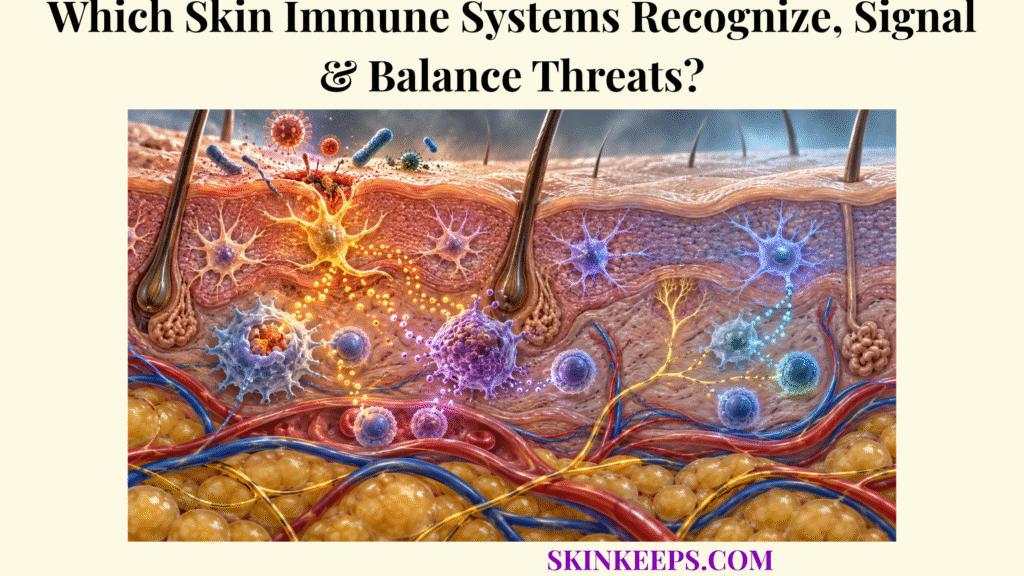
Skin immunity protects the body through an integrated surveillance system in which the physical barrier limits exposure, keratinocytes and resident immune cells detect danger, signaling molecules coordinate local defense, recruited leukocytes control threats, adaptive lymphocytes add specificity and memory, and regulatory mechanisms restore immune balance. Skin is therefore both a physical barrier and an active immune organ.
This article explains how skin detects microbial or tissue danger, converts recognition into signaling and inflammation, builds targeted adaptive responses, links acute defense with tissue repair, and then limits unnecessary immune activity after a threat is controlled.
How Does the Skin’s Immune Defense Monitor for Threats?
The skin’s immune defense monitors threats continuously through a combination of barrier integrity, danger-sensing keratinocytes, resident immune cells, antimicrobial molecules, and local host–microbe interactions that detect change before widespread tissue invasion occurs. This layered defense is one of the body’s core skin functions, and it works before, during, and after visible inflammation.
How Do Keratinocytes Participate in Immune Surveillance?
Keratinocytes participate in immune surveillance by using pattern-recognition and stress-sensing pathways to detect microbial products or tissue damage and then changing their production of cytokines, chemokines, antimicrobial peptides, and alarmins. This keratinocyte immune surveillance adds active immune coordination to their structural barrier role.
Pattern-recognition receptors, or PRRs, identify broad classes of microbial or damage-associated molecular patterns. Toll-like receptors and NOD-like receptor systems are among the pathways keratinocytes can use to translate barrier disturbance or cellular stress into local signaling.
How Do Resident Immune Cells Monitor the Epidermis and Dermis?
Resident cutaneous immune cells monitor different skin compartments, with Langerhans cells positioned in the epidermis and dendritic cells, macrophages, mast cells, lymphocytes, and other immune populations distributed through the dermis and appendage-associated niches. Their location allows different cell types to sample epidermal, perivascular, stromal, and appendage-associated environments.
How Does the Physical Barrier Reduce the Threats Skin Immunity Must Handle?
The physical barrier reduces immune burden by restricting microbial and chemical penetration, limiting the number of foreign signals that reach viable epidermal and dermal cells and therefore lowering the need for full inflammatory escalation. Barrier defense and immune defense are sequential and interactive rather than competing explanations of skin protection.
| Defense layer | Main components | Surveillance / defense role |
|---|---|---|
| Physical barrier | Stratum corneum, lipids, junctions | Restricts entry and reduces antigen exposure. |
| Epidermal immune interface | Keratinocytes, Langerhans cells | Detects microbial and damage signals and communicates danger. |
| Dermal immune network | Dendritic cells, macrophages, mast cells, lymphocytes | Samples tissue, coordinates local responses, and connects with systemic immunity. |
| Molecular innate defense | Antimicrobial peptides, cytokines, chemokines | Provides direct antimicrobial effects plus immune communication. |
Which Cutaneous Immune Cells Recognize Microbes and Tissue Damage?
Cutaneous immune cells recognize microbes and tissue damage through complementary systems: keratinocytes detect broad danger patterns, Langerhans and dendritic cells sample and process antigen, macrophages phagocytose material and coordinate local responses, and mast cells rapidly release inflammatory mediators when activated. These overlapping roles are distributed across many skin immune cells rather than assigned to one dominant cell population.
How Do Keratinocytes Recognize Microbial and Damage-Associated Signals?
Keratinocytes recognize microbial and tissue-damage signals through pattern-recognition systems that detect conserved microbial molecules and danger-associated cellular changes rather than identifying pathogens by species at first contact. A PAMP or MAMP is a conserved microorganism-associated molecular pattern, a DAMP is a signal associated with cellular stress or injury, and a PRR is a cellular receptor that recognizes classes of these signals.
This early skin microorganism recognition can alter local gene expression and mediator release without automatically producing clinically visible inflammation.
How Do Langerhans Cells and Dermal Dendritic Cells Detect and Process Antigens?
Langerhans cells and dermal dendritic cells capture environmental or tissue antigens, process them into peptide fragments, and can present those fragments to T lymphocytes locally or after migration toward skin-draining lymphoid tissue. Processed peptides are displayed on major histocompatibility complex, or MHC, molecules together with contextual signals that shape the T-cell response.
Langerhans cells are not the only cutaneous antigen-presenting cells. Dermal dendritic-cell subsets also perform major antigen-presentation and T-cell-priming functions, and the final outcome can be inflammatory, regulatory, or tolerogenic depending on antigen and tissue context.
How Do Macrophages and Mast Cells Respond to Tissue Injury and Invading Organisms?
Macrophages and mast cells support rapid cutaneous defense through different mechanisms: macrophages phagocytose microbes and damaged material while producing regulatory mediators, whereas mast cells release preformed and newly synthesized mediators that rapidly alter local vascular and immune activity.
Macrophages participate in debris clearance, cytokine signaling, efferocytosis, repair, and resolution across changing activation states rather than a rigid two-state M1/M2 model. Mast cells can release histamine, proteases, lipid mediators, and cytokines and therefore function in defense and vascular regulation as well as allergy.
| Cell / group | Typical location | Recognition / surveillance role | Main response |
|---|---|---|---|
| Keratinocytes | Epidermis | Sense microbial and damage-associated signals. | Cytokines, chemokines, AMPs, alarmins. |
| Langerhans cells | Epidermis | Sample and process antigen. | Antigen presentation, migration, activation or tolerance. |
| Dermal dendritic cells | Dermis | Sample tissue antigens. | T-cell priming and adaptive-response coordination. |
| Macrophages | Dermal / perivascular niches | Detect and phagocytose microbes and damaged cells. | Clearance, signaling, repair and resolution. |
| Mast cells | Dermis, often near vessels and nerves | Detect immune and tissue signals. | Rapid mediator release and vascular modulation. |
| Tissue-resident T cells | Epidermis and dermis | Local antigen-specific surveillance. | Rapid memory or effector response after re-exposure. |
| Recruited neutrophils | Enter from blood during inflammation | Respond rapidly to strong inflammatory or infectious signals. | Microbial killing and acute inflammatory defense. |
How Do Skin Immune Signals Coordinate a Defense Response?
Skin immune signals coordinate defense when threat-recognition events trigger cytokines, chemokines, antimicrobial peptides, and other mediators that communicate danger, alter local tissue behavior, and recruit additional immune cells to the affected site. This converts recognition into a coordinated skin immune response rather than leaving each cell to react in isolation.
How Do Cytokines and Chemokines Transmit Danger Signals Between Skin Cells?
Cytokines coordinate immune-cell activation and tissue behavior, while chemokines provide directional signals that help immune cells migrate toward sites where inflammatory or microbial signals are strongest. Chemokines are a specialized subset of cytokines, so the two terms are related but not interchangeable.
Signals such as IL-1, TNF, IL-6, CXCL8, and CCL-family chemokines can participate in skin cytokines and inflammation, but not every cytokine promotes inflammation. Some cytokines restrain, redirect, or help resolve immune responses.
How Do These Signals Recruit Additional Immune Cells to Affected Tissue?
Inflammatory signaling recruits circulating leukocytes by activating nearby blood vessels, increasing endothelial adhesion signals, and creating chemokine gradients that guide neutrophils and monocytes from the circulation into affected skin. This multi-step skin immune-cell recruitment involves leukocyte adhesion, extravasation across the vascular wall, and chemotaxis through tissue.
How Do Antimicrobial Peptides Contribute to Rapid Local Defense?
Antimicrobial peptides contribute to rapid innate defense by disrupting or inhibiting selected microorganisms while also influencing inflammatory signaling and communication between epithelial and immune cells. β-defensins, cathelicidin, and S100-family proteins are examples of host molecules that can contribute to this local defense network.
Antimicrobial peptides do not sterilize healthy skin. Normal skin supports a resident microbiome, and antimicrobial activity is integrated with barrier chemistry, microbial competition, and immune regulation.
How Does Cutaneous Inflammation Contain Infection and Tissue Damage?
Cutaneous inflammation contains infection and tissue damage by increasing local blood flow and vascular permeability, recruiting defensive leukocytes, promoting microbial and debris clearance, and generating signals that transition injured tissue toward repair when the threat is controlled. Acute regulated inflammation can therefore be protective without implying that persistent inflammation is harmless.
How Does Increased Blood Flow Support an Inflammatory Response?
Inflammatory vasodilation increases local perfusion while endothelial activation and increased vascular permeability help immune cells and plasma proteins move from the circulation toward injured or infected tissue. Redness and warmth reflect vascular change, but those visible features do not directly kill microbes.
How Do Neutrophils and Macrophages Help Remove Microbes and Damaged Material?
Neutrophils provide rapid antimicrobial activity during acute inflammation, while recruited monocytes and resident or recruited macrophages phagocytose microbes and cellular debris and coordinate later phases of inflammatory control and repair. Their functions overlap in time, but macrophages are not simply “later neutrophils.”
Macrophage efferocytosis—the engulfment of dying cells—helps clear spent inflammatory cells and can support a shift toward resolution. Macrophages also release regulatory and repair-associated signals across multiple phases of tissue recovery.
How Does Inflammation Connect Immune Defense With Wound Healing?
Inflammation connects immune defense with wound healing because the same recruited and resident immune cells that clear microbes and damaged material also release signals that influence keratinocyte migration, fibroblast activity, angiogenesis, and the transition toward tissue reconstruction. Controlled acute inflammation supports repair; prolonged or poorly resolved inflammation can delay healing and damage tissue.
How Does the Skin’s Adaptive Immune System Build Targeted Defense?
The skin’s adaptive immune system builds targeted defense when antigen-presenting cells process specific antigens and communicate them to T lymphocytes, generating antigen-specific effector responses and memory populations that can respond more rapidly during later encounters. Adaptive immunity adds specificity and memory to the rapid, broader recognition mechanisms of innate immunity.
How Do Antigen-Presenting Cells Communicate With T Cells?
Skin antigen-presenting cells communicate with T cells by processing antigens into peptide fragments and displaying them on MHC molecules together with contextual co-stimulatory and cytokine signals that help determine the resulting T-cell response. Skin-derived dendritic cells can migrate toward draining lymph nodes, where naïve T-cell priming commonly occurs.
Antigen presentation does not automatically trigger an inflammatory attack. The maturation state of the antigen-presenting cell, co-stimulation, cytokine environment, antigen type, and tissue context influence whether the response becomes strongly effector, regulatory, memory-forming, or tolerant.
How Do Resident Memory T Cells Support Faster Responses to Previously Encountered Threats?
Tissue-resident memory T cells, or TRM cells, remain within skin after selected immune responses and can react rapidly when matching antigen is encountered again, producing local cytokine or cytotoxic responses without first requiring a full naïve-cell priming process.
Human epidermis and dermis contain heterogeneous resident T-cell populations, including conventional and regulatory subsets. TRM cells can accelerate useful recall responses, but persistent antigen-specific memory can also participate in chronic inflammatory or autoimmune disease when directed toward inappropriate targets.
How Does Adaptive Immunity Complement Innate Skin Defense?
Adaptive immunity complements innate skin defense by adding antigen-specific lymphocyte responses and immunological memory to the rapid but broader recognition and antimicrobial mechanisms supplied by keratinocytes and innate immune cells. Innate immunity is fast and pattern-based; adaptive immunity can clonally expand selected lymphocytes and preserve antigen-specific memory.
How Does Skin Immune Regulation Balance Defense With Tolerance?
Skin immune regulation balances defense with tolerance by reducing inflammatory activity once danger is controlled, limiting collateral tissue injury, maintaining regulatory lymphocyte activity, and allowing many self and commensal antigens to coexist with the immune system without continuous destructive inflammation. This active regulation is central to skin immune tolerance.
How Does the Skin Reduce Immune Activity After a Threat Is Controlled?
After a threat is controlled, declining danger signals, clearance of dying inflammatory cells, regulatory cytokines, macrophage-mediated efferocytosis, and changes in lymphocyte activity help shift the tissue away from acute inflammation and toward repair and homeostasis. Resolution is therefore an active biological process rather than inflammation simply switching off.
How Do Regulatory Signals Prevent Excessive Tissue Damage?
Regulatory pathways prevent excessive tissue damage by restraining leukocyte activation, reducing pro-inflammatory signaling when it is no longer required, and promoting repair-oriented and tolerance-supporting immune states. FOXP3-positive regulatory T cells, or Tregs, are important brakes on cutaneous inflammation and help maintain tolerance toward self and selected commensal antigens.
How Does the Skin Tolerate Harmless Microbes and Environmental Exposures?
Healthy skin tolerates many harmless or commensal exposures by interpreting antigens in their biological context and maintaining regulatory pathways that prevent routine microbial contact from producing continuous destructive inflammation. The immune system can detect and interact with commensals without needing to eliminate them.
How Does the Microbiome Contribute to Skin Immune Balance?
The skin microbiome contributes to immune balance through continuous molecular interactions with epithelial and immune cells that can influence antimicrobial-peptide production, barrier function, T-cell development, inflammatory tone, and tolerance to resident organisms. These effects vary by microbial species and strain, body site, barrier condition, host genetics, and immune context.
Host–microbe interactions range from mutualistic to pathogenic and should not be reduced to “good” and “bad” bacteria. Microbial imbalance may accompany disease, but dysbiosis alone does not prove a single causal pathway.
| Immune state | Dominant pattern | Biological result | Main risk if persistent |
|---|---|---|---|
| Effective regulated defense | Appropriate recognition, signaling, recruitment, clearance | Threat controlled with limited tissue damage | Low if resolution succeeds |
| Insufficient defense | Weak recognition, recruitment, or microbial control | Threat persists or spreads | Greater infection susceptibility |
| Excessive inflammation | Strong or prolonged inflammatory signaling | Collateral barrier and tissue injury | Chronic inflammatory damage |
| Misguided adaptive response | Response directed toward harmless or self antigen | Persistent antigen-specific inflammation | Hypersensitivity or autoimmune-type disease |
| Tolerance / homeostasis | Regulatory control with managed microbial interaction | Self and commensals coexist without destructive inflammation | Protection may fail if regulation becomes inappropriate or defense is suppressed |
What Happens When Cutaneous Immune Balance Breaks Down?
Cutaneous immune balance breaks down when defense becomes insufficient, excessive, persistent, or misdirected, allowing microorganisms to escape control or causing immune responses themselves to damage otherwise healthy tissue. These are mechanistic patterns, not diagnoses.
What Happens When Immune Defense Is Too Weak to Control Microbes?
Insufficient immune defense can allow microorganisms to persist, multiply, penetrate deeper tissue, or spread when barrier protection, antimicrobial signaling, leukocyte function, or systemic immunity cannot adequately contain the threat. Infection susceptibility can also depend on organism virulence, wound severity, circulation, systemic disease, medications, and barrier disruption.
How Can Excessive Immune Activation Cause Persistent Inflammation?
Excessive or poorly resolved immune activation can sustain cytokine production, leukocyte recruitment, vascular change, and barrier disruption after the original threat has disappeared or become insufficient to justify the continuing response. Persistent inflammation can become self-amplifying as damaged tissue generates new inflammatory signals.
Chronic inflammatory disease should not be reduced to “too much immunity.” Different disorders involve distinct cellular pathways, environmental triggers, barrier changes, genetic factors, and adaptive or innate mechanisms.
How Can Abnormal Immune Responses Contribute to Hypersensitivity or Inflammatory Skin Disease?
Abnormal cutaneous immune responses can contribute to disease when adaptive or innate pathways react excessively to harmless environmental antigens, target self-associated structures, or maintain inappropriate inflammatory memory after earlier exposures. Allergic contact dermatitis, atopic dermatitis, psoriasis, and autoimmune skin disease illustrate different immune-dysregulation patterns without sharing one single mechanism.
Persistent or severe inflammatory findings require diagnosis of the actual disorder rather than being labeled simply as “immune imbalance.”
| Immune imbalance | Mechanistic pattern | Possible outcome |
|---|---|---|
| Insufficient antimicrobial defense | Inadequate barrier, innate, or adaptive control | Recurrent or spreading infection susceptibility |
| Excessive innate inflammation | Persistent cytokine and leukocyte activation | Tissue irritation and barrier injury |
| Hypersensitivity | Disproportionate response to an otherwise limited environmental antigen | Allergic inflammatory reaction |
| Loss of tolerance | Inappropriate recognition of self or tolerated antigens | Autoimmune or inflammatory disease pathways |
| Dysregulated immune memory | Persistent local antigen-responsive lymphocyte populations | Recurrent inflammation at previously affected sites |
These patterns describe mechanisms, not diagnoses. Similar visible skin findings can arise from very different immune pathways.
What Are the Key Takeaways About the Skin’s Immune Systems?
The key fact about the skin’s immune systems is that effective defense requires both activation and restraint: skin must recognize danger quickly enough to contain it while limiting the response once the threat is controlled.
- Barrier surveillance: The physical barrier limits exposure while viable skin cells monitor microbial and tissue-damage signals.
- Keratinocyte surveillance: Keratinocytes sense danger and produce cytokines, chemokines, antimicrobial peptides, and alarmins.
- Antigen sampling: Langerhans cells and dermal dendritic cells capture and process antigens.
- Immune signaling: Cytokines regulate cell behavior, while chemokines help direct leukocyte recruitment.
- Rapid innate defense: Antimicrobial peptides, neutrophils, macrophages, mast cells, and other innate mechanisms help contain immediate threats.
- Inflammation: Regulated acute inflammation increases local defense and helps connect threat clearance with tissue repair.
- Adaptive defense: T lymphocytes add antigen specificity and immunological memory.
- Resident memory: TRM cells can remain in skin and provide rapid local recall responses.
- Resolution: Active regulatory pathways reduce inflammation after control is achieved.
- Tolerance: Tregs and other mechanisms prevent destructive responses toward self and many harmless commensal antigens.
- Microbiome balance: Resident microbial communities interact continuously with cutaneous immunity and can influence immune homeostasis.
- Immune dysregulation: Defense that is too weak, excessive, persistent, or misdirected can contribute to infection susceptibility or inflammatory disease.
What Common Questions Do People Ask About Skin Immunity?
Common questions about skin immunity focus on whether keratinocytes are immune cells, what Langerhans cells do, whether inflammation is beneficial, how the microbiome interacts with immunity, and why the immune system normally tolerates harmless skin organisms.
Are Keratinocytes Part of the Skin Immune System?
Yes. Keratinocytes are structural epidermal cells rather than leukocytes, but they actively participate in immunity by sensing danger and releasing cytokines, chemokines, antimicrobial peptides, and other immune mediators.
What Do Langerhans Cells Do in the Skin?
Langerhans cells are epidermal immune sentinels that sample and process antigens and can communicate antigen information to T cells, although dermal dendritic-cell populations also perform major antigen-presenting functions.
Is Skin Inflammation Always Harmful?
No. Controlled acute inflammation helps contain microbes, remove damaged material, and initiate repair, whereas excessive, persistent, or poorly regulated inflammation can injure tissue and disrupt the skin barrier.
Does the Skin Microbiome Strengthen Immunity?
Skin microbial communities can support immune homeostasis, antimicrobial defense, barrier function, and immune education, but their effects depend on microbial strain, body site, barrier condition, and host immune context.
Why Does the Immune System Not Attack Normal Skin Microbes Continuously?
Healthy skin maintains regulatory interactions with many resident microbes through immune-tolerance mechanisms, regulatory T cells, epithelial–microbial signaling, and context-dependent antigen presentation that prevent routine commensal contact from producing destructive chronic inflammation.
Journal of Investigative Dermatology / PMC — The Central Roles of Keratinocytes in Coordinating Skin Immunity: keratinocyte microbial sensing, barrier–immune crosstalk, cytokines, chemokines, antimicrobial peptides, and communication with immune and nonimmune cells.
JCI Insight / PMC — Cutaneous immune responses mediated by dendritic cells and mast cells: epidermal Langerhans cells, dermal dendritic cells, mast-cell signaling, cellular immune networks, and adaptive immune interactions.
Journal of Dermatological Science / PMC — Human skin dendritic cells in health and disease: human dermal dendritic-cell heterogeneity, antigen presentation, lymph-node migration, and distinction between DC and macrophage populations.
Frontiers in Immunology / PMC — Human epidermal resident memory T cells: beyond the dermal perspective: 2026 review of human epidermal conventional and regulatory TRM populations, local surveillance, heterogeneity, and immune homeostasis.
Frontiers in Immunology / PMC — Pathophysiology of Skin Resident Memory T Cells: long-lived local memory, antigen-specific recall, peripheral immune surveillance, and disease-context caution.
Nature Immunology / PMC — Crosstalk between skin microbiota and immune system in health and disease: reciprocal microbiome–immune communication, barrier function, immunological tolerance, antimicrobials, and host–microbe homeostasis.
Dermatologic Clinics / PMC — Establishing tolerance to commensal skin bacteria: timing is everything: regulatory T cells, developmental immune education, and tolerance toward commensal skin bacteria.
Cells / PMC — Role of the Skin Immune System in Wound Healing: immune surveillance, pathogen and debris clearance, inflammatory signaling, keratinocyte and stromal coordination, and the transition from defense to tissue repair.
Advances in Wound Care / PMC — Immunology of Wound Healing: early neutrophil recruitment, macrophage clearance and efferocytosis, inflammation-to-repair transitions, and consequences of persistent inflammation.
Medical note: This article explains cutaneous immune physiology for education and does not diagnose infection, eczema, psoriasis, allergy, contact dermatitis, autoimmune disease, or immune deficiency. Recurrent unexplained inflammatory rashes, persistent severe itching, repeated skin infections, chronic non-healing inflammation, recurrent unexplained swelling, or persistent lesions deserve medical evaluation. Spreading redness, increasing warmth or pain, pus, rapidly worsening swelling, fever associated with a skin lesion, red streaking, non-healing sores, or significant tissue breakdown warrant prompt assessment. Facial, lip, or tongue swelling with difficulty breathing, a severe systemic allergic reaction, rapidly progressive severe infection symptoms, confusion, or serious systemic illness associated with a skin infection requires urgent medical care.



