
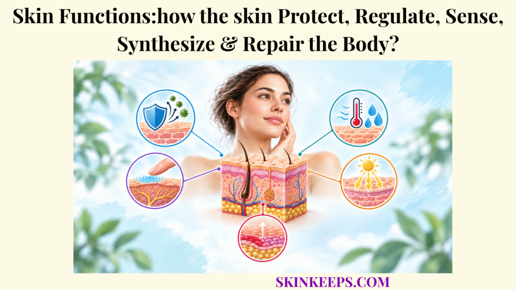
Skin functions protect the body’s internal environment by forming a barrier against external threats, limiting water loss and UV injury, regulating heat exchange, detecting sensory stimuli, coordinating immune defense, initiating vitamin D synthesis, and repairing damaged tissue. Skin is therefore not simply an external covering. It is a living interface between the body and the environment, combining structural protection with sensory detection, physiological responses, biochemical activity, and tissue restoration.
These functions depend on coordinated work across the epidermis, dermis, subcutaneous tissue, cutaneous blood vessels, sweat glands, sensory nerves, immune cells, and repair cells. Understanding which structures perform each function—and where the skin’s role connects with the nervous, immune, endocrine, and circulatory systems—turns a list of facts into a functional model of skin homeostasis, the maintenance of a relatively stable internal physiological environment.
How Do Skin Protective Functions Defend the Body From External Threats?
Skin protective functions defend the body through overlapping physical, chemical, microbial, immunological, UV-protective, and water-retaining barriers that separate vulnerable internal tissues from the external environment. Protection is therefore a layered system rather than one wall. The outer epidermis limits entry, surface chemistry and resident microbes influence microbial competition, immune cells respond to danger signals, pigmentation reduces part of the UV burden, and the permeability barrier slows excessive water escape from inside the body.
The physical and permeability components are especially important because maintaining the outer boundary solves two problems at once: it restricts harmful penetration from outside while limiting uncontrolled loss of water from within. This broader concept of skin barrier protection includes structural, chemical, microbial, and immunological elements that function together rather than independently.
UV defense belongs within protection but uses a partly different set of mechanisms. Melanin, antioxidant systems, epidermal responses, and DNA-repair pathways can reduce radiation-related injury, yet endogenous defenses are incomplete. Skin protection is strongest when these overlapping mechanisms are understood as a coordinated boundary system rather than as isolated features.
How Does the Skin Barrier Block Microbes, Chemicals, and Physical Damage?
The skin barrier limits microbes, irritants, chemicals, and mechanical stress through the tightly organized stratum corneum, epidermal cell junctions, extracellular lipids, acidic surface conditions, antimicrobial molecules, and interactions with resident microbes. The stratum corneum is the outermost epidermal layer. Its flattened corneocytes provide structural resilience, while the spaces between them contain organized lipid lamellae rich in ceramides, cholesterol, and free fatty acids that strongly influence permeability.
Keratinocytes do more than build the epidermis. They form structural layers, participate in tight-junction organization, release antimicrobial molecules, sense tissue stress and microbial signals, and communicate with immune cells. The mildly acidic skin surface—the acid mantle—also supports barrier enzyme activity and influences microbial growth. None of these defenses makes skin impermeable; the protective effect comes from multiple systems lowering the chance that external threats can cross into vulnerable tissue.
The skin microbiome is another interacting component. Resident organisms compete for space and nutrients, communicate with host cells, and can influence both antimicrobial defense and barrier homeostasis. That relationship is selective and context-dependent: not every skin microbe is beneficial, and surface microbial defense does not replace the physical or immune barriers.
How Do Melanin and Other Skin Systems Defend Against UV Radiation and Solar Injury?
Skin limits ultraviolet injury through melanin absorption and scattering, antioxidant defenses, cellular damage responses, epidermal adaptation, and DNA-repair mechanisms, although these natural defenses do not make UV exposure harmless. Melanocytes synthesize melanin and transfer pigment-containing melanosomes to keratinocytes. Melanin can absorb and scatter part of incoming ultraviolet radiation, helping reduce how much radiation reaches vulnerable cellular targets.
Eumelanin generally provides stronger photoprotective effects than pheomelanin, but pigmentation is only one part of skin UV defense. Antioxidant systems help manage reactive species, damaged cells activate stress responses, and DNA-repair pathways attempt to correct radiation-induced injury. These processes reduce damage; they do not eliminate it.
Safety point: Natural pigmentation does not replace sun protection. Ultraviolet radiation can damage cellular DNA across all skin tones.
How Does the Epidermal Barrier Prevent Excessive Water Loss?
The epidermal barrier limits excessive water loss by combining water-containing corneocytes with highly organized extracellular lipids that restrict passive diffusion of water through the stratum corneum. Transepidermal water loss is the passive movement of water through the epidermis into the external environment. Some water loss is physiologic; the barrier’s task is to prevent that passive loss from becoming excessive.
Corneocytes contain water-binding substances collectively described as natural moisturizing factors, while the extracellular matrix around them contains ceramides, cholesterol, and free fatty acids arranged into lipid layers. This organization creates the principal permeability barrier and is central to skin water-loss prevention. When barrier organization is disrupted, water escapes more readily and the outer skin can become less effective at maintaining hydration.
| Protective mechanism | Primary skin structure | Threat or loss controlled | Main protective result |
|---|---|---|---|
| Physical barrier | Stratum corneum and keratinized epidermis | Mechanical stress and penetration | Separates internal tissue from the environment. |
| Permeability barrier | Corneocytes and extracellular lipids | Excessive water loss and chemical penetration | Supports hydration and internal fluid balance. |
| Chemical and microbial barrier | Acidic surface, lipids, antimicrobial molecules, and microbiome | Pathogenic microbial growth | Supports surface defense through interacting chemical and biological mechanisms. |
| UV defense | Melanin plus cellular antioxidant and repair responses | Ultraviolet radiation | Reduces, but does not eliminate, photodamage. |
How Do Skin Functions Regulate Body Temperature and Sense the Environment?
Skin functions support environmental adaptation by changing sweat production and cutaneous blood flow to modify heat exchange while specialized sensory receptors detect touch, pressure, vibration, temperature, and potentially damaging stimuli. These two roles share an important sequence: the skin can detect conditions at the body surface, neural systems process that information, and skin structures can then act as effectors within broader homeostatic responses.
The distinction matters because detection, central processing, and response are not the same event. Temperature signals from the skin contribute to information reaching the hypothalamus, while sweat glands and cutaneous blood vessels help execute responses. Sensory receptors similarly transduce a local stimulus into neural signals, but conscious perception depends on processing within the nervous system.
How Do Sweating, Vasodilation, and Vasoconstriction Regulate Body Temperature?
Skin contributes to temperature regulation by evaporating sweat to dissipate heat, increasing cutaneous blood flow during heat exposure, and reducing skin blood flow during cold exposure to conserve core heat. The hypothalamus provides central thermoregulatory control. Peripheral thermoreceptors in skin contribute information about surface temperature, while central sensors provide information about internal temperature. Those inputs help the nervous system coordinate responses appropriate to heat or cold.
During heat exposure, eccrine sweat glands release sweat onto the skin surface. Cooling occurs when that sweat evaporates, transferring heat away from the body; sweat that cannot evaporate does not provide the same cooling effect. At the same time, increased cutaneous blood flow can move more internal heat toward the skin surface for exchange with the environment. These mechanisms are central to skin temperature regulation, but they operate under central neural control rather than making the skin an independent thermostat.
During cold exposure, cutaneous vasoconstriction narrows skin blood vessels and reduces blood flow near the surface. This primarily limits heat loss; it does not itself create meaningful new heat. Whole-body thermoregulation also involves behavior, skeletal muscle, endocrine responses, and other systems beyond the skin.
How Do Skin Sensory Receptors Detect Touch, Pressure, Temperature, and Pain?
Skin sensory receptors detect physical deformation, temperature change, and potentially damaging stimuli by converting those stimuli into electrical signals within sensory nerve endings. This conversion of stimulus energy into an electrical response is sensory transduction. Functionally, cutaneous receptors can be grouped into mechanoreceptors for physical deformation, thermoreceptors for temperature change, and nociceptors for potentially tissue-damaging stimuli.
Different receptor structures favor different forms of information. Merkel-associated receptors contribute to fine spatial detail and sustained pressure; Meissner corpuscles respond strongly to dynamic light touch; Pacinian corpuscles are highly sensitive to rapid deformation and vibration; free nerve endings include many thermal and nociceptive sensors. These receptor systems overlap rather than behaving as perfectly isolated switches, which is why skin sensation is best understood as population coding across multiple nerve endings and pathways.
| Stimulus | Receptor or structure | Typical information detected | Functional result |
|---|---|---|---|
| Light touch or movement across skin | Meissner corpuscles and other low-threshold mechanoreceptors | Dynamic light tactile input | Helps detect contact and object movement. |
| Texture or sustained pressure | Merkel-associated receptors | Fine spatial detail and sustained indentation | Supports discrimination of surface features. |
| Deep vibration or rapid pressure change | Pacinian corpuscles | High-frequency vibration and rapid deformation | Detects deeper mechanical changes. |
| Temperature | Thermosensitive free nerve endings | Heat or cold change | Supports thermal perception and protective responses. |
| Potential tissue-damaging stimulus | Nociceptive free nerve endings | Noxious mechanical, thermal, or chemical input | Initiates protective pain signaling. |
Receptor functions overlap. The table shows dominant functional associations rather than perfectly exclusive sensory channels.
How Are Sensory Signals Carried From the Skin to the Nervous System?
Sensory signals leave the skin through afferent nerve fibers and travel through peripheral nerves into the spinal cord, where specialized ascending pathways relay information toward higher brain regions for conscious perception. A stimulus first changes the electrical state of a sensory ending, creating a receptor potential. If that response reaches the necessary threshold in the sensory neuron, action potentials carry information along afferent fibers.
Different mechanical, thermal, and nociceptive signals use different fiber classes and ascending pathways after leaving the skin. The spinal cord and brainstem relay and organize these signals, while thalamic and cortical regions contribute to conscious somatosensory perception. Skin begins detection; the nervous system interprets what the signal means.
How Do Skin Functions Support Immune Defense and Vitamin D Synthesis?
Skin functions extend beyond passive protection because epidermal and dermal cells actively detect biological threats, coordinate local immune responses, and initiate the UVB-dependent biochemical pathway that produces vitamin D3. Immune defense and vitamin D synthesis are separate functions, but both show that skin is a metabolically and biologically active organ rather than an inert covering.
The immune system of skin links structural cells with resident and recruited immune cells. Vitamin D synthesis begins through a photochemical reaction in epidermal molecules and then depends on later processing outside the skin. In both cases, the skin performs an important part of a larger body-wide system without acting alone.
How Do Skin Immune Cells Recognize, Signal, and Respond to Threats?
Skin detects biological threats through keratinocytes and resident immune cells that recognize danger signals and release cytokines, chemokines, antimicrobial molecules, and other mediators that coordinate local defense. Keratinocytes are active immune participants as well as barrier-forming cells. They can respond to microbial products and tissue stress, produce signaling molecules, and communicate with nearby immune and structural cells.
Langerhans cells and other dendritic cells sample the local environment and participate in antigen handling. Macrophages contribute to surveillance, debris clearance, and inflammatory regulation; mast cells rapidly release mediators in response to selected signals; T lymphocytes provide adaptive immune functions. These interacting cells make skin immune defense a network rather than a function controlled by one cell type.
How Does the Skin Balance Inflammation With Immune Control?
Healthy skin balances defense with immune regulation by activating inflammatory responses when tissue is threatened while using regulatory signals to limit unnecessary or prolonged immune activation. Acute inflammation can be protective because it recruits cells and mediators needed to contain microbes, clear damaged material, and initiate repair. The problem is not inflammation itself but inflammation that is excessive, persistent, poorly controlled, or activated without an appropriate trigger.
Cytokines and chemokines help recruit and instruct immune cells, while local regulatory pathways reduce activation as the threat resolves. Restoring homeostasis therefore requires both the capacity to mount a response and the capacity to restrain it.
How Does UVB Exposure Trigger Vitamin D Synthesis in the Epidermis?
UVB radiation initiates vitamin D synthesis in the skin by converting 7-dehydrocholesterol into previtamin D3, which then undergoes heat-dependent conversion to vitamin D3. The epidermis performs these photochemical starting steps. Vitamin D3 then enters a broader metabolic pathway that includes processing in the liver and kidneys before the biologically active hormonal form is produced.
This mechanism is the basis of skin vitamin D synthesis, but physiology should not be turned into a recommendation for deliberate unprotected UV exposure. UV radiation also damages skin and increases skin-cancer risk, so vitamin D needs should be addressed through safe dietary, supplemental, and medical guidance rather than tanning or intentional UV injury.
How Do Skin Functions Repair Damaged Tissue After Injury?
Skin functions repair tissue through four overlapping phases—hemostasis, inflammation, proliferation, and remodeling—that stop bleeding, remove damaged material, rebuild the epidermis and dermis, restore blood supply, and reorganize the repaired extracellular matrix. These phases overlap rather than operating as four isolated blocks, and cells active in one stage can continue signaling into the next.
The goal of skin wound repair is to restore tissue continuity and barrier function after injury. Adult repair often ends with scar tissue rather than perfect regeneration, so a healed wound can be strong and functional without becoming structurally identical to uninjured skin.
How Does Hemostasis Stop Bleeding After Skin Injury?
Hemostasis begins immediately after skin injury as local vascular responses, activated platelets, and the coagulation system form a temporary fibrin-rich plug that limits blood loss and creates an initial wound matrix. Early vasoconstriction reduces local blood flow, while platelet activation and coagulation stabilize the injured area with a fibrin network.
The clot is not completed repair. It is a temporary protective structure and provisional matrix that limits bleeding, helps reduce immediate exposure of deeper tissue, and provides a surface through which later repair cells and signals can operate.
How Does Inflammation Clear Damaged Tissue and Control Microbes?
The inflammatory phase protects the wound by recruiting immune cells that remove microorganisms and damaged material while releasing signals that prepare the tissue for rebuilding. Neutrophils enter early and contribute to microbial control and debris clearance. Macrophages then perform phagocytic and regulatory functions while releasing mediators that influence fibroblasts, keratinocytes, endothelial cells, and other repair participants.
Controlled acute inflammation is a normal part of healing. Excessive, persistent, or dysregulated inflammation can interfere with repair, but inflammation should not be described as inherently harmful because normal tissue restoration depends on a properly timed inflammatory response.
How Do Keratinocytes, Fibroblasts, Collagen, and New Blood Vessels Rebuild Damaged Skin?
During proliferation, keratinocytes restore epidermal coverage, fibroblasts deposit extracellular matrix and collagen, and endothelial cells form new blood vessels that support metabolically active repair tissue. Keratinocyte migration and proliferation drive re-epithelialization, the restoration of an epidermal cover over the wound. Fibroblasts build a provisional extracellular matrix and collagen framework, while angiogenesis supplies the developing repair tissue with oxygen and nutrients.
This phase also produces granulation tissue, a vascular reparative tissue containing new vessels, matrix, and active repair cells. Granulation tissue is not the same as a mature scar; it is an earlier reconstruction stage that later changes substantially during remodeling.
How Does Tissue Remodeling Strengthen the Wound and Form a Scar?
Wound remodeling strengthens repaired skin by reorganizing and replacing the provisional extracellular matrix, altering collagen composition and alignment, reducing excess vascular tissue, and gradually producing a mature scar. Collagen type III laid down earlier is progressively reorganized and replaced in part by type I collagen, while matrix metalloproteinases and their regulators reshape extracellular material.
Myofibroblasts can contribute to wound contraction, and cellularity and vascularity decline as the scar matures. Remodeling can continue for months or longer. The result is repaired tissue that restores continuity and substantial strength, but adult scar tissue does not fully reproduce the architecture or mechanical properties of unwounded skin.
| Healing phase | Major cells or components | Main process | Biological outcome |
|---|---|---|---|
| Hemostasis | Platelets, coagulation proteins, fibrin | Stops bleeding and creates a temporary matrix. | Stabilizes the wound. |
| Inflammation | Neutrophils, macrophages, inflammatory mediators | Removes microbes and damaged material and coordinates repair signaling. | Prepares tissue for reconstruction. |
| Proliferation | Keratinocytes, fibroblasts, endothelial cells | Re-epithelialization, matrix deposition, granulation tissue formation, and angiogenesis. | Rebuilds tissue continuity and vascular support. |
| Remodeling | Fibroblasts, myofibroblasts, collagen, matrix-remodeling enzymes | Reorganizes extracellular matrix and matures collagen. | Produces stronger repaired tissue and scar maturation. |
What Should You Remember About Skin Functions?
The most important point about skin functions is that the skin operates as an integrated interface: it keeps damaging influences out, keeps essential water in, detects environmental change, modifies heat exchange, participates in immunity and vitamin D metabolism, and restores tissue after injury. Each function uses specialized structures, but none should be understood in complete isolation from the wider physiology of the body.
- Barrier protection: Skin separates internal tissues from microbes, chemicals, mechanical stress, and other environmental threats.
- UV defense: Melanin and cellular defense systems reduce UV injury but do not make ultraviolet exposure harmless.
- Water control: The stratum corneum and its specialized lipids limit excessive transepidermal water loss.
- Temperature regulation: Sweat evaporation and changes in cutaneous blood flow help the body exchange or conserve heat.
- Sensation: Mechanoreceptors, thermoreceptors, and nociceptors detect mechanical, thermal, and potentially damaging stimuli.
- Immune defense: Keratinocytes and resident immune cells detect threats and coordinate local immune signaling.
- Vitamin D synthesis: UVB initiates conversion of 7-dehydrocholesterol toward vitamin D3 production in skin.
- Wound repair: Hemostasis, inflammation, proliferation, and remodeling work together to restore damaged tissue.
What Common Questions Do People Ask About Skin Functions?
Common questions about skin functions usually focus on which functions are most important, how skin communicates with the nervous and immune systems, and where skin’s role ends and broader body systems take over.
What Are the Main Functions of the Skin?
The major skin functions include barrier protection, water conservation, UV defense, temperature regulation, sensory detection, immune surveillance, vitamin D3 synthesis, and wound repair.
Is the Skin the Organ That Controls Body Temperature?
No. Skin is a major thermoregulatory effector and sensory organ, but the hypothalamus provides central temperature control. Sweating and changes in skin blood flow help the body lose or conserve heat.
Does Melanin Completely Protect the Skin From UV Damage?
No. Melanin contributes important photoprotection, but ultraviolet radiation can still damage cells and DNA across all skin tones, so natural pigmentation does not replace sun protection.
Does the Skin Produce Fully Active Vitamin D?
No. Skin initiates vitamin D3 synthesis after UVB converts 7-dehydrocholesterol to previtamin D3, but later liver and kidney processing is required to produce the biologically active hormone.
Why Does Healed Skin Form a Scar?
Healed adult skin often forms a scar because wound repair rebuilds tissue through collagen deposition and extracellular-matrix remodeling rather than perfectly recreating the original skin architecture.
NCBI Bookshelf / StatPearls — Anatomy, Skin (Integument): overview of skin as a protective organ with thermoregulatory, sensory, immune-surveillance, fluid-loss, and homeostatic functions.
Cells — Skin Barrier Function: The Interplay of Physical, Chemical, and Immunologic Properties: stratum-corneum organization, corneocytes, extracellular lipids, skin acidity, permeability, and interacting physical, chemical, and immune barrier mechanisms.
International Journal of Molecular Sciences / PubMed — Skin Barrier Function and the Microbiome: relationships between resident microbes, epidermal barrier integrity, chemical conditions, and cutaneous immune homeostasis.
Photochemistry and Photobiology / PMC — The Protective Role of Melanin Against UV Damage in Human Skin: melanin absorption and scattering of UV radiation, eumelanin/pheomelanin differences, and the incomplete nature of endogenous photoprotection.
NCBI Bookshelf / StatPearls — Physiology, Temperature Regulation: hypothalamic central control, peripheral thermoreceptors, sweating, cutaneous blood-flow changes, vasoconstriction, and heat exchange.
NCBI Bookshelf — Cutaneous and Subcutaneous Somatic Sensory Receptors: mechanoreceptors, nociceptors, thermoreceptors, sensory transduction, receptor potentials, and action-potential signaling.
Journal of Investigative Dermatology / PubMed — The Central Roles of Keratinocytes in Coordinating Skin Immunity: keratinocytes as active immune sentinels that communicate with microbes, immune cells, neurons, fibroblasts, and other cutaneous cells.
NCBI Bookshelf — Overview of Vitamin D: UVB-mediated conversion of 7-dehydrocholesterol to previtamin D3 and subsequent vitamin D3 formation in skin.
American Academy of Dermatology — Vitamin D and UV Exposure: safety guidance favoring dietary or supplemental vitamin D rather than intentional unprotected UV exposure.
NCBI Bookshelf / StatPearls — Physiology, Wound Healing: the overlapping hemostasis, inflammation, proliferation, and remodeling phases and the major cells involved in tissue reconstruction.
Medical note: This article explains normal skin physiology for educational purposes and does not diagnose disease or replace medical care. Severe burns, spreading redness or pus, fever associated with a wound, major or persistent sensory loss, rapidly worsening tissue injury, or a wound that does not heal as expected should receive appropriate medical evaluation.



