
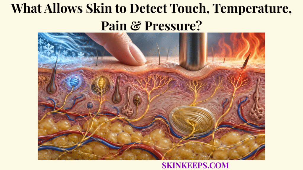
Skin detects touch, temperature, pressure, vibration, stretch, and potentially damaging stimuli through specialized sensory nerve endings and mechanosensory structures that convert physical or chemical changes into electrical signals carried toward the spinal cord and brain. Mechanoreceptors, thermoreceptive afferents, and nociceptors begin the process, but receptor activation itself is not the same as conscious sensation.
This page explains which receptor systems detect different stimulus features, how sensory transduction creates electrochemical nerve signals, how those signals travel through peripheral and central pathways, how spinal reflexes can protect the body before full conscious perception, and why disruption anywhere along the sensory pathway can alter sensation.
How Do Skin Sensory Receptors Detect Changes in the Environment?
Skin sensory receptors detect environmental change when mechanical force, temperature change, or potentially damaging chemical or physical stimuli alter ion-channel activity in sensory endings and generate electrical signals that can trigger action potentials. This sensory role is one of the body’s major skin functions, turning events at the body surface into neural information rather than conscious perception by the skin itself.
How Do Specialized Nerve Endings Respond to Mechanical, Thermal, and Potentially Damaging Stimuli?
Specialized sensory endings respond selectively because their membrane proteins, surrounding tissue structures, depth, and nerve-fiber properties make them more sensitive to particular mechanical, thermal, or noxious stimulus features. A receptor’s threshold, receptive-field size, and local end-organ structure influence which aspects of a stimulus are encoded most strongly.
A receptive field is the area of skin in which stimulation can alter the activity of a particular sensory neuron. Receptors with small receptive fields can support precise spatial localization, while larger receptive fields sample broader areas and often emphasize other stimulus features.
How Does Sensory Transduction Convert a Stimulus Into an Electrical Nerve Signal?
Sensory transduction converts a stimulus into neural activity when receptor deformation, temperature change, or chemical stimulation alters ion flow across the sensory membrane, generating a receptor potential that can initiate action potentials. A receptor potential is graded, meaning its size varies with local stimulation, whereas action potentials are propagated signals that travel along the sensory axon.
When depolarization reaches the appropriate threshold, action-potential firing begins and the afferent neuron carries an electrochemical neural signal away from the receptor region. Stimulus intensity can be represented partly through firing frequency and recruitment of additional sensory fibers.
Why Are Different Skin Sensory Receptors Specialized for Different Types of Information?
Different cutaneous receptors are specialized because their end-organ architecture, adaptation rate, receptive-field size, ion-channel expression, and nerve-fiber properties filter different features of the same physical environment. Receptors that are slowly adapting continue firing during a sustained stimulus, while rapidly adapting receptors emphasize changes, movement, onset, offset, or vibration.
Some protective reflexes branch through spinal circuitry before cortical perception is complete.
Which Cutaneous Mechanoreceptors Detect Touch, Pressure, Vibration & Stretch?
Cutaneous mechanoreceptors detect touch, pressure, vibration, and skin deformation through distinct low-threshold mechanosensory afferents whose response patterns differ by depth, receptive-field size, and adaptation rate. The family of skin receptors for touch and pressure therefore works as an overlapping population rather than a collection of isolated one-receptor/one-sensation switches.
How Do Merkel Receptors Detect Sustained Light Touch, Edges, and Texture?
Merkel cell–neurite complexes encode sustained indentation and fine spatial features through slowly adapting type-I mechanosensory responses that remain active while a mechanical stimulus persists. Their superficial location and relatively small receptive fields support discrimination of edges, curvature, texture, and detailed form.
Merkel-associated responses work alongside Meissner-mediated dynamic sensing as part of the broader network of skin light-touch receptors. Calling Merkel systems simple “pressure receptors” misses their strong role in sustained spatial detail.
How Do Meissner Corpuscles Detect Light Touch and Low-Frequency Movement?
Meissner corpuscles respond strongly to changing mechanical deformation in superficial glabrous skin, making their rapidly adapting afferents well suited to dynamic light touch, motion across the skin, grip slip, and relatively low-frequency vibration. They are especially abundant in glabrous regions specialized for tactile discrimination rather than being distributed equally across all hairy skin.
How Do Pacinian Corpuscles Respond to Deep Pressure and Vibration?
Pacinian corpuscles respond most strongly to rapid mechanical changes and high-frequency vibration because their deep location and layered capsule mechanically filter sustained deformation while transmitting transient vibration to the sensory axon. Their very rapidly adapting responses and broad receptive fields make them particularly effective for detecting vibration and distant mechanical transients.
Within the broader family of skin pressure and vibration receptors, Pacinian afferents should not be reduced to generic “deep pressure” detectors because their defining strength is rapid mechanical change and high-frequency vibration.
How Do Ruffini Endings Contribute to Detecting Skin Stretch and Sustained Deformation?
Slowly adapting type-II mechanosensory activity contributes to detection of sustained skin stretch and deformation and has traditionally been associated with Ruffini endings, although typical Ruffini corpuscles are relatively sparse in human digital skin. Human histology confirms that Ruffini corpuscles exist, but the anatomical source of all recorded SA-II responses is less straightforward than classic textbook diagrams suggest.
| Receptor / afferent system | Typical mechanical feature | Adaptation | Typical location / depth | Main contribution |
|---|---|---|---|---|
| Merkel cell–neurite / SA-I | Sustained indentation, edges, texture, curvature | Slowly adapting | Basal epidermal region / superficial | Fine spatial detail and sustained touch |
| Meissner corpuscle / RA-I | Dynamic light touch, slip, lower-frequency vibration | Rapidly adapting | Dermal papillae of glabrous skin | Movement across skin and grip changes |
| Pacinian corpuscle / RA-II | Rapid deformation and high-frequency vibration | Very rapidly adapting | Deep dermis / subcutaneous tissue | Vibration and distant mechanical transients |
| SA-II stretch-sensitive system / traditionally Ruffini-associated | Sustained stretch and skin deformation | Slowly adapting | Deeper dermal structures | Stretch/tension coding; anatomical assignment is nuanced in humans |
These receptor systems overlap functionally; tactile perception emerges from population activity and central processing rather than one receptor acting alone.
How Do Skin Thermoreceptors Detect Heat and Cold?
Skin thermoreceptors detect warming and cooling through temperature-sensitive free sensory nerve endings whose ion channels alter neuronal excitability as local skin temperature changes. These skin temperature signals provide continuous thermal information for perception, thermoregulation, and protective behavior.
Many thermoreceptive terminals are skin free nerve endings rather than encapsulated sensory corpuscles. Warm-sensitive and cold-sensitive afferent populations overlap in their response ranges, and extreme temperatures increasingly recruit high-threshold nociceptive pathways.
How Do Temperature-Sensitive Nerve Endings Respond to Warming and Cooling?
Temperature-sensitive sensory endings change their firing as skin warms or cools, providing neural information about both the direction and magnitude of local thermal change. Dynamic responses can be especially strong during rapid changes, while ongoing firing helps represent more stable thermal conditions.
How Do Ion Channels in Sensory Neurons Respond to Different Temperatures?
Temperature-sensitive TRP ion channels alter cation flow in sensory neurons across different thermal ranges, depolarizing the nerve ending and contributing to warm, cold, or noxious thermal signaling. TRPM8 contributes strongly to cool/cold-associated signaling, while TRPV1 participates in noxious heat and capsaicin-associated signaling.
These channels are important transducers but do not operate as rigid universal temperature switches. Channel expression, other membrane proteins, tissue conditions, and central processing influence the final sensory response.
How Does Temperature Sensing Support Thermoregulation and Protective Behavior?
Cutaneous temperature sensing supports thermoregulation by providing peripheral thermal information to central circuits and supports protection by driving conscious avoidance and nociceptive responses when thermal conditions become potentially damaging. The sensory system therefore contributes information that the nervous system uses for both homeostatic regulation and protective behavior.
| Thermal stimulus | Predominant sensory response | Functional outcome |
|---|---|---|
| Mild cooling | Increased activity in cold-sensitive afferent populations | Cold perception and thermoregulatory input |
| Mild warming | Increased activity in warm-sensitive afferent populations | Warmth perception and thermoregulatory input |
| Potentially damaging heat or cold | Recruitment of high-threshold nociceptive pathways | Pain-related signaling, avoidance, and protective behavior |
This overview avoids rigid temperature cutoffs because receptor activation and thermal perception vary with physiological and experimental context.
How Do Cutaneous Nociceptors Detect Pain and Potential Tissue Damage?
Cutaneous nociceptors detect potentially tissue-damaging mechanical, thermal, and chemical stimuli through high-threshold sensory endings that generate neural activity capable of triggering protective reflexes and contributing to conscious pain. This distinction is central to skin pain detection: nociception encodes tissue threat, while pain is a conscious sensory and emotional experience created by nervous-system processing.
How Do Nociceptors Respond to Extreme Heat, Cold, Pressure, or Damaging Chemicals?
Nociceptors respond when mechanical force, temperature extremes, or chemical conditions reach potentially damaging ranges and activate high-threshold ion channels or receptors on peripheral sensory endings. Different nociceptor populations can be preferentially responsive to high mechanical force, thermal extremes, injury-related chemicals, or combinations of these stimuli.
How Do Inflammatory Chemicals Increase Nociceptor Sensitivity After Injury?
Inflammatory mediators released after tissue injury can sensitize nociceptors by lowering their activation threshold or increasing their response to stimulation, making injured skin more tender or painful. Prostaglandins, bradykinin, histamine, protons, ATP, and cytokines can participate in this local signaling environment.
Peripheral sensitization means increased responsiveness of peripheral nociceptors after tissue injury or inflammation. It can contribute to hyperalgesia, an increased pain response to a normally painful stimulus, without implying that one inflammatory molecule explains all inflammatory pain.
Why Can Pain Continue After the Original Stimulus Has Been Removed?
Pain can persist after the original stimulus ends because injured tissue may continue releasing sensitizing mediators and because repeated nociceptive input can alter excitability in peripheral and central neural circuits. Peripheral sensitization and central sensitization can therefore extend the response beyond the brief initiating event, although persistent pain has many possible mechanisms and requires clinical context.
Protective route: spinal interneurons → withdrawal response · Ascending route: anterolateral pathways → thalamic/brain networks → pain perception.
How Does the Skin’s Sensory Network Send Signals to the Brain?
The skin’s sensory network sends signals toward the brain when receptor-generated action potentials travel along peripheral afferent neurons into the spinal cord or brainstem and then ascend through modality-specific sensory pathways to thalamic and cortical processing regions. Peripheral nerves carry signals; they do not themselves decide what the stimulus consciously means.
How Do Peripheral Sensory Nerves Carry Signals Away From the Skin?
Peripheral sensory neurons carry action potentials from receptor endings in the skin along axons whose cell bodies lie in sensory ganglia, including dorsal root ganglia for most body sensation. These neurons are typically pseudounipolar, with a peripheral axonal branch connected to the skin and a central branch that enters the spinal cord.
Large myelinated Aβ fibers carry much low-threshold mechanosensory information. Thinly myelinated Aδ fibers and unmyelinated C fibers carry overlapping categories of thermal, nociceptive, itch, and other sensory information, so they should not be reduced to universal “fast” and “slow” channels for every modality.
How Are Sensory Signals Relayed and Processed in the Spinal Cord?
Sensory signals enter the spinal cord through dorsal roots, but their subsequent route depends on modality: discriminative touch and vibration primarily ascend through the dorsal column system, whereas pain and temperature information primarily enters the anterolateral system after spinal synaptic processing. These pathways are major organizational routes rather than perfectly exclusive channels.
Fine touch, vibration, and two-point discrimination from the body are strongly associated with the dorsal column–medial lemniscus system. Pain and temperature signals commonly synapse in the dorsal horn, cross within the spinal cord, and ascend in anterolateral pathways such as the spinothalamic tract.
How Does the Brain Distinguish Stimulus Location, Intensity, and Type?
The nervous system distinguishes stimulus type, strength, and location by combining which sensory neurons are active, their receptive fields, their firing patterns, the pathways they enter, and the spatial organization of their cortical targets. Stimulus intensity is represented partly by firing rate and recruitment, while localization depends strongly on receptive-field organization and somatotopic mapping.
Texture and complex tactile pattern recognition depend on population coding and cortical processing rather than a single peripheral receptor. Conscious perception emerges from distributed neural processing in the thalamus, somatosensory cortex, and related networks.
How Do Reflex Pathways Produce Rapid Protective Responses Before Conscious Perception?
Protective withdrawal can begin before full conscious perception because nociceptive sensory fibers activate spinal interneuron circuits that recruit flexor muscles while ascending sensory signals continue toward the brain. This is one way skin sensory signaling in body protection can produce fast behavior before cortical processing is complete.
The withdrawal response is polysynaptic: sensory afferents activate spinal interneurons, which recruit motor neurons driving flexor activity and often inhibit opposing extensor muscles through reciprocal circuitry. The cortex does not have to complete conscious pain perception before the first protective movement begins.
What Can Disrupt Skin Sensation and Alter Touch, Temperature, Pain or Pressure Detection?
Skin sensation can become reduced, absent, painful, or exaggerated when receptors, peripheral sensory axons, nerve roots, spinal pathways, or central sensory networks are compressed, injured, inflamed, metabolically impaired, or otherwise disrupted. Numbness, tingling, burning, or hypersensitivity therefore cannot by themselves identify where the problem is located.
How Can Nerve Compression or Injury Reduce Sensory Signaling?
Compression or injury can reduce normal sensation when mechanical damage, ischemia, demyelination, or axonal injury interferes with action-potential conduction along sensory fibers. Depending on which fibers and pathways are affected, symptoms can include reduced touch or vibration, impaired temperature sensation, numbness, tingling, or pain.
How Can Inflammation Increase Sensitivity and Pain?
Inflammation can increase sensitivity when inflammatory mediators sensitize peripheral nociceptors and lower the amount of stimulation needed to generate strong nociceptive signaling. This can produce tenderness and hyperalgesia during acute tissue inflammation without meaning that every inflamed area has developed a neuropathic pain disorder.
Why Can Damaged Sensory Nerves Cause Numbness, Tingling, Burning, or Abnormal Sensations?
Damaged sensory nerves can produce both sensory loss and abnormal spontaneous activity, so the same injured pathway may cause numbness in one region while producing tingling, electric, burning, or painful sensations in another. Paresthesia describes abnormal sensations such as tingling or pins and needles; dysesthesia describes unpleasant abnormal sensation.
Small-fiber dysfunction illustrates how Aδ and C-fiber abnormalities can produce burning, pain, numbness, and tingling, but burning alone is not enough to diagnose small-fiber neuropathy or any other specific disorder.
When Can Persistent Sensory Changes Indicate a Problem Beyond the Skin Itself?
Persistent or progressive sensory change may reflect a peripheral nerve, nerve-root, spinal-cord, metabolic, vascular, or central neurological problem rather than a disorder confined to the skin surface. Persistent numbness, recurrent unexplained tingling, progressive sensory loss, reduced temperature sensation, or sensory symptoms that interfere with function or sleep deserve medical evaluation.
Sudden one-sided numbness or weakness, new facial droop, new speech difficulty, rapidly progressive weakness or numbness, loss of bladder or bowel control with sensory change, or major trauma followed by sensory loss requires prompt or urgent medical assessment.
| Sensory change | Possible mechanistic category | Affected pathway level | Boundary |
|---|---|---|---|
| Reduced touch or vibration | Impaired mechanoreceptor or large-fiber conduction | Receptor, peripheral nerve, or DCML pathway | Does not identify the cause by itself |
| Numbness | Reduced sensory afferent transmission | Peripheral or central pathway | Requires context and examination |
| Tingling / pins and needles | Abnormal sensory-fiber activity | Often peripheral, but may be central | Do not equate automatically with neuropathy |
| Burning pain | Small-fiber/nociceptive dysfunction or inflammation | Peripheral nociceptors, small fibers, or central pathways | Many causes are possible |
| Increased pain after injury | Peripheral or central sensitization | Nociceptive pathways | Acute sensitization differs from chronic pain syndromes |
| Reduced heat/cold detection | Thermal-afferent dysfunction | Aδ/C fibers or central pathway | Persistent change warrants assessment |
What Are the Key Takeaways About the Skin’s Sensory Systems?
The key fact about the skin’s sensory systems is that different receptor populations detect different features of mechanical, thermal, and potentially damaging stimuli, while peripheral nerves and central pathways transform those signals into conscious sensation and protective responses.
- Sensory transduction: Skin receptors convert mechanical, thermal, or chemical stimulus energy into electrical neural signals.
- Merkel-associated receptors: Slowly adapting responses encode sustained touch and fine spatial features such as edges and texture.
- Meissner corpuscles: Rapidly adapting superficial receptors support dynamic light touch, motion, and slip detection.
- Pacinian corpuscles: Deep rapidly adapting receptors are highly sensitive to transient mechanical change and high-frequency vibration.
- Ruffini-associated signaling: Slowly adapting type-II responses contribute to stretch and sustained deformation, but the anatomical relationship is more nuanced in humans than simplified diagrams suggest.
- Thermoreceptors: Temperature-sensitive free nerve endings use specialized ion channels to detect warming and cooling.
- Nociceptors: High-threshold sensory endings detect potentially tissue-damaging mechanical, thermal, and chemical stimuli.
- Peripheral nerves: Sensory afferents carry action potentials from skin toward the spinal cord and brain.
- Ascending pathways: Fine touch/vibration and pain/temperature use different major spinal pathways.
- Protective reflexes: Spinal circuits can initiate withdrawal before conscious perception is complete.
- Perception: The brain interprets neural population activity as touch, pressure, vibration, temperature, and pain.
What Common Questions Do People Ask About Skin Sensation?
Common questions about skin sensation focus on which receptors detect touch, whether pain starts in the skin or brain, why numbness and tingling occur, and how sensory information reaches conscious awareness.
Which Receptor Detects Light Touch Best?
Light touch is encoded by multiple mechanoreceptor systems rather than one receptor alone, with Merkel-associated receptors contributing fine sustained spatial detail and Meissner corpuscles contributing strongly to dynamic light touch and movement across glabrous skin.
Which Skin Receptors Detect Vibration?
Meissner and Pacinian corpuscles both contribute to vibration sensing, but Pacinian corpuscles are particularly sensitive to rapid, high-frequency vibration while Meissner receptors respond more strongly to lower-frequency dynamic skin motion.
Are Nociceptors the Same as Pain Receptors?
Nociceptors are better described as high-threshold sensory neurons that detect potentially damaging stimuli; conscious pain arises only after nociceptive signals are processed by the nervous system.
Why Can Skin Feel Numb and Painful at the Same Time?
A damaged or irritated sensory pathway can lose normal signaling while also generating abnormal spontaneous or exaggerated activity, allowing reduced sensation and burning, tingling, or pain to coexist.
Does the Brain Have to Feel Pain Before the Body Pulls Away?
No. A nociceptive withdrawal reflex can begin through spinal-cord circuits before full conscious pain perception is completed in the brain.
NCBI Bookshelf — Cutaneous and Subcutaneous Somatic Sensory Receptors: mechanoreceptor, thermoreceptor, and nociceptor classification; sensory transduction; receptor potentials; adaptation; and firing patterns.
Nature Reviews Neuroscience / PMC — The Mechanosensory Neurons of Touch and Their Mechanisms of Activation: Merkel, Meissner, and Pacinian systems, low-threshold mechanoreceptors, adaptation, glabrous-skin organization, and mechanotransduction.
Journal of Clinical Medicine / PMC — The Human Cutaneous Sensory Corpuscles: An Update: human Meissner, Pacinian, and Ruffini anatomy and the distribution of sensory corpuscles in glabrous skin.
Journal of Anatomy / PMC — Verification and characterisation of human digital Ruffini’s sensory corpuscles: evidence that typical Ruffini corpuscles occur in human digital skin at low density and the anatomical nuance of SA-II assignments.
Channels / PMC — TRP ion channels in thermosensation, thermoregulation and metabolism: temperature-sensitive TRP channels, peripheral thermosensory neurons, and thermal transduction.
Journal of Clinical Investigation — Nociceptors: the sensors of the pain pathway: high-threshold mechanical, thermal, and chemical nociception and conversion of potentially damaging stimuli into afferent neural signals.
NCBI Bookshelf — The Somatic Sensory System: mechanosensory transmission, dorsal column–medial lemniscus organization, thalamic and cortical processing, and trigeminal qualification for facial sensation.
NCBI Bookshelf — Central Pain Pathways: The Spinothalamic Tract: pain and temperature signaling through dorsal-horn and anterolateral/spinothalamic pathways.
NCBI Bookshelf — Physiology, Withdrawal Response: polysynaptic nociceptive withdrawal, sensory afferents, spinal interneurons, motor responses, and rapid protection before full conscious processing.
Mayo Clinic — Peripheral nerve injuries: sensory nerve injury as a cause of numbness, tingling, pain, and altered pain or temperature detection, with medical-evaluation guidance.
Medical note: This article explains normal cutaneous sensory physiology and broad sensory-dysfunction concepts for education. Numbness, tingling, burning, hypersensitivity, or altered temperature sensation do not by themselves diagnose peripheral neuropathy, nerve entrapment, spinal disease, stroke, multiple sclerosis, or another neurological disorder. Persistent or progressive sensory changes deserve medical assessment. Sudden one-sided numbness or weakness, new facial droop, new speech difficulty, loss of bladder or bowel control with sensory change, rapidly progressive neurological symptoms, or major trauma followed by sensory loss requires prompt or urgent medical evaluation.



