
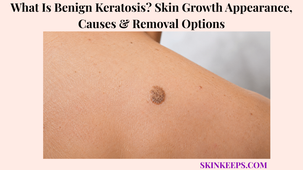
Benign keratosis is usually a noncancerous keratin-based skin growth that can look waxy, rough, scaly, warty, or stuck onto the skin.
Benign keratosis often overlaps clinically with seborrheic keratosis-type growths. The sections below cover appearance, causes, diagnosis, removal options, aftercare, mistakes, and warning signs that need review before any removal attempt.
What Is Benign Keratosis and Is It the Same as Seborrheic Keratosis?
Benign keratosis is usually used to describe a noncancerous keratin-based skin growth, most often a seborrheic keratosis-type lesion that can look waxy, scaly, rough, or stuck onto the skin.
Keratin is a structural protein in the outer skin layer, and keratinocytes are the main cells that form that outer layer. A benign keratin-based growth means the lesion is not cancer by definition, but that does not mean every rough or dark growth can be safely self-labeled.
Many people use benign keratosis as a patient-friendly label for seborrheic keratosis or a similar seborrheic-keratosis-type growth. The safer approach is diagnosis first, especially when a lesion is new, changing, bleeding, painful, ulcerated, very dark, irregular, or unlike other spots.
Why Is Seborrheic Keratosis the Main Condition People Mean by Benign Keratosis?
Seborrheic keratosis is the main condition people often mean by benign keratosis because it is a common noncancerous keratinocyte growth that can look stuck-on, waxy, scaly, or warty.
These growths commonly appear in adulthood and become more common with age. Some people develop just one, while others develop many lesions over time.
Removal may be chosen for irritation, catching on clothing or jewelry, appearance concerns, bleeding from friction, or diagnostic uncertainty. Removal is not automatically needed when the lesion is typical, stable, and not bothersome.
Why Is Benign Keratosis Different From Actinic Keratosis?
Benign keratosis is different from actinic keratosis because benign keratosis is noncancerous, while actinic keratosis is a sun-damage-related precancerous lesion.
Both can feel rough or scaly, which is why a rough sun-exposed patch should not automatically be treated as benign. Actinic keratosis belongs to a different risk pathway and needs a different diagnostic and treatment approach.
Practical rule: A typical stable benign keratosis may not need removal, but a new, changing, bleeding, painful, ulcerated, very dark, or irregular growth should be checked before cosmetic treatment.
What Does Benign Keratosis Look Like on Skin?
Benign keratosis often looks like a tan, brown, black, yellowish, gray, or skin-colored growth with a waxy, wart-like, rough, scaly, or stuck-on surface.
The growth may be raised or flatter, round or oval, sharply demarcated or irregular-looking, and sometimes crumbly or greasy-looking. Appearance can vary enough that texture alone should not be used as a diagnosis.
What Surface Texture Can Benign Keratosis Have?
Benign keratosis can feel waxy, rough, warty, scaly, crumbly, greasy-looking, raised, flat, pasted-on, or stuck onto the skin.
A classic clue is a stuck-on or pasted-on appearance, as if the growth sits on top of the skin surface. Some lesions look like a waxy plaque, a rough wart-like plaque, or a scaly crusted spot.
A sudden texture change, ulceration, non-healing crust, or repeated bleeding should be checked because cancer and precancerous lesions can overlap visually.
What Colors Can Benign Keratosis Have?
Benign keratosis can be tan, light brown, dark brown, black, gray, yellowish, skin-colored, or mixed in color.
Dark color does not automatically mean cancer, but a new very dark lesion, irregular color pattern, changing border, or different-looking spot should be evaluated. Pigmented benign keratosis can resemble melanoma, so uncertain dark lesions should not be removed at home.
What Symptoms Can Benign Keratosis Cause?
Benign keratosis is usually painless, but it can itch, catch on clothing or jewelry, become irritated, or bleed if rubbed, scratched, shaved, or picked.
Friction from collars, bra straps, belts, razors, waistbands, necklaces, or combing can irritate a raised lesion. Bleeding after clear friction is different from sudden bleeding without an obvious cause.
Pain, tenderness, persistent inflammation, ulceration, repeated crusting, or bleeding without clear trauma should be checked rather than treated as ordinary irritation.
| Feature | Common Benign Keratosis Clue | When It Becomes Concerning |
|---|---|---|
| Texture | Waxy, rough, warty, scaly, stuck-on | Rapid texture change or ulceration |
| Color | Tan, brown, black, gray, yellowish, skin-colored | New very dark lesion or irregular color pattern |
| Shape | Round, oval, irregular, sharply demarcated | Changing border or different-from-others lesion |
| Surface | Raised, flat, crumbly, greasy-looking | Non-healing crust or bleeding without friction |
| Symptoms | Usually painless; may itch or irritate | Pain, tenderness, persistent inflammation |
| Friction | Catches on clothing or jewelry | Recurrent bleeding or open sore |
Where Does Benign Keratosis Usually Appear?
Benign keratosis can appear on many body areas, especially the face, scalp, neck, chest, back, shoulders, abdomen, arms, and legs, but palms and soles are not typical sites.
Common irritation sites include the neck, bra line, waistline, scalp, beard area, collar line, and areas rubbed by clothing, belts, straps, razors, combs, or jewelry.
Location alone cannot diagnose the growth. Genital, mucosal, palm, sole, rapidly changing, or unusual keratosis-like lesions should be checked rather than assumed benign.
| Location | Common Irritation Trigger | Removal Consideration |
|---|---|---|
| Face / scalp | Shaving, combing, sun-exposed visibility | Cosmetic or diagnostic concern |
| Neck | Collars, necklaces, friction | Irritation or catching |
| Chest / back | Clothing, bra straps, sweating | Multiple lesions common; check changing ones |
| Shoulders | Straps, sun exposure, friction | Rule out sun-damage lesions if rough or changing |
| Abdomen | Belts, waistbands | Irritation from clothing |
| Arms / legs | Scratching, shaving, friction | Check if bleeding or painful |
| Palms / soles | Not typical for seborrheic-keratosis-type lesions | Dermatology review if keratosis-like growth appears |
What Causes Benign Keratosis?
The exact cause of benign keratosis is not fully understood, but age, genetic tendency, skin growth patterns, and long-term skin changes appear to contribute.
The growth comes from the epidermis and involves keratinocyte growth, which helps explain the waxy, rough, stuck-on, or warty surface. Still, the exact trigger is not the same for every person.
Why Do Benign Keratoses Become More Common With Age?
Benign keratoses often become more common with age because these noncancerous keratinocyte growths tend to appear in adulthood and may increase in number over time.
Many people first notice them in middle age or later. Some people develop several lesions over the years, but a new or different-looking growth still deserves attention instead of automatic reassurance.
Why Can Family Tendency Matter?
Family tendency can matter because some people are genetically prone to developing multiple benign keratotic growths.
A family history of similar stuck-on growths can support the pattern, but family history does not prove that every new growth is harmless. A changing, bleeding, painful, irregular, or very dark lesion still needs assessment.
Does Sun Exposure Cause Benign Keratosis?
Sun exposure may influence some keratosis-like lesions, but benign keratosis can also appear on less sun-exposed skin and should not be confused with actinic keratosis.
Some lesions appear on sun-exposed areas such as the face, scalp, shoulders, and arms. Others appear on the trunk or covered areas, so sun exposure should not be framed as the only cause.
| Factor | What It May Explain | What It Does Not Prove |
|---|---|---|
| Age | More lesions over time | New lesions are not automatically harmless |
| Genetics | Family tendency and multiple growths | Every growth is not guaranteed benign |
| Keratinocyte growth | Waxy or stuck-on keratin surface | Does not exclude cancer mimic |
| Friction | Irritation or bleeding after rubbing | Does not explain rapid growth or ulceration |
| Sun exposure | Some overlap with sun-damaged areas | Does not make it actinic keratosis |
| Skin tone | Pigment visibility and treatment-risk differences | Color alone cannot diagnose |
How Is Benign Keratosis Different From Other Skin Growths?
Benign keratosis can resemble warts, skin tags, moles, actinic keratosis, basal cell carcinoma, squamous cell carcinoma, or melanoma, so changing or suspicious growths should be checked.
Correct diagnosis matters because a benign growth may need no treatment, while a cancer-like or precancerous lesion needs a different plan. It is also why undiagnosed growths should not be burned, frozen, scraped, picked, or treated with wart remover at home.
How Is Benign Keratosis Different From a Wart?
Benign keratosis is different from a wart because warts are viral growths caused by HPV, while benign keratosis is a nonviral keratin-based growth.
Both can look rough or warty. Location, surface pattern, number of lesions, dermoscopy, and clinical history can help separate them. Wart-remover products should not be used on an undiagnosed pigmented or changing growth.
How Is Benign Keratosis Different From a Skin Tag?
Benign keratosis is different from skin tags because skin tags are usually soft and hanging, while benign keratoses are often waxy, scaly, or stuck onto the skin.
Both can become irritated by clothing or jewelry, but the removal method and diagnostic concern can differ. A dark, rough, or changing growth should not be treated like an ordinary soft tag without confirmation.
How Is Benign Keratosis Different From Actinic Keratosis?
Benign keratosis is different from actinic keratosis because actinic keratosis is usually a rough sun-damaged precancerous lesion, while benign keratosis is noncancerous.
The overlap is texture: both may feel rough or scaly. A rough lesion on chronically sun-exposed skin should be examined when the diagnosis is uncertain.
How Is Benign Keratosis Different From Melanoma or Skin Cancer?
Benign keratosis is noncancerous, but pigmented benign keratosis can resemble melanoma, and some basal cell or squamous cell cancers can look waxy, scaly, crusted, or bleeding.
Basal cell carcinoma can look pearly, waxy, bleeding, or non-healing. Squamous cell carcinoma can look scaly, crusted, tender, or non-healing. Biopsy is needed when the diagnosis is uncertain.
| Condition | Common Clue | Why Confusion Happens | Safer Next Step |
|---|---|---|---|
| Benign keratosis | Waxy, stuck-on, scaly growth | Can be dark, rough, or irregular | Check if new or changing |
| Wart | Rough viral growth | Warty texture overlaps | Confirm before wart treatment |
| Skin tag | Soft hanging growth | Both can irritate | Remove only if diagnosed |
| Actinic keratosis | Rough sun-damaged patch | Scaly texture overlaps | Evaluate sun-exposed lesions |
| Basal cell carcinoma | Pearly, waxy, bleeding, non-healing growth | Can mimic benign growth | Biopsy if suspicious |
| Squamous cell carcinoma | Scaly, crusted, tender, non-healing lesion | Keratin/scaling overlaps | Prompt evaluation |
| Melanoma | New or changing pigmented lesion | Pigmented keratosis can mimic melanoma | Urgent review if evolving |
How Is Benign Keratosis Diagnosed or Checked?
Benign keratosis is often diagnosed by skin examination, sometimes with dermoscopy, but biopsy or removal may be recommended if the growth looks suspicious or the diagnosis is uncertain.
Dermoscopy is a tool that lets a clinician examine surface patterns more closely. It can help identify features of seborrheic-keratosis-type growths, but it does not make self-diagnosis safe for suspicious lesions.
What Does a Dermatologist Check With Benign Keratosis?
A dermatologist checks benign keratosis by looking at the growth’s shape, color, surface texture, stuck-on appearance, border, size, symptoms, and change over time.
The exam may also review number of lesions, irritation or bleeding, skin-cancer history, family history, immune suppression, transplant history, high-risk location, and whether the lesion looks different from other spots.
When Might Biopsy Be Needed?
Biopsy may be needed when a keratosis-like growth is rapidly growing, bleeding without clear trauma, painful, ulcerated, irregular in color, melanoma-like, cancer-like, or difficult to identify.
A biopsy or diagnostic removal allows tissue to be examined under a microscope. This may be recommended before cosmetic removal when the growth resembles melanoma, basal cell carcinoma, squamous cell carcinoma, actinic keratosis, or another lesion.
- Visual exam.
- Dermoscopy if useful.
- Change, symptoms, and risk history reviewed.
- Biopsy or removal if suspicious.
- Pathology confirmation when needed.
What Removal Options Are Used for Benign Keratosis?
Benign keratosis usually does not need treatment, but removal may be chosen if the growth is irritated, itchy, catching on clothing, cosmetically bothersome, bleeding from friction, or difficult to distinguish from skin cancer.
The best method depends on lesion thickness, location, skin tone, suspicion level, cosmetic goal, aftercare ability, and whether tissue should be sent for pathology.
How Does Cryotherapy Remove Benign Keratosis?
Cryotherapy removes selected benign keratoses by freezing the growth with liquid nitrogen so the treated tissue crusts and falls away.
Cryotherapy is often used for thinner lesions when the diagnosis is clear. Repeat treatment may be needed, and it may not work as well for raised or thicker growths.
Light or dark pigment change can occur after freezing, and permanent pigment loss is a particular concern for Black or brown skin. Cryotherapy should not be used on a suspicious lesion when tissue diagnosis is needed.
How Do Curettage and Electrocautery Remove Benign Keratosis?
Curettage and electrocautery remove benign keratosis by scraping or heat-treating the growth after the area is numbed.
Curettage uses a clinician-held instrument to scrape the growth. Electrocautery or electrodesiccation uses heat or electrical current to destroy tissue and can help control bleeding.
These methods may be used for raised or thicker lesions. Crusting, redness, tenderness, scarring, and pigment change are possible, so aftercare and diagnosis matter.
How Do Shave Removal or Laser Treatment Help Benign Keratosis?
Shave removal or laser treatment may be used for selected benign keratoses, especially when the growth is raised, cosmetically bothersome, or needs tissue sent for pathology.
Shave removal can remove a raised lesion and provide tissue for microscopic examination when diagnosis is uncertain. Laser may be used in selected cosmetic cases, but it does not provide pathology unless tissue is sampled separately.
| Removal Option | Best-Fit Use | Main Benefit | Key Caution |
|---|---|---|---|
| Cryotherapy | Thin, clearly benign lesions | Fast office treatment | Pigment loss/change risk; may not suit thick growths |
| Curettage | Raised or thicker lesions | Removes bulk | Needs trained clinician |
| Electrocautery / electrodesiccation | Raised, irritated, bleeding-prone lesions | Destroys tissue and helps control bleeding | Scar and pigment risk |
| Shave removal | Raised or uncertain lesion | Can send tissue to pathology | Minor procedure risks |
| Laser | Selected cosmetic cases | Precise cosmetic approach | Cost, pigment change, and no pathology unless tissue is sampled |
| No removal | Typical, stable, asymptomatic lesion | Avoids unnecessary procedure | Monitor for change |
What Should You Expect After Benign Keratosis Removal?
After benign keratosis removal, the treated area may become red, crusted, tender, lighter, darker, or temporarily irritated while it heals.
Crusting is common after freezing, cautery, or scraping. The area should be cared for according to the clinician’s instructions, and picking the crust can increase bleeding, infection, pigment change, or scar risk.
New benign keratoses can still appear elsewhere over time. If tissue was sent for pathology, the result should be reviewed and followed up if delayed, unclear, or unexpected.
| What Happens | Usually Expected | Concerning Sign |
|---|---|---|
| Redness/tenderness | Temporary after procedure | Worsening pain, swelling, pus, fever |
| Crusting | Common after freezing or cautery | Picking causes bleeding or infection |
| Lighter mark | Possible pigment loss | Permanent change possible |
| Darker mark | Post-inflammatory pigmentation possible | More likely after irritation in some skin tones |
| Small scar | Possible after scraping, shaving, or cautery | Raised, painful, or infected scar needs review |
| New lesions elsewhere | Can occur over time | New or different lesion should still be checked |
| Pathology result | Needed when lesion was sampled | Follow up if result is delayed or unclear |
What Benign Keratosis Mistakes Should You Avoid?
The biggest benign keratosis mistake is removing, burning, freezing, cutting, or chemically treating a skin growth at home before confirming that it is truly benign.
Do not use wart remover on an undiagnosed pigmented growth. Do not use strong acids, caustic mixtures, oils, bleaching products, home freezing tools, or scraping methods.
DIY removal can scar, infect, change pigment, worsen irritation, and delay diagnosis of a skin cancer mimic. Cosmetic removal should not happen before diagnosis when the lesion is suspicious.
| Mistake | Why It Is Risky | Better Action |
|---|---|---|
| DIY freezing or burning | Scarring, pigment change, missed cancer | Diagnose first |
| Wart remover on dark lesion | Chemical burn and delayed diagnosis | Dermatology review |
| Picking irritated lesion | Bleeding and infection | Protect and assess |
| Assuming “benign” online | Skin cancers can mimic benign keratosis | Check suspicious changes |
| Cosmetic removal first | No pathology if cancer-like | Biopsy if uncertain |
| Ignoring rapid growth | Could be a mimic or inflamed lesion | Prompt evaluation |
| Skipping aftercare | Infection and scarring risk | Follow clinician instructions |
When Should Benign Keratosis Be Checked by a Dermatologist?
A keratosis-like growth should be checked by a dermatologist when it is new, rapidly growing, bleeding, painful, ulcerated, changing color, irregular, inflamed, very dark, unlike your other spots, or difficult to identify.
People with immune suppression, organ transplant history, personal skin-cancer history, or strong family skin-cancer history should use a lower threshold for review.
Which Benign-Keratosis Warning Signs Need Faster Review?
Faster review is needed when a keratosis-like growth rapidly enlarges, bleeds without clear friction, ulcerates, hurts, develops irregular colors, becomes very dark, changes border, stays inflamed, or forms a non-healing crust.
A single lesion that looks different from the rest should also be checked. Do not wait months on a changing, bleeding, painful, ulcerated, or melanoma-like growth.
What Should You Bring to a Dermatology Appointment?
A useful benign keratosis appointment starts with photos, timing, change history, symptoms, treatments tried, skin-cancer history, immune status, and whether removal is wanted for irritation or appearance.
Bring details about when the growth appeared, whether it changed, whether it itches, hurts, bleeds, catches, or becomes irritated, and whether any home products were tried.
Seek dermatology review if a growth is:
What Should You Remember About Benign Keratosis?
The most important thing to remember about benign keratosis is that it is usually harmless, but safe care depends on knowing when a growth is typical, when it should be checked, and which removal method fits the lesion.
Diagnosis should come before removal when the growth is suspicious, uncertain, dark, changing, painful, bleeding, ulcerated, or different from other spots.
Frequently Asked Questions About Benign Keratosis
Is benign keratosis cancer?
No. Benign keratosis is noncancerous, but some skin cancers can mimic benign keratosis, so suspicious or changing growths should be checked.
Is benign keratosis the same as seborrheic keratosis?
In many patient contexts, benign keratosis usually refers to seborrheic keratosis or a similar benign keratin-based growth, but it should not be confused with actinic keratosis.
What does benign keratosis look like?
Benign keratosis may look waxy, scaly, rough, warty, tan, brown, black, yellowish, gray, skin-colored, stuck-on, or pasted-on.
What causes benign keratosis?
The exact cause is not fully understood, but age, genetic tendency, and keratinocyte growth patterns appear to contribute.
Does benign keratosis need removal?
Usually no. Removal may be chosen if the lesion is irritated, itchy, catching on clothing or jewelry, cosmetically bothersome, bleeding from friction, or suspicious-looking.
What is the best removal option for benign keratosis?
There is no one best removal option for every lesion. Cryotherapy, curettage, electrocautery, electrodesiccation, shave removal, or laser may be used depending on lesion thickness, location, skin tone, cosmetic goal, and whether pathology is needed.
Can I remove benign keratosis at home?
No. Home cutting, burning, freezing, scraping, acids, wart removers, and caustic products can scar, infect, change pigment, or delay cancer diagnosis.
When should a benign keratosis-like growth be biopsied?
Biopsy may be needed for unclear diagnosis, rapid growth, bleeding, ulceration, pain, irregular color, melanoma-like appearance, skin-cancer history, immune suppression, or clinician concern.
Sources & Evidence About Benign Keratosis
Cleveland Clinic — Seborrheic Keratosis was used for the common benign/noncancerous growth framing, middle-age and older-age tendency, clinical diagnosis caution for new growths, biopsy when uncertain, removal reasons, and removal methods including cryotherapy, electrodessication, curettage, shave excision, and laser.
DermNet — Seborrhoeic Keratosis was used for stuck-on, well-demarcated warty plaque appearance, color range, palms/soles exception, resemblance to basal cell carcinoma, squamous cell carcinoma, and melanoma, dermoscopy context, biopsy if doubt remains, treatment options, and pigment-loss caution in dark-skinned patients.
Mayo Clinic — Seborrheic Keratosis Diagnosis and Treatment was used for diagnosis by appearance, microscopic examination when diagnosis is uncertain, treatment usually not being needed, removal for irritation, bleeding, look or feel, cryotherapy limitations for raised thicker growths, and pigment-loss risk especially on Black or brown skin.
American Academy of Dermatology — Seborrheic Keratoses Overview was used for harmless noncancerous nature, wart-like or waxy appearance, color range, chest/back/head/neck distribution, palms/soles exception, and non-contagious framing.
American Academy of Dermatology — Seborrheic Keratoses Diagnosis and Treatment was used for dermatologist diagnosis, removal if cancer-like, irritated, catching on clothing or jewelry, or cosmetically bothersome, and procedure options such as cryosurgery, electrosurgery, curettage, and shave/scrape removal for microscopic examination.
American Academy of Dermatology — Seborrheic Keratoses Tips for Managing was used for warning signs such as fast growth, black color change, itching, bleeding, different appearance, rough scaly actinic-keratosis-like lesions, irritation, and avoiding self-removal because of infection risk.
American Academy of Dermatology — Seborrheic Keratoses Who Gets and Causes was used for cause uncertainty, family tendency, middle-age or later onset, increase in number with age, sun-exposure uncertainty, and occurrence across skin tones.
Educational Disclaimer: This SkinKeeps article is for educational purposes only and does not diagnose or replace medical care. A new, rapidly growing, bleeding, painful, ulcerated, irregularly colored, very dark, inflamed, non-healing, different-looking, immune-risk, or difficult-to-identify skin growth should be checked by a qualified healthcare professional or dermatologist before removal.



