
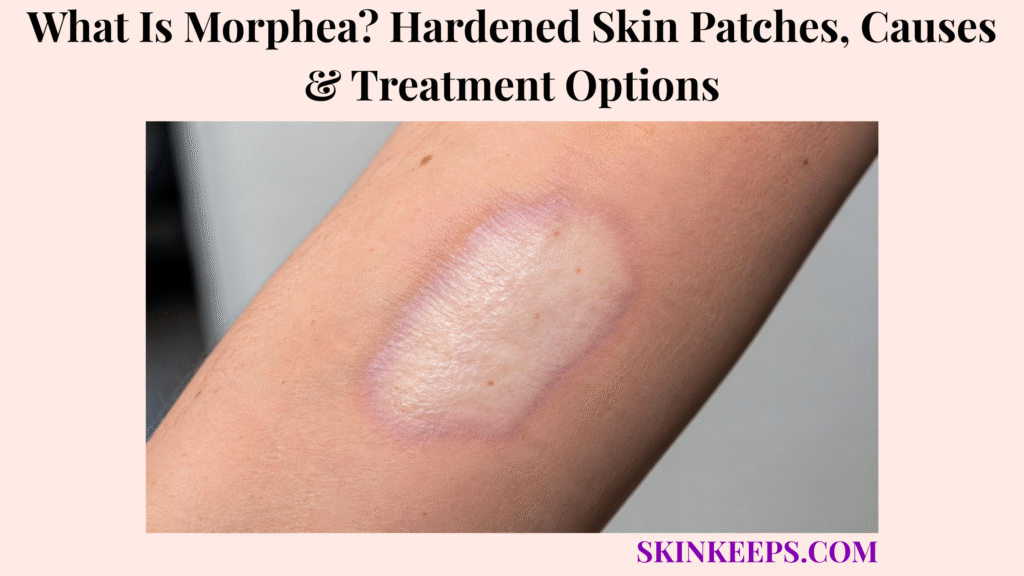
Morphea, also called localized scleroderma, is an inflammatory and fibrosing disorder that causes localized areas of skin to become thickened, hardened and less flexible. Inflammation activates fibroblasts to produce excess collagen, so an early pink or purple patch can progressively become indurated, sclerotic and bound down.
Some morphea remains limited to superficial plaques, while linear, generalized or deep disease can extend into subcutaneous fat, fascia, muscle, joints or bone. Early recognition matters most when disease crosses a joint, affects a growing limb or involves the face or scalp because active fibrosis can leave permanent atrophy, contracture or asymmetry.
This article is for educational purposes only and does not constitute medical advice, diagnosis, or treatment. Rapidly expanding or increasingly hardened skin patches, linear lesions crossing a joint, progressive limb asymmetry, restricted movement, deep pain, facial or scalp involvement, significant tissue loss, or new neurological, visual, or other unusual symptoms should be evaluated by a licensed dermatologist or qualified healthcare professional.
How Can You Recognize Morphea?
Morphea often progresses from an early pink or violaceous inflammatory patch into firmer, bound-down sclerosis and later residual pigmentation or atrophy.
What Does Early Active Morphea Look Like?
Early active morphea may appear as a pink, red-purple or bruise-like patch before obvious skin hardening develops.
The lesion may expand at its edge while the centre becomes firmer. A violaceous or lilac-coloured rim can mark ongoing inflammation, although not every active lesion displays a visible lilac ring.
What Does a Mature Morphea Plaque Look and Feel Like?
A mature morphea plaque typically feels hard, thickened, less flexible and bound down compared with surrounding skin.
The surface may look smooth or shiny and can develop an ivory centre or altered pigmentation. Local follicular damage can reduce hair growth or sweating within the plaque.
What Happens After Active Morphea Settles?
Inactive morphea may soften but leave permanent hyperpigmentation, hypopigmentation, skin thinning or deeper tissue atrophy.
Fat loss can leave a visible depression, and deeper disease may leave muscle or bone asymmetry. Residual appearance does not automatically mean inflammation is still active.
Is Morphea Itchy or Painful?
Morphea may cause little discomfort, but active or deeper disease can cause itching, pain, tingling or movement restriction.
Superficial plaques can be asymptomatic. Deep fibrosis affecting fascia, muscle, nerves or joints can cause pain even when the surface colour looks modest.
What Does Linear Morphea Look Like?
Linear morphea forms a band of abnormal hardened skin that can extend deeply along an arm, leg, trunk or craniofacial region.
This subtype is especially important in children because it can cross joints and involve muscle or bone in a growing limb, leading to restricted motion, contracture or limb asymmetry.
What Is Morphea En Coup de Sabre?
En coup de sabre is craniofacial linear morphea that typically forms a depressed or sclerotic band across the forehead or scalp.
Deeper involvement can affect subcutaneous fat, muscle, bone and scalp follicles, producing permanent alopecia or facial asymmetry. Neurological or ocular abnormalities occur in some patients and justify specialist evaluation.
Figure 1. Morphea may begin as inflammatory colour change, progress through active sclerosis and later leave pigmentation or atrophy; deep extension creates a separate structural-damage pathway.
Why Does Morphea Develop, and Which Types Matter Most?
Morphea appears to result from immune dysregulation and excessive collagen production, while its clinical severity depends strongly on subtype, depth and anatomical location.
What Causes Morphea?
The exact initiating cause of morphea is unknown, but current evidence links it to genetic susceptibility, immune dysregulation, vascular changes and excessive fibroblast-driven collagen production.
A useful mechanism is susceptibility → inflammation → fibroblast activation → excess collagen → fibrosis. No single trigger explains every case.
Can Injury or Radiation Trigger Morphea?
Local trauma, friction, surgery or radiotherapy may trigger morphea in susceptible people, but these events do not explain every case.
The association is thought to involve localized inflammatory signalling after tissue injury. Morphea is not contagious.
Is Morphea an Autoimmune Disease?
Morphea has strong autoimmune features, although no single autoantibody defines or proves the diagnosis.
Autoantibodies can be detected in some patients, and other autoimmune diseases may coexist, but laboratory positivity is not required for morphea.
What Is Limited or Plaque Morphea?
Limited plaque morphea usually causes one or a few oval areas of superficial sclerosis and generally carries less functional risk than deep or linear disease.
It is a common adult pattern and often remains mainly cutaneous. It can still leave long-lasting pigmentation or texture change.
What Is Generalized Morphea?
Generalized morphea affects multiple body regions and can form numerous or confluent areas of sclerosis.
The wider disease burden can make treatment more demanding, and deeper extension may occur. Severity should therefore not be judged by visible surface area alone.
Why Does the Depth of Morphea Matter?
Morphea depth matters because fibrosis can extend beyond the skin into fat, fascia, muscle, joints and bone, where permanent functional damage becomes more likely.
The practical sequence is skin → fat → fascia → muscle → joint → bone. Increasing depth raises the risk of pain, atrophy, contracture, restricted motion and growth disturbance.
| Pattern | Typical Distribution | Depth / Main Risk | Main Monitoring Concern |
|---|---|---|---|
| Limited / plaque | One or a few plaques | Mainly cutaneous damage | Activity and residual atrophy |
| Linear | Limb, trunk or head band | Joint, muscle, bone and growth risk | Range of motion and asymmetry |
| Generalized | Multiple body regions | Large burden ± deeper disease | Progression and treatment response |
| Deep disease | Variable | Fat, fascia, muscle, joint or bone | Functional impairment |
| En coup de sabre | Forehead / scalp | Craniofacial tissue risk | Alopecia, asymmetry, ocular/neurologic context |
How Is Morphea Distinguished and Diagnosed?
Morphea is usually diagnosed clinically, but assessment must also determine disease activity, depth and whether another sclerosing disorder better explains the findings.
How Is Morphea Different From Systemic Sclerosis?
Morphea causes localized skin and deeper-tissue fibrosis but does not progress into systemic sclerosis or scleroderma or produce its characteristic internal-organ fibrosis.
Morphea can extend locally into muscle, joints or bone without becoming systemic sclerosis. Systemic sclerosis instead has a distinct vascular and systemic pattern that can involve the fingers, lungs, heart, kidneys and gastrointestinal tract.
Which Skin Conditions Can Resemble Morphea?
Morphea can resemble inflammatory, annular, atrophic or deeper fibrosing disorders depending on its stage and tissue depth.
Granuloma annulare can mimic early annular inflammatory morphea but does not produce the same progressive bound-down sclerosis.
Lichen sclerosus can resemble pale atrophic morphea but has its own characteristic morphology and genital disease context.
Erythema migrans or Lyme disease skin rash is an expanding Borrelia-associated rash rather than a fibrosing plaque.
Deep fibrosing disease can also require distinction from eosinophilic fasciitis.
Can Morphea Usually Be Diagnosed From the Skin Examination?
Yes; typical morphea is diagnosed mainly from lesion colour, firmness, distribution, progression and estimated tissue depth.
Clinical examination should include palpation, subtype recognition and functional assessment. Marking lesion borders, serial photographs and documenting range of motion can make progression easier to identify.
When Is a Skin Biopsy Needed?
Skin biopsy is most useful when morphea is clinically uncertain or another inflammatory or fibrosing disorder needs to be excluded.
Histology can show collagen alteration, inflammation and adnexal entrapment or loss. Biopsy depth should match the suspected disease depth rather than assuming a superficial sample answers every deep-tissue question.
Do Blood Tests Diagnose Morphea?
No; there is no single blood test or autoantibody that establishes a diagnosis of morphea.
ANA and other autoantibodies can be positive, but testing is guided by disease severity, extracutaneous symptoms and alternative autoimmune concerns. Routine broad autoantibody testing solely to prove morphea is not the diagnostic model.
Routine Borrelia testing is also not recommended without an independent clinical reason.
When Is MRI Useful in Morphea?
MRI is useful when morphea may extend into deeper tissues or when craniofacial disease raises concern about structures beneath the skin.
For extremity disease, MRI can assess muscle, joints and bone when deep or extensive involvement is suspected. Expert review guidance supports special MRI consideration in craniofacial disease because intracranial abnormalities can occur even when neurological symptoms are limited.
MRI is not routine for every superficial plaque.
Why Must Active Morphea Be Separated From Permanent Damage?
Active morphea requires treatment to stop progression, whereas established atrophy or pigmentation may represent residual damage after inflammation has already become inactive.
Expanding erythema, a violaceous border, increasing induration or new lesions suggest activity. Longstanding pigmentation, thinning, fixed asymmetry, fat loss or permanent alopecia can represent established damage.
The practical rule is treat inflammation—not colour alone.
Figure 2. Morphea assessment moves from morphology to disease activity, subtype, depth and function, with biopsy or MRI added only when the clinical question requires them.
How Is Morphea Treated?
Morphea treatment is chosen according to disease activity, depth, subtype and functional risk, with the main goal of stopping inflammation before irreversible fibrosis develops.
What Is the Main Goal of Morphea Treatment?
The main treatment goal is to suppress active inflammation and prevent additional fibrosis before permanent tissue damage occurs.
Treatment can stop progression, but it may not fully restore destroyed fat, muscle, bone, hair follicles or a fixed contracture. Early treatment matters most in linear, deep, facial and joint-crossing disease.
How Is Limited Superficial Morphea Treated?
Active limited superficial morphea can often be treated with local therapy such as topical corticosteroids.
Selected topical tacrolimus or other local therapies can also be considered. Emollients can reduce dryness or itch but do not suppress significant active fibrosis by themselves.
When Is Phototherapy Used for Morphea?
Phototherapy is mainly used for superficial or more widespread cutaneous morphea when light can adequately reach the active tissue.
Options include narrowband UVB and UVA1, with UVA1 penetrating more deeply into skin. Phototherapy alone is less appropriate when rapidly progressive disease threatens deep tissue, joints or craniofacial structures.
When Is Methotrexate Used for Morphea?
Methotrexate is a guideline-supported systemic treatment for severe, active linear, generalized or deep morphea with meaningful risk of functional or structural damage.
This includes severe skin disease, musculoskeletal involvement and potentially disfiguring or disabling juvenile disease. Treatment belongs under specialist supervision rather than self-directed dosing.
Why Are Systemic Corticosteroids Sometimes Added?
Systemic corticosteroids may be added temporarily during rapidly active severe morphea to suppress inflammation while a slower-acting treatment such as methotrexate takes effect.
This is a bridge strategy for active progression. Systemic corticosteroids should not be presented as preferred long-term monotherapy.
What If Methotrexate Is Not Effective or Cannot Be Used?
Mycophenolate mofetil is an established systemic alternative for selected patients who do not respond to, tolerate or qualify for methotrexate.
Other biologic or targeted approaches remain specialist or emerging options and should not be placed on equal footing with guideline-established first-line therapy.
Why Are Physical and Occupational Therapy Important?
Physical and occupational therapy help preserve joint movement and reduce contracture when morphea involves deeper tissue or crosses a joint.
Range-of-motion work and rehabilitation complement medical treatment. Stopping inflammation does not guarantee reversal of an already fixed contracture.
| Disease Pattern | Main Treatment Logic |
|---|---|
| Limited superficial active disease | Topical therapy |
| Widespread superficial disease | Phototherapy ± systemic assessment |
| Active linear / generalized / deep disease | Methotrexate-based systemic treatment |
| Rapid severe progression | Methotrexate + temporary systemic corticosteroid where appropriate |
| Methotrexate unsuitable or ineffective | Selected systemic alternative such as mycophenolate |
| Joint / deep involvement | Medical therapy + physical / occupational therapy |
What Problems Can Morphea Cause, and How Should It Be Followed?
Morphea can become inactive but still leave permanent structural damage, and some patients experience relapse that requires renewed assessment of activity and function.
Does Morphea Eventually Stop Progressing?
Many milder morphea cases eventually become inactive, but the disease can remain active for years or relapse after treatment.
Relapse is especially relevant in childhood-onset, linear and generalized disease. Potentially disabling active disease should not simply be left untreated in the hope that it will eventually stop.
Can Morphea Leave Permanent Skin Changes?
Yes; morphea can leave permanent pigmentation changes, skin thinning, fat loss, depressions or alopecia after active inflammation stops.
Hyperpigmentation, hypopigmentation, atrophy and asymmetry may remain even after activity is controlled. Complete normalization cannot always be achieved.
Can Linear Morphea Affect Growth or Joint Movement?
Yes; deep linear morphea in children can restrict joint movement and interfere with normal muscle or bone growth.
Possible consequences include limb-length or circumference differences, contracture and mobility loss. Joint-crossing disease therefore receives more aggressive assessment and treatment than one small superficial plaque.
Which Complications Can Craniofacial Morphea Cause?
Craniofacial morphea can cause scalp hair loss, soft-tissue loss, facial asymmetry and underlying bone involvement, with neurological or ocular abnormalities in some patients.
Possible manifestations include headache, seizures, visual problems or dental/maxillofacial effects, but not every patient with en coup de sabre develops these complications.
Does Morphea Damage the Heart, Lungs or Kidneys?
Morphea does not progress to systemic sclerosis or produce the characteristic heart, lung, kidney or gastrointestinal fibrosis associated with systemic sclerosis.
It can still cause important localized extracutaneous complications, especially musculoskeletal, neurological and ocular problems.
When Should Morphea Be Reassessed Promptly?
Morphea needs earlier reassessment when lesions continue expanding, new inflammatory borders appear, movement declines, deep pain develops or facial, scalp, eye or neurological symptoms emerge.
New plaques, increasing induration, declining range of motion, limb asymmetry, visual symptoms, severe headache or seizures can indicate activity or complications that change treatment intensity.
What Should You Remember About Morphea?
Morphea is localized scleroderma that can remain superficial or extend into deeper tissues, and the most important management questions are whether disease is active, how deep it extends and whether permanent functional damage is developing.
- Morphea and localized scleroderma describe the same disease spectrum.
- Morphea is distinct from systemic sclerosis.
- Early lesions may be pink or violaceous.
- A lilac border can indicate activity.
- Mature disease becomes hard and bound down.
- Inactive disease can leave pigment change and atrophy.
- Linear disease can affect deeper tissues and growing limbs.
- En coup de sabre affects the craniofacial region.
- Disease depth matters as much as visible surface area.
- Diagnosis is usually clinical.
- Biopsy is selective.
- MRI is useful for relevant deep or craniofacial disease.
- No single blood test diagnoses morphea.
- Treatment targets active inflammation.
- Limited superficial disease can use local therapy.
- Severe active linear, generalized or deep disease often needs systemic treatment.
- Rehabilitation matters when joints are involved.
- Permanent damage may remain after inflammation stops.
- Relapse can occur.
Recognize activity → determine subtype and depth → distinguish systemic sclerosis → treat according to damage risk → preserve function → monitor relapse.
Frequently Asked Questions About Morphea
The most important morphea questions concern its cause, distinction from systemic sclerosis, depth, reversibility and treatment of active disease.
What Causes Morphea to Develop?
The exact cause of morphea is unknown, but immune dysregulation, genetic susceptibility, vascular changes and excessive collagen production appear to contribute. Fibroblast activation drives the fibrosis that hardens affected tissue.
Is Morphea the Same as Systemic Scleroderma?
No; morphea is localized scleroderma and does not progress into systemic sclerosis or cause its characteristic internal-organ fibrosis. Morphea can still extend deeply within a localized anatomical region.
Can Morphea Spread Deeper Than the Skin?
Yes; morphea can extend into subcutaneous fat, fascia, muscle, joints and bone, particularly in linear or deep disease. Greater depth raises the risk of structural and functional damage.
Can Hardened Skin From Morphea Become Normal Again?
Some skin hardness can improve when active morphea is controlled, but established atrophy, tissue loss, contracture or destroyed hair follicles may be permanent. Activity and fixed damage therefore need to be separated.
What Treatments Are Used for Active Morphea?
Active morphea treatment ranges from topical therapy for limited superficial disease to phototherapy or systemic treatment such as methotrexate for more extensive, deep or function-threatening disease. Rehabilitation is added when movement or joints are affected.
Which Sources Support This Morphea Guidance?
2024 S2k Guideline — Diagnosis and Therapy of Localized Scleroderma — Primary guidance source for localized-scleroderma terminology, lack of progression to systemic sclerosis, depth, topical corticosteroids, phototherapy, methotrexate, systemic glucocorticoids and second-line systemic treatment.
DermNet — Morphoea / Localised Scleroderma — Used for inflammatory morphology, lilac border, mature sclerosis, pigmentation and atrophy, linear/generalized disease, depth and natural history.
DermNet — Morphoea En Coup de Sabre — Used specifically for craniofacial linear morphea, scalp/forehead involvement, alopecia, soft-tissue and bone involvement and neurological/visual complications.
Morphea: The 2023 Update — Used for pathogenesis, autoantibody limitations, Borrelia-testing boundary, MRI of deep extremity disease, craniofacial MRI context and activity-versus-damage assessment.
American Academy of Dermatology — Scleroderma: Diagnosis and Treatment — Used for patient-facing treatment context, local treatment, phototherapy and physical/occupational therapy principles.
SHARE Recommendations — Juvenile Localised Scleroderma — Used for systemic treatment of active potentially disabling or disfiguring juvenile disease, methotrexate, temporary systemic corticosteroids, UVA1 and mycophenolate.
Mycophenolate for Morphea — Multicenter Cohort Study — Used specifically for mycophenolate as a selected alternative in severe, recalcitrant, methotrexate-intolerant or contraindicated disease.



