
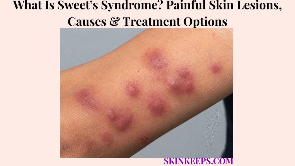
Sweet’s syndrome, also called acute febrile neutrophilic dermatosis, is a rare inflammatory skin disorder in which neutrophils accumulate in the skin and cause the sudden appearance of painful swollen plaques, nodules or blister-like lesions. Fever, malaise, joint pain, eye inflammation or other systemic symptoms may occur, but the lesions themselves are driven by sterile inflammation rather than ordinary bacterial invasion.
The condition can arise without a clear cause, follow infection or inflammatory disease, accompany malignancy, or be triggered by a medicine. Because Sweet’s syndrome can resemble cellulitis, abscesses and other painful eruptions, diagnosis relies on the clinical pattern together with skin biopsy and exclusion of infection; treatment usually controls inflammation while clinicians also address any associated trigger or underlying disease.
This article is for educational purposes only. Sudden painful skin lesions with persistent fever, eye symptoms, ulceration, significant systemic illness or abnormal blood findings should be medically evaluated.
What Is Sweet’s Syndrome and What Do Its Skin Lesions Look and Feel Like?
Sweet’s syndrome is a sterile neutrophilic dermatosis that usually begins abruptly with painful swollen plaques, nodules or pseudovesicular lesions, often accompanied by fever or malaise.
What Happens in the Skin During Sweet’s Syndrome?
Abnormal inflammatory signaling recruits large numbers of neutrophils into the dermis, producing intense inflammation, tissue swelling and tender skin lesions.
The exact molecular pathway is not fully settled, but cytokine dysregulation and abnormal neutrophil activation are central to current disease models. This produces a dense inflammatory infiltrate without making Sweet’s syndrome a routine bacterial skin infection.
Why Is It Called Acute Febrile Neutrophilic Dermatosis?
The name describes its typical pattern: abrupt onset, frequent fever, neutrophil-rich inflammation and prominent skin disease.
“Acute” refers to sudden development, “febrile” to the common association with fever, “neutrophilic” to the white blood cells that accumulate in affected tissue, and “dermatosis” to the skin manifestation.
What Do Sweet’s Syndrome Lesions Look Like?
Typical Sweet’s syndrome lesions appear suddenly as tender swollen papules, nodules or plaques that may look red, pink, purple, violaceous or dusky and can sometimes resemble blisters or pustules.
The face, neck and upper extremities are common sites, although lesions can develop elsewhere. Some eruptions are few and discrete, while others are numerous or form larger edematous plaques.
Why Can Sweet’s Syndrome Look Blistered?
Marked swelling in the upper dermis can create a pseudovesicular surface that looks blistered even when the central process is neutrophilic inflammation rather than a classic blistering disease.
True vesicles, pustules, bullous lesions, erosions or ulcers can occur in selected variants, so morphology alone is not enough to confirm the diagnosis.
What Symptoms Can Occur With the Skin Lesions?
Fever, malaise, headache, aching joints or muscles, eye inflammation and mouth ulcers can accompany the cutaneous eruption.
Sweet’s syndrome can occasionally involve organs outside the skin, which is one reason substantial systemic symptoms or eye complaints should not be dismissed as part of an ordinary rash.
How Can Sweet’s Syndrome Look on Darker Skin?
On darker skin, Sweet’s syndrome may appear purple, violaceous, dusky, dark brown or subtly red rather than bright red, and post-inflammatory pigment change can be more noticeable after the eruption settles.
Persistent dark discoloration after inflammation is better understood in the context of hyperpigmentation; the important distinction is that color change after Sweet’s syndrome reflects the aftermath of inflammation rather than the core neutrophilic disease itself.
What Causes Sweet’s Syndrome and Which Conditions or Medicines Can Be Associated With It?
Sweet’s syndrome can be classical or idiopathic, associated with malignancy or inflammatory disease, or triggered by a medication, so the broader clinical context matters as much as the skin eruption itself.
What Are the Three Main Clinical Types of Sweet’s Syndrome?
Sweet’s syndrome is commonly organized into classical or idiopathic, malignancy-associated and drug-induced forms.
These categories help clinicians decide what history, examinations or investigations are most relevant, but the categories can overlap with infection, pregnancy or inflammatory disease as contributing contexts.
Which Infections or Inflammatory Conditions Can Be Associated With Classical Sweet’s Syndrome?
Classical Sweet’s syndrome can occur after infections or alongside inflammatory disease, pregnancy or selected autoimmune conditions without the skin lesions themselves being infected.
Upper respiratory and gastrointestinal infections are recognized triggers, and inflammatory bowel disease is an important association. The correct mechanism is an inflammatory response to a trigger, not bacteria necessarily invading each Sweet’s lesion.
How Is Sweet’s Syndrome Associated With Cancer?
Sweet’s syndrome can occur with malignancy, particularly hematologic cancers such as acute myeloid leukemia, but the syndrome does not automatically mean cancer is present.
Malignancy-associated Sweet’s syndrome can appear before, during or after a cancer diagnosis. The importance of this association is that unexplained, recurrent or clinically atypical disease may justify broader evaluation rather than assuming every patient needs the same cancer work-up.
Which Medicines Can Trigger Sweet’s Syndrome?
Drug-induced Sweet’s syndrome has been associated with several medicines, with granulocyte colony-stimulating factor among the best-recognized triggers.
Other reported associations include trimethoprim-sulfamethoxazole, selected anti-inflammatory medicines, azathioprine and tretinoin, but a temporal association alone does not prove causality in every case.
Should a Suspected Triggering Medicine Be Stopped Immediately at Home?
No; suspected medication-related Sweet’s syndrome should be reviewed with the treating clinician because the implicated medicine may be essential for another serious condition.
Clinicians weigh the probability of drug causation, the medicine’s necessity, disease severity and available alternatives before making a treatment change. Rapidly progressive illness still requires medical assessment rather than unsupervised medication decisions.
How Is Sweet’s Syndrome Diagnosed and Distinguished From Infection or Other Painful Skin Disorders?
Sweet’s syndrome is diagnosed from its abrupt clinical pattern plus skin biopsy showing dense neutrophilic inflammation, while infection and other painful inflammatory disorders must be excluded.
Why Is Skin Biopsy Important?
Skin biopsy is a key diagnostic test because Sweet’s syndrome typically shows a dense dermal neutrophilic infiltrate with marked edema and classically without a primary leukocytoclastic vasculitis pattern.
Biopsy findings must still be interpreted with the clinical picture because neutrophil-rich inflammation can also occur in infection and other disorders. When infection is a serious possibility, clinicians may obtain appropriate cultures or other microbiologic testing alongside tissue evaluation.
What Blood Tests Can Support the Diagnosis?
Leukocytosis, neutrophilia and elevated inflammatory markers such as C-reactive protein or erythrocyte sedimentation rate can support the diagnosis but are not specific enough to confirm Sweet’s syndrome.
Some biopsy-confirmed patients do not have neutrophilia, particularly in malignancy-associated disease, so normal or atypical blood counts do not automatically exclude the diagnosis.
How Is Sweet’s Syndrome Different From Cellulitis?
Cellulitis is a bacterial infection of the skin and subcutaneous tissue, while Sweet’s syndrome is sterile neutrophilic inflammation that can mimic bacterial infection clinically.
Cellulitis is treated according to an infectious mechanism. Sweet’s syndrome instead requires confirmation of the inflammatory pattern and exclusion of infection before clinicians assume antibiotics are the correct primary treatment.
How Is Sweet’s Syndrome Different From a Skin Abscess?
A skin abscess is a localized pus-filled bacterial collection that often has a drainable cavity, whereas Sweet’s lesions are inflammatory plaques or nodules without the typical anatomy of a bacterial abscess.
The skin abscess page owns drainage-based management of a true pus collection. A tender nodule in Sweet’s syndrome should not be cut or drained merely because it is painful and swollen.
What Other Conditions Can Resemble Sweet’s Syndrome?
Pyoderma gangrenosum, erythema multiforme, erythema nodosum, drug eruptions, lupus, Behçet disease and selected infections can resemble part of the Sweet’s syndrome picture.
Pyoderma gangrenosum is another neutrophilic dermatosis, but it is particularly associated with painful ulcerative disease and has a different clinical management problem from the edematous plaques and nodules typical of Sweet’s syndrome.
Erythema multiforme is a separate inflammatory disorder characterized more by target or targetoid lesions, illustrating why lesion architecture and biopsy context matter more than simply grouping all red or purple eruptions together.
When Do Doctors Look for an Underlying Disease?
Further investigation becomes more important when Sweet’s syndrome is recurrent, severe, unexplained, associated with abnormal blood counts or accompanied by systemic features suggesting malignancy or inflammatory disease.
There is no single universally accepted investigation panel for every patient. Clinicians tailor additional testing to age, history, examination, blood abnormalities and the likelihood of specific inflammatory, malignant or medication-related associations.
How Is Sweet’s Syndrome Treated?
Sweet’s syndrome is usually treated with anti-inflammatory therapy—most often systemic corticosteroids for significant disease—while the associated trigger or underlying condition is addressed at the same time.
Why Are Systemic Corticosteroids the Main Treatment?
Systemic corticosteroids rapidly suppress the neutrophil-driven inflammatory response and often improve fever, pain and skin lesions within days.
They remain the usual first-line treatment for substantial disease, but dose, duration and tapering are individualized because severity, comorbidities and recurrence risk vary.
Can Localized Sweet’s Syndrome Use Topical or Injected Steroids?
Yes; selected localized lesions can be treated with potent topical or intralesional corticosteroids, while widespread or systemic disease usually requires broader therapy.
Local treatment is not automatically appropriate for every lesion, particularly when diagnosis is uncertain, lesions are extensive or extracutaneous disease is present.
What Can Be Used When Corticosteroids Are Unsuitable or Ineffective?
Specialists may use alternative anti-inflammatory or immunomodulatory treatments when corticosteroids are contraindicated, poorly tolerated or ineffective.
Reported options include colchicine, potassium iodide, dapsone, indomethacin, ciclosporin and selected biologic or targeted therapies. Evidence quality varies, so these treatments should not be presented as interchangeable self-treatment alternatives.
Why Must the Underlying Trigger Also Be Treated?
Sweet’s syndrome management is more complete when the associated trigger is addressed, whether that means controlling inflammatory disease, treating an underlying malignancy or reviewing a causative medicine.
Skin-directed treatment alone may suppress the eruption without resolving the factor that helped provoke it. This is especially relevant in malignancy-associated or drug-induced disease and when episodes recur.
Are Antibiotics the Standard Treatment?
No; antibiotics do not treat the core sterile neutrophilic inflammation of Sweet’s syndrome and are reserved for a true concurrent or secondary bacterial infection.
Because fever, high neutrophil counts and tender erythematous plaques can mimic infection, clinicians may initially investigate both possibilities. Once Sweet’s syndrome is established, reflex antibiotic treatment without infectious evidence is not the treatment model.
Can Sweet’s Syndrome Come Back, and When Does It Need Further Medical Evaluation?
Sweet’s syndrome can recur, and recurrent or unusually severe episodes may require reassessment for persistent inflammation, medication exposure, malignancy or another underlying association.
Does Sweet’s Syndrome Usually Heal Without Scarring?
Many Sweet’s syndrome lesions heal without permanent scarring, although temporary discoloration may persist and ulcerated or otherwise complicated lesions can leave scars.
Residual color change may last for months after the active inflammation has settled, especially in darker skin, even when the inflammatory disease itself is no longer active.
Can Sweet’s Syndrome Recur?
Yes; recurrence is well recognized and can occur in idiopathic, drug-associated and malignancy-associated disease.
DermNet notes that repeated episodes occur in roughly half of patients overall, with recurrence particularly important in some idiopathic and cancer-associated cases. The figure is a population estimate, not a prediction for an individual patient.
Why Does Recurrence Need Reassessment?
A recurrent episode may indicate persistent inflammatory disease, re-exposure to a triggering medicine, ongoing malignancy or an association that was not identified during the first episode.
Recurrence therefore deserves more than cosmetic retreatment of the skin. The timing, medication history, blood counts, systemic symptoms and status of known associated disease may all need to be reviewed again.
When Should Suspected Sweet’s Syndrome Be Assessed Promptly?
Prompt assessment is appropriate for sudden painful plaques with fever, rapidly increasing lesions, ulceration or blistering, eye symptoms, persistent systemic illness, abnormal blood findings or recurrent Sweet-like episodes.
Particular concern is warranted when the eruption occurs during cancer treatment, follows introduction of a possible culprit medicine, involves eye pain or visual symptoms, or is accompanied by high fever and rapidly worsening systemic illness.
Can Secondary Infection Occur on Top of Sweet’s Syndrome?
Yes; although Sweet’s syndrome itself is sterile, damaged or ulcerated lesions can develop a separate bacterial infection that requires its own diagnosis and treatment.
The correct sequence is to treat the inflammatory eruption, confirm healing, identify relevant associations, monitor for recurrence and reassess the underlying context when the course no longer fits an uncomplicated episode.
What Should You Remember About Sweet’s Syndrome?
Sweet’s syndrome is a rare sterile neutrophilic dermatosis that causes abrupt painful inflammatory lesions and often fever, with biopsy, association assessment and anti-inflammatory treatment forming the core management pathway.
- Sweet’s syndrome is also called acute febrile neutrophilic dermatosis.
- The core process is sterile neutrophilic inflammation, not routine bacterial invasion.
- Cytokine dysregulation and abnormal neutrophil recruitment are central to current disease models.
- Onset is often sudden and lesions are characteristically painful or tender.
- Papules, plaques and nodules may look swollen, pseudovesicular, pustular or occasionally bullous.
- Fever, malaise, joint pain, eye inflammation and mouth ulcers can accompany the skin eruption.
- Darker skin may show purple, dusky or dark-brown lesions rather than bright redness.
- Classical or idiopathic, malignancy-associated and drug-induced forms are recognized.
- Infection can trigger Sweet’s syndrome without making the skin lesions themselves infected.
- Acute myeloid leukemia is an important association, but Sweet’s syndrome does not automatically mean leukemia.
- G-CSF is one of the best-recognized medication triggers.
- Suspected medication causation should be reviewed by the treating clinician rather than managed by unsupervised drug withdrawal.
- Skin biopsy is central to diagnosis.
- Neutrophilia, CRP and ESR can support the diagnosis but are not specific.
- Cellulitis and abscesses are infectious mimics with different treatment logic.
- Pyoderma gangrenosum and erythema multiforme are relevant noninfectious differentials.
- Systemic corticosteroids are usual first-line therapy for significant disease.
- Selected localized disease can use topical or intralesional corticosteroids.
- Specialist steroid-sparing or immunomodulatory alternatives exist when needed.
- Antibiotics are not standard therapy for the sterile inflammatory process.
- Any underlying malignancy, inflammatory disease or drug trigger should be addressed appropriately.
- Many lesions heal without permanent scars, although pigment change can remain.
- Recurrence can occur and may justify renewed assessment of the underlying association.
Frequently Asked Questions About Sweet’s Syndrome
The main Sweet’s syndrome questions concern infection, cancer association, painful lesion formation, corticosteroid response and recurrence.
Is Sweet’s Syndrome an Infection?
No; Sweet’s syndrome is a sterile neutrophilic inflammatory disorder, although an infection elsewhere in the body can sometimes trigger it.
Is Sweet’s Syndrome Associated With Leukemia or Other Cancers?
Yes; Sweet’s syndrome can occur with hematologic malignancies such as acute myeloid leukemia, but many people with Sweet’s syndrome do not have cancer.
Why Does Sweet’s Syndrome Cause Painful Red or Purple Skin Lesions?
Dense neutrophilic inflammation and dermal swelling produce the tender plaques and nodules characteristic of Sweet’s syndrome.
How Quickly Can Sweet’s Syndrome Respond to Corticosteroid Treatment?
Many significant cases improve markedly within days after appropriate corticosteroid therapy, although response depends on severity and the underlying association.
Can Sweet’s Syndrome Come Back After Treatment?
Yes; recurrence can occur and may justify renewed evaluation for an inflammatory disease, medicine trigger, malignancy or another associated condition.
DermNet — Acute febrile neutrophilic dermatosis (Sweet syndrome). Supports current terminology, sterile neutrophilic mechanism, morphology, darker-skin presentation, clinical subtypes, biopsy findings, treatment options and recurrence.
British Association of Dermatologists — Sweet syndrome. Supports noninfectious framing, painful red/purple lesions, systemic symptoms, malignancy and medication associations, biopsy, corticosteroid treatment and typical healing without scars.
Cleveland Clinic — Sweet Syndrome. Supports sudden painful rash and fever, common associations, clinician-directed medication review, treatment of underlying disease and recurrence context.
Nelson et al. — Neutrophilic Dermatoses: a Clinical Update. Supports Sweet’s syndrome as a sterile neutrophilic dermatosis, diagnostic criteria, associated-disease assessment, corticosteroid-first treatment and individualized steroid-sparing therapy.



